
Condition
Nephritic Syndrome
Nephritic syndrome is inflammation of the kidney's glomeruli causing blood in the urine, high blood pressure, and reduced kidney function.

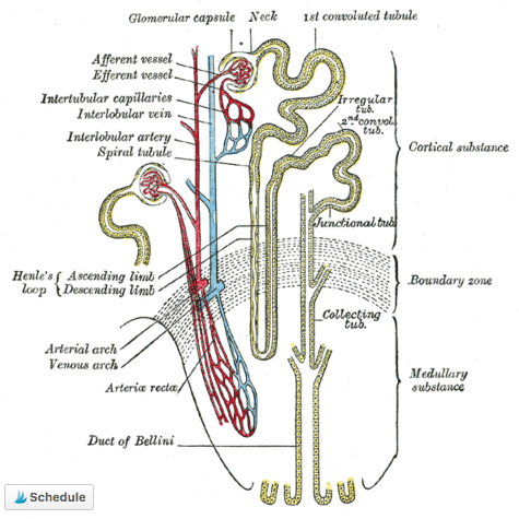
Overview
Aetiology
Nephritic syndrome is the clinical picture produced by inflammation of the glomeruli. The distinction from nephrotic syndrome is the most useful thing to hold onto: nephritic disease is inflammatory and the glomerulus leaks blood; nephrotic disease is a defect of the filtration barrier and it leaks protein.
The mechanism is immune-mediated. Immune complexes deposit in the glomerulus — or form there in situ — and activate complement. Neutrophils and macrophages are recruited, and the inflammatory response damages the capillary wall, letting red cells escape into the urinary space. As they pass down the tubule they are deformed and moulded into red cell casts, which are the pathognomonic finding. Inflammatory proliferation simultaneously narrows the capillary lumen, so the glomerular filtration rate falls — producing oliguria, sodium and water retention, hypertension and edema, and a rising urea and creatinine.
The causes divide usefully by age and by complement level:
- Post-infectious glomerulonephritis — classically follows a group A streptococcal throat or skin infection by one to three weeks. The commonest cause in children, and complement C3 is low.
- IgA nephropathy (Berger’s disease) — the commonest primary glomerulonephritis worldwide. Haematuria appears during an upper respiratory infection rather than weeks after it, which distinguishes it from post-infectious disease. Complement is normal.
- Lupus nephritis — with other features of SLE; both C3 and C4 are low.
- ANCA-associated vasculitis — granulomatosis with polyangiitis, microscopic polyangiitis. Often with systemic and pulmonary features.
- Anti-GBM disease (Goodpasture’s) — antibodies against the glomerular basement membrane, frequently with pulmonary haemorrhage.
- Membranoproliferative glomerulonephritis, and Henoch-Schönlein purpura, the systemic form of IgA disease.
Rapidly progressive glomerulonephritis is the term for nephritic syndrome with renal function deteriorating over days to weeks, with crescents on biopsy. It is a nephrological emergency: without prompt immunosuppression the damage becomes irreversible.
Presentation
Symptoms
The classic pentad is haematuria, hypertension, edema, oliguria and azotaemia.
Haematuria is the presenting feature and is characteristically smoky, tea- or cola-coloured rather than bright red, reflecting its glomerular origin. Edema is typically periorbital and worst on waking, in contrast to the dependent ankle edema of other causes, and it reflects salt and water retention rather than the low albumin of nephrotic syndrome. Hypertension is common and may be severe enough to cause headache, visual disturbance or encephalopathy.
Urine output falls. Non-specific features — malaise, nausea, anorexia and flank ache from capsular stretch — are usual. Where the cause is systemic, its own features are present: rash, arthralgia, haemoptysis, sinus disease or fever.
Assessment
Diagnostic Considerations
Urinalysis is the decisive investigation and should be done immediately. Dysmorphic red cells and red cell casts on microscopy establish a glomerular origin and separate nephritic syndrome from every other cause of blood in the urine. Proteinuria is present but usually modest — below 3.5 g per day, above which the picture becomes nephrotic.
Blood tests establish severity and cause: urea, creatinine and eGFR; electrolytes; full blood count; and albumin. The serological panel is directed by the differential — complement C3 and C4 (low C3 with normal C4 suggests post-infectious or C3 glomerulopathy; both low suggests lupus), ANA and anti-dsDNA, ANCA, anti-GBM antibodies, antistreptolysin O titre and anti-DNase B, cryoglobulins, and hepatitis B and C serology. Renal ultrasound assesses size and excludes obstruction.
Renal biopsy is the definitive investigation and is indicated in most adults, and in children where the course is atypical, function is declining, or complement stays low beyond eight weeks. It establishes the diagnosis, grades activity against chronicity, and determines treatment — and in rapidly progressive disease it should not be delayed.
Assessment
Differential Diagnosis
- Nephrotic syndrome — heavy proteinuria, hypoalbuminaemia, no red cell casts
- Urinary tract infection — dysuria, pyuria, no casts
- Urinary tract malignancy — painless visible haematuria in an older adult
- Renal or ureteric calculus — colicky loin-to-groin pain
- Pre-renal acute kidney injury from hypovolaemia
- Obstructive uropathy — hydronephrosis on ultrasound
- Interstitial nephritis — often drug-induced, with white cell casts and eosinophilia
- Benign familial haematuria or exercise-induced haematuria
Context
Comorbidities
Hypertension and acute kidney injury are intrinsic to the syndrome, and fluid overload may progress to pulmonary edema. Post-infectious glomerulonephritis in children usually resolves completely; IgA nephropathy, lupus nephritis and vasculitis carry a real risk of progression to chronic kidney disease and end-stage renal failure, and require long-term follow-up of blood pressure, proteinuria and renal function. Where systemic disease is the cause, its extrarenal manifestations — pulmonary, cutaneous, articular — need managing alongside, and immunosuppressive treatment brings its own infection risk.