
Condition
Kidney Stones
Kidney stones are hard mineral deposits that form in the kidneys when excretory chemicals become too concentrated or fall out of solution.

Overview
Aetiology
Excretory chemicals can fall out of solution if the pH changes too much, or becomes too concentrated to stay in solution. This can occur due to metabolic conditions like hyperparathyroidism, gout, or sarcoidosis.
Hypertension, hypercalcemia, hyperparathyroidism, hyperoxaluria, and phosphaturia are also known risk factors.
In industrialized countries, the majority of kidney stones are composed of calcium salts 2Reference 2Kidney stones, while the remainder involve uric acid, struvite, or cystine. Stone formation is promoted by a combination of increased urinary calcium and reduced water intake, which concentrates the urine further.
Increased Uric Acid Concentrations
Uric acid levels rise with high alcohol intake, which both competes with uric acid for elimination through the kidneys and supplies purines that are metabolized into uric acid. A high intake of animal protein has a similar effect, increasing uric acid production through its purine and sulfate load.
Increased Calcium Concentrations
A fall in blood pH from acidic metabolites such as uric, lactic, and acetic acid prompts the parathyroid to release calcium and phosphate from bone to buffer the blood, raising circulating calcium. Calcium concentrations are pushed higher by reduced renal calcium reabsorption, increased absorption from the digestive tract (which may be genetic or dietary), and reduced biliary excretion with reabsorption of excreted calcium 3Reference 3Impaired bile flow and disordered hepatic calcium homeostasis are early features of halothane‐induced liver injury in guinea pigs. Lack of weight-bearing exercise lowers calcium storage in bone, while perimenopause increases osteoclast activity, and dehydration concentrates calcium in the blood and urine. Parathyroid or thyroid tumors and metabolic syndrome can also contribute, and uric acid salts out calcium, increasing the likelihood of both uric acid and calcium stones 6Reference 6In vitroDissolved urate salts out calcium oxalate in undiluted human urine in vitro: implications for calcium oxalate stone genesis.
Increased Oxalate Intake
Oxalate handling is impaired by a deficiency of the gut bacterium Oxalobacter formigenes, which normally dissolves dietary oxalate (Bone & Mills, 2013).
Presentation
Symptoms
The classic presentation is renal colic — severe, cramping pain in the flank that radiates to the groin and comes in waves as the stone moves. It is often accompanied by blood in the urine, nausea and vomiting, urinary urgency, and pain on urination. Small stones may pass unnoticed, while a stone that obstructs the ureter can cause intense pain and, if infection develops, fever.
Assessment
Diagnostic Considerations
- Kidney ultrasound
- Kidney function testing
- Parathyroid function testing
- Vitamin D testing
- Blood pH testing
- Genetic testing for predispositions of mineral transport changes
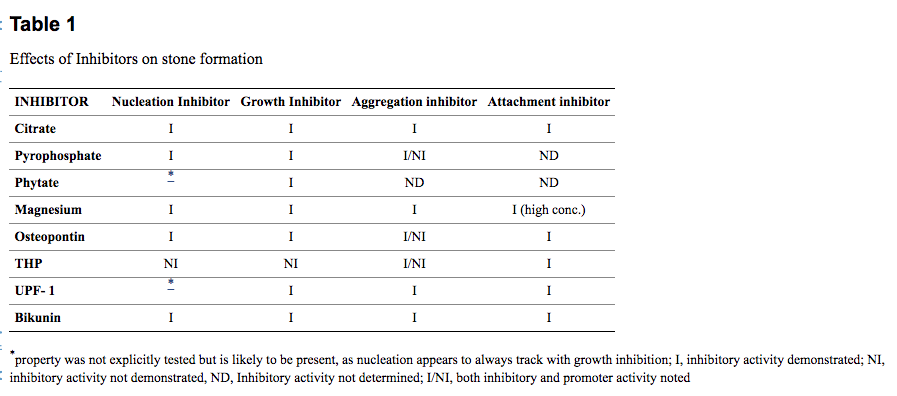
Inhibitors of Kidney Stone Formation
Several urinary factors normally inhibit crystallization and help prevent stones, most importantly citrate and magnesium, along with adequate fluid intake to keep the urine dilute.
Assessment
Differential Diagnosis
- Kidney infection
- Polycystic Kidney Disease (PKD)
- Kidney cancer
- Hypercalcemia
- Tumors on the parathyroid or thyroid gland
- Hyperparathyroidism
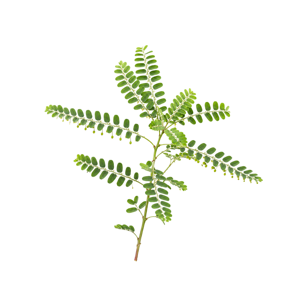
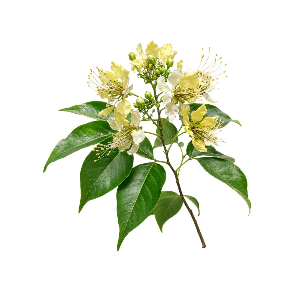
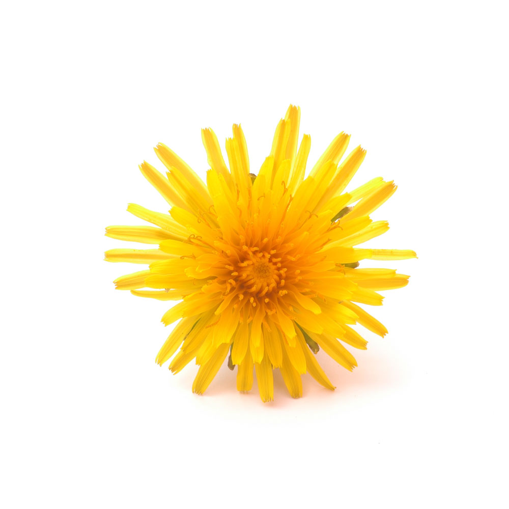
Herbs for Kidney Stones
- Verbena officinalis
- Lithospermum officinale
- Taraxacum officinale
- Arctium lappa
- Silene saxifraga
- Crataeva nurvala
- Cascara sagrada
- Yellow dock
The Herbs
Materia Medica (5)
Herbs traditionally or clinically used for kidney stones.
References:
- Sarris, J., & Wardle, J. (2014). Clinical naturopathy: an evidence-based guide to practice. Elsevier Health Sciences.
- Pak, C. Y. (1998). Kidney stones. The lancet, 351(9118), 1797-1801.
- Frost, L., Mahoney, J., Field, J., & Farrell, G. C. (1996). Impaired bile flow and disordered hepatic calcium homeostasis are early features of halothane‐induced liver injury in guinea pigs. Hepatology, 23(1), 80-86.
- Jilka, R. L., Hangoc, G., Girasole, G., Passeri, G., Williams, D. C., Abrams, J. S., & Manolagas, S. C. (1992). Increased osteoclast development after estrogen loss: mediation by interleukin-6. Science, 257(5066), 88-91.
- Bone, K., & Mills, S. (2013). Principles and Practice of Phytotherapy-E-Book: Modern Herbal Medicine. Elsevier Health Sciences.
- Grover, P. K., Marshall, V. R., & Ryall, R. L. (2003). Dissolved urate salts out calcium oxalate in undiluted human urine in vitro: implications for calcium oxalate stone genesis. Chemistry & biology, 10(3), 271-278.