Compound Monograph
Ginsenoside F1
Ginsenoside F1 is a minor protopanaxatriol-type saponin of Asian ginseng — the triol-branch structural counterpart to compound K, and the ginsenoside most associated with skin research: keratinocyte photoprotection and skin-whitening isolate work. Native levels are trace, and there is no human trial of the isolated molecule.

Sources · 3
Where Does It Come From?
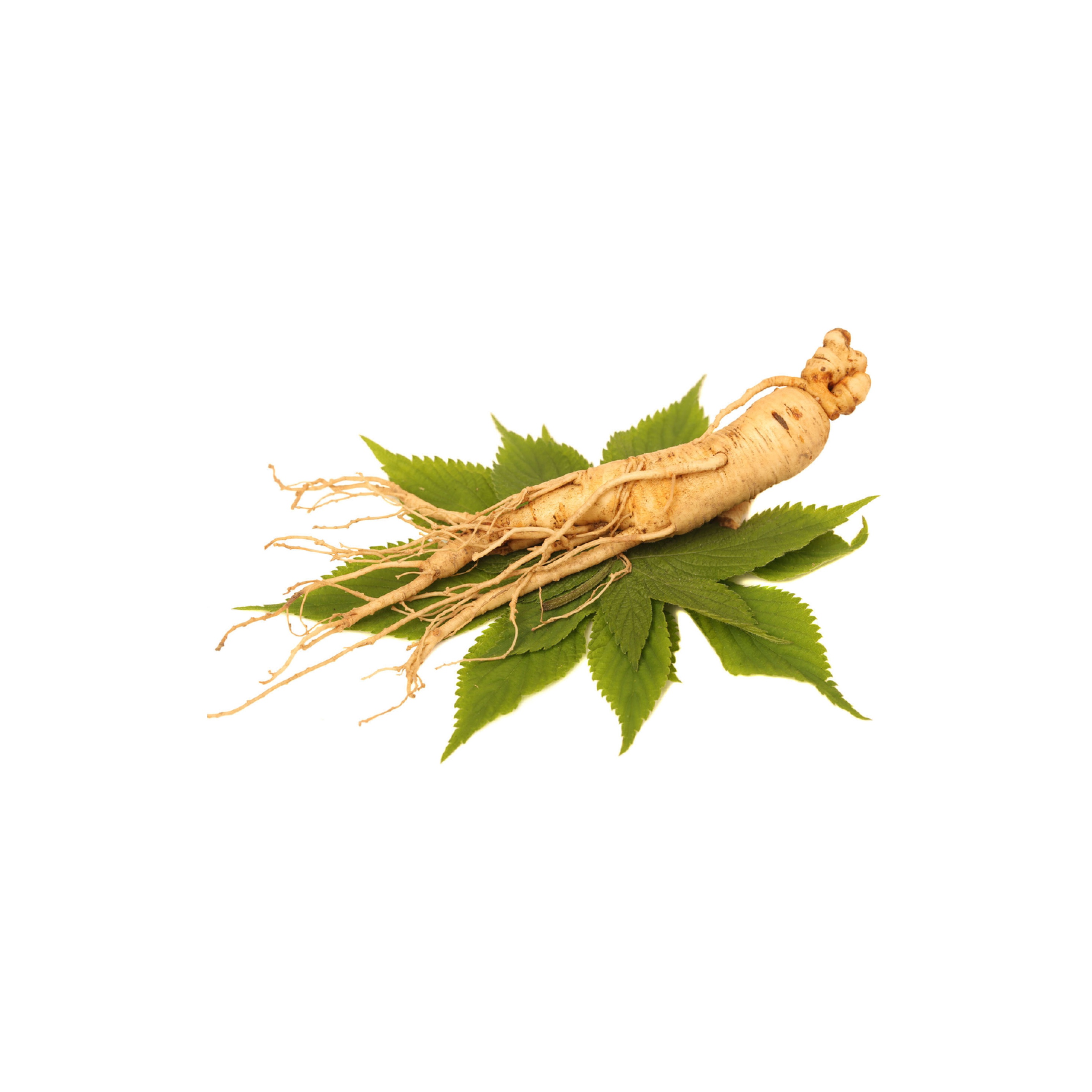
Ginsenoside F1 is a naturally occurring dammarane triterpenoid saponin (protopanaxatriol group), found in Asian ginseng and 2 other sources. It is well tolerated orally (low toxicity).
Concentration
Content by Source
Concentrations not reported. No percent-dry-weight or mg/g figures have been published for this compound in its source plants.
Ginsenoside F1 at a Glance
Ginsenoside F1 is a minor protopanaxatriol (PPT)-type dammarane saponin of Asian ginseng (Panax ginseng). Structurally it is a single-glucose saponin — 20-O-β-D-glucopyranosyl-20(S)-protopanaxatriol — which makes it the PPT-branch structural twin of compound K, the well-known protopanaxadiol (PPD) metabolite 6Reference 6Detection of 13 ginsenosides (Rb1, Rb2, Rc, Rd, Re, Rf, Rg1, Rg3, Rh2, F1, compound K, 20(S)-protopanaxadiol, and 20(S)-protopanaxatriol) in human plasma and application of the analytical method to human pharmacokinetic studies following two-week repeated administration of red ginseng extract — human PKView study →.
Unlike the two headline ginseng saponins Rg1 and Rb1, F1 is barely present in the raw root — it is chiefly a metabolite formed when gut bacteria (or enzymatic processing) strip sugars from Re and Rg1 7,
The honest posture throughout is that F1’s evidence is entirely preclinical and isolate-based — cell cultures, reconstructed-skin models and mice — and that native F1 levels are too low for ordinary ginseng intake to deliver it in any quantity. There is no human trial of the isolated molecule.
The PPT-Side Metabolite (Re/Rg1 → F1)
Ginseng’s dammarane saponins split into two families defined by their aglycone (sapogenin): the protopanaxadiol (PPD) series (Rb1, Rb2, Rc, Rd…) and the protopanaxatriol (PPT) series (Re, Rg1, Rf…), the latter carrying an extra hydroxyl at C-6. Gut bacteria progressively deglycosylate both families, and the two branches have famous near-terminal monoglucosides:
- PPD branch → compound K = 20-O-β-D-glucopyranosyl-20(S)-protopanaxadiol.
- PPT branch → ginsenoside F1 = 20-O-β-D-glucopyranosyl-20(S)-protopanaxatriol.
The two molecules are the same C-20 monoglucoside on the two different skeletons — F1 is, quite literally, “compound K with the extra triol hydroxyl.” That parallel is the cleanest way to place F1 6Reference 6Detection of 13 ginsenosides (Rb1, Rb2, Rc, Rd, Re, Rf, Rg1, Rg3, Rh2, F1, compound K, 20(S)-protopanaxadiol, and 20(S)-protopanaxatriol) in human plasma and application of the analytical method to human pharmacokinetic studies following two-week repeated administration of red ginseng extract — human PKView study →.
The honest complication is metabolic prominence. Compound K is the dominant PPD metabolite that actually reaches the blood. On the PPT branch, the analogous headline human metabolites are ginsenoside Rh1 (the C-6 monoglucoside) and free protopanaxatriol — F1 (the C-20 monoglucoside) is the minor, more rat-associated route off the same tree 7,
What gives F1 its own identity is where it has been studied. Its best-developed literature is dermatological isolate work — protecting keratinocytes from UV damage and interfering with skin pigmentation 1,
What It's Used For
F1’s evidence base is small, recent and consistent in one respect: nearly every study uses the isolated (often enzymatically prepared) molecule in cells, reconstructed skin or mice. The strongest lines are dermatological; a separate immunology thread reports NK-cell enhancement.
Support is an experimental score — weighted by study type (human > animal > in vitro) and volume, tempered for isolate-vs-plant match and bioavailability. A beta, not a validated metric. Every row rests on isolated F1 in cells, reconstructed skin or mice (no human data), so scores are capped accordingly. Each application links to its write-up.
F1’s flagship result, all on isolated F1 (enzymatically prepared from purified ginseng saponin). In human HaCaT keratinocytes, F1 significantly reduced UVB-induced cell death and apoptosis and prevented UVB-induced cleavage of PARP; the mechanism is specific — F1 does not itself raise Bcl-2, but it blocks the UVB-driven downregulation of Bcl-2 (and the neuronal transcription factor Brn-3a), keeping the anti-apoptotic brake in place 1Reference 1In vitroGinsenoside F1 protects human HaCaT keratinocytes from ultraviolet-B-induced apoptosis by maintaining constant levels of Bcl-2 — in vitroView study →.
This sits within a broader ginseng-in-skin literature where many saponins show photoprotective and barrier effects 9Reference 9ReviewThe regulatory role of Korean ginseng in skin cells — reviewView study →.
Gap: the work is in an immortalised keratinocyte line, not human skin in vivo; it establishes a mechanism, not a clinical photoprotective effect, and says nothing about oral ginseng (which delivers negligible F1) 1,
The non-skin thread. Screening 15 ginsenosides, isolated F1 was the most potent enhancer of NK-cell cytotoxicity against diverse activating-receptor and cancer-cell triggers; F1-treated NK cells upregulated cytotoxic mediators, and F1 improved tumour clearance in NK-dependent mouse models of lymphoma and metastatic melanoma, acting through an insulin-like growth factor-1 (IGF-1)-dependent mechanism 4Reference 4In vitroGinsenoside F1 promotes cytotoxic activity of NK cells via insulin-like growth factor-1-dependent mechanism — in vitro and animal modelView study →.
Reviews of “rare” ginsenosides fold in additional immunostimulatory reports — macrophage phagocytosis and MAPK/NF-κB activation 5Reference 5ReviewImmunomodulatory activities of emerging rare ginsenosides F1, Rg5, Rk1, Rh1, and Rg2: from molecular mechanisms to therapeutic applications — reviewView study →.
Gap: isolated F1 in cultured NK cells and mice only; the pro-immune/pro-IGF-1 direction is a double-edged property (IGF-1 signalling is not uniformly desirable), and none of it has been tested as F1 in humans 4,
A second dermatological line, again on isolated F1. Rather than simply blocking pigment synthesis, F1 inhibits the transfer of melanin from melanocytes to surrounding keratinocytes — shown in a melanocyte–keratinocyte coculture and confirmed in a three-dimensional human-skin equivalent 2Reference 2In vitroWhitening efficacy of ginsenoside F1 through inhibition of melanin transfer in cocultured human melanocytes–keratinocytes and three-dimensional human skin equivalent — in vitroView study →.
Separately, F1 combined with ginsenoside Rh3 synergistically suppressed bFGF-induced melanocyte proliferation by inhibiting ERK-mediated upregulation of MITF, the master melanogenesis transcription factor 3Reference 3In vitroInhibitory effects of ginsenosides on basic fibroblast growth factor-induced melanocyte proliferation — in vitroView study →.
Gap: reconstructed-skin and cell models, not a controlled human depigmentation trial; the melanin-transfer and anti-proliferative effects are promising cosmetic mechanisms rather than established clinical whitening 2,
Mechanisms
| Target / pathway | Effect | Relevant to |
|---|---|---|
| Bcl-2 (anti-apoptotic) | blocks UVB-induced downregulation of Bcl-2 (does not itself induce it); prevents PARP cleavage | skin photoprotection |
| Brn-3a | preserves expression under UVB, supporting keratinocyte survival | skin photoprotection |
| Melanosome / melanin transfer (melanocyte → keratinocyte) | inhibits pigment transfer to keratinocytes → depigmenting effect | skin whitening |
| ERK → MITF | with Rh3, suppresses bFGF-driven ERK phosphorylation and MITF, curbing melanocyte proliferation | skin whitening |
| NK-cell cytotoxic program via IGF-1 | raises cytotoxic mediators / activation signals → enhanced tumour-cell killing | immune surveillance |
| MAPK / NF-κB (macrophage) | reported enhancement of phagocytosis / immune activation | immune surveillance |
The dermatological mechanisms are the load-bearing ones — F1 is applied topically as an isolate, which is why its skin data are more interpretable than an oral-ginseng inference. The IGF-1/NK axis is elegant but preclinical and double-edged.
Pharmacokinetics
F1 is a monoglucosyl PPT saponin — one sugar on the protopanaxatriol aglycone — so it is smaller and more lipophilic than the tri-glycosylated parents Rg1/Re, and correspondingly better able to cross membranes than they are. The practical problem is supply, not permeability: native F1 is trace in ginseng, and on the PPT deglycosylation branch the human gut preferentially generates Rh1 and free protopanaxatriol rather than F1 (F1 is more prominent in rat intestine) 7,
Clinical Trials
There are no registered trials of isolated ginsenoside F1. All human ginseng evidence is at the whole-extract level, where F1 is a negligible constituent, so no clinical outcome can be attributed to it. The F1-specific literature is entirely preclinical (cells, reconstructed skin, mice).
| Completed | Planned | Terminated | Preclinical |
|---|---|---|---|
| None(isolated F1) | None identified | None | Skin(photoprotection, whitening) + NK/immune |
Last checked: July 2026.
Isolate vs. Plant Studies
F1 is an unusually clean example of isolate-only evidence. Every result above uses the purified molecule — often enzymatically biotransformed F1 — in HaCaT keratinocytes, melanocyte cocultures, 3D skin equivalents or mice 1,
Prevalence in Nature
In unprocessed ginseng root, F1 is trace to near-absent — it is not one of the stored marker saponins like Rb1, Rg1 or Re. Instead it arises as a deglycosylation metabolite of the protopanaxatriol saponins Re and Rg1, produced by intestinal bacteria in vivo and by the β-glucosidase activity used in fermentation and enzymatic processing of ginseng 7,
Discovery & Synthesis
F1 was characterised as one of the minor protopanaxatriol ginsenosides / deglycosylation products of the ginseng saponin pool; its scientific profile is defined less by a first-isolation milestone than by the 2003 keratinocyte photoprotection study of Lee and colleagues, which used enzymatically prepared F1 from purified ginseng saponin and put the molecule on the dermatology map 1Reference 1In vitroGinsenoside F1 protects human HaCaT keratinocytes from ultraviolet-B-induced apoptosis by maintaining constant levels of Bcl-2 — in vitroView study →. It carries CAS number 53963-43-2.
Manufacture today is overwhelmingly by biotransformation: because native yields are so low, F1 is produced by treating the abundant parent saponins (Re/Rg1) or crude ginseng saponin with β-glucosidase enzymes or microbial fermentation to strip sugars down to the monoglucoside — the same enzymatic strategy used across the “rare ginsenoside” ingredient sector. This is why F1 shows up as a cosmetic/nutraceutical isolate rather than a plant extract fraction.
Patents: not yet researched.
Toxicity & Safety
F1 carries a low flag, consistent with the ginsenoside class and with the favourable tolerability noted in the NK-cell work 4Reference 4In vitroGinsenoside F1 promotes cytotoxic activity of NK cells via insulin-like growth factor-1-dependent mechanism — in vitro and animal modelView study →. It is a minor metabolite that humans are already exposed to at trace levels via ginseng, and its poor native abundance keeps ordinary systemic exposure minimal. The honest qualifier is that no dedicated toxicology package (repeat-dose, genotoxicity, reproductive) exists for the isolated molecule, so “low” reflects class behaviour and the absence of adverse signals rather than a completed safety dossier.
- Class-typical low toxicity. A minor ginsenoside metabolite with no adverse signals reported in the available preclinical work; favourable tolerability in the NK-cell study.
- All benefits are isolate + preclinical. Skin and immune effects come from cells, reconstructed skin and mice — not human trials; do not read them as proven in people.
- Pro-IGF-1 / pro-immune direction is double-edged. F1’s NK/IGF-1 mechanism is not uniformly desirable, and its interaction profile in humans is uncharacterised.
- No dedicated isolate toxicology. No repeat-dose, genotoxicity or reproductive dataset for pure F1; safety here is inferred from the class.
Dosage
There is no established human dose of isolated ginsenoside F1. The figures in the literature are experimental: micromolar concentrations applied to keratinocytes, melanocytes and 3D skin models in the dermatological studies 1,
These are concentrations and doses studied in research, and are not a personal recommendation.
References
- Lee EH, Cho SY, Kim SJ, Shin ES, Chang HK, Kim DH, Yeom MH, Woe KS, Lee J, Sim YC, Lee TR. (2003). Ginsenoside F1 protects human HaCaT keratinocytes from ultraviolet-B-induced apoptosis by maintaining constant levels of Bcl-2 — in vitro. Journal of Investigative Dermatology, 121(3), 607–613. https://pubmed.ncbi.nlm.nih.gov/12925222/
- Lee CS, Nam G, Bae IH, Park J. (2019). Whitening efficacy of ginsenoside F1 through inhibition of melanin transfer in cocultured human melanocytes–keratinocytes and three-dimensional human skin equivalent — in vitro. Journal of Ginseng Research, 43(2), 300–304. https://pubmed.ncbi.nlm.nih.gov/30962737/
- Lee JE, Park JI, Myung CH, Hwang JS. (2017). Inhibitory effects of ginsenosides on basic fibroblast growth factor-induced melanocyte proliferation — in vitro. Journal of Ginseng Research, 41(3), 268–276. https://pubmed.ncbi.nlm.nih.gov/28701866/
- Kwon HJ, Lee H, Choi GE, Kwon SJ, Song AY, Kim SJ, Choi WS, Hwang SH, Kim SC, Kim HS. (2018). Ginsenoside F1 promotes cytotoxic activity of NK cells via insulin-like growth factor-1-dependent mechanism — in vitro and animal model. Frontiers in Immunology, 9, 2785. https://pubmed.ncbi.nlm.nih.gov/30546365/
- Hong CE, Lyu SY. (2025). Immunomodulatory activities of emerging rare ginsenosides F1, Rg5, Rk1, Rh1, and Rg2: from molecular mechanisms to therapeutic applications — review. Pharmaceuticals, 18(10), 1529. https://pubmed.ncbi.nlm.nih.gov/41155644/
- Jin S, Jeon JH, Lee S, Kang WY, Seong SJ, Yoon YR, Choi MK, Song IS. (2019). Detection of 13 ginsenosides (Rb1, Rb2, Rc, Rd, Re, Rf, Rg1, Rg3, Rh2, F1, compound K, 20(S)-protopanaxadiol, and 20(S)-protopanaxatriol) in human plasma and application of the analytical method to human pharmacokinetic studies following two-week repeated administration of red ginseng extract — human PK. Molecules, 24(14), 2618. https://pubmed.ncbi.nlm.nih.gov/31323835/
- He C, Feng R, Sun Y, Chu S, Chen J, Ma C, Fu J, Zhao Z, Huang M, Shou J, Li X, Wang Y, Hu J, Wang Y, Zhang J. (2016). Simultaneous quantification of ginsenoside Rg1 and its metabolites by HPLC-MS/MS: Rg1 excretion in rat bile, urine and feces — animal model. Acta Pharmaceutica Sinica B, 6(6), 595–602. https://pubmed.ncbi.nlm.nih.gov/27818927/
- Won HJ, Kim HI, Park T, Kim H, Jo K, Jeon H, Ha SJ, Hyun JM, Jeong A, Kim JS, Park YJ, Eo YH, Lee J. (2019). Non-clinical pharmacokinetic behavior of ginsenosides — review. Journal of Ginseng Research, 43(3), 354–360. https://pubmed.ncbi.nlm.nih.gov/31308806/
- You L, Cho JY. (2021). The regulatory role of Korean ginseng in skin cells — review. Journal of Ginseng Research, 45(3), 363–370. https://pubmed.ncbi.nlm.nih.gov/34025129/