Compound Monograph
Salvianolic Acid A
The minor-but-more-potent half of the danshen salvianolic pair — a water-soluble caffeic-acid trimer that is far scarcer in the root than salvianolic acid B but generally the stronger performer per unit in antioxidant, antiplatelet and cardioprotective assays. Two facts govern the page: it is largely a product of drying rather than a compound of the living root, and its oral absolute bioavailability is under 1% in rats. It has reached a first-in-human phase 1 study in 116 volunteers; it has never been shown to treat anything in a person.

Sources · 3
Where Does It Come From?
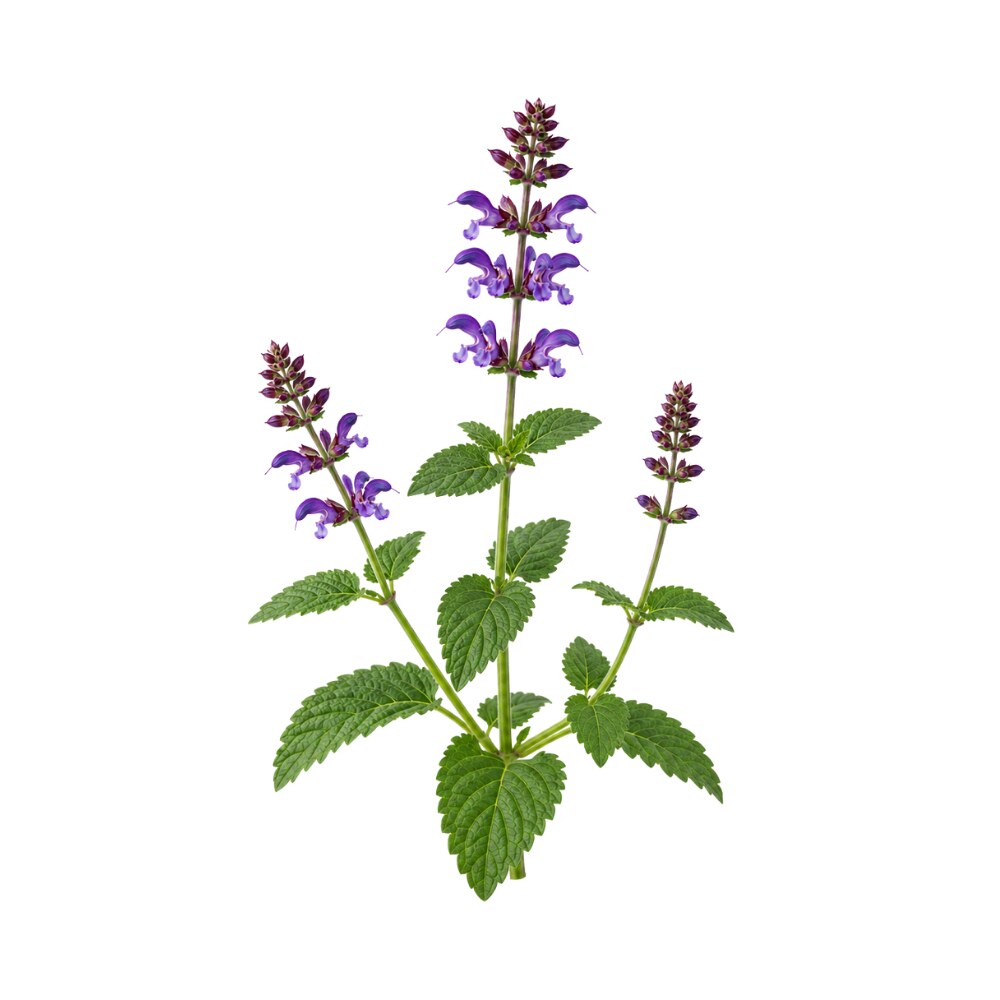
Salvianolic Acid A is a naturally occurring polyphenol (caffeic-acid trimer / phenolic acid; c26h22o10, mw 494.4), found in Dan shen / Chinese red sage and 2 other sources. It is well tolerated orally (low toxicity).
Concentration
Content by Source
Concentrations not reported. No percent-dry-weight or mg/g figures have been published for this compound in its source plants.
Salvianolic Acid A at a Glance
Salvianolic acid A (Sal A) is one of the two water-soluble phenolic acids that carry the hydrophilic half of danshen’s pharmacology. It is a caffeic-acid trimer — a molecule of danshensu (3,4-dihydroxyphenyllactic acid) esterified to a doubled caffeic-acid unit, catechol groups everywhere, and correspondingly a very good antioxidant on paper.
Its whole identity in the literature is a comparison. Salvianolic acid B is the abundant one; salvianolic acid A is the potent one — the standard formulation, stated in almost exactly those words in the field’s most-cited review of the pair 1Reference 1Salvianolic acids — small compounds with multiple mechanisms for cardiovascular protectionView study →. Sal B is the compound the Chinese Pharmacopoeia sets a specification for, the compound the injectable products are built around, and the compound with roughly three times as much published literature. Sal A is the one that keeps outperforming it per unit in assays, and the one that a Beijing laboratory has spent thirty years trying to turn into a drug.
Two facts should be read before anything below. First, the salvianolic acids are mostly not in the fresh root. They are formed during post-harvest drying 14Reference 14Post-harvest induced production of salvianolic acids and significant promotion of antioxidant properties in roots of Salvia miltiorrhiza (Danshen)View study →, and salvianolic acid A can be made from salvianolic acid B under heat, pressure and humidity — a reaction used deliberately in Chinese manufacturing to produce Sal-A-rich material, because its native content in the root is so low 32Reference 32Conversion of salvianolic acid B into salvianolic acid A in tissues of Radix Salviae Miltiorrhizae using high temperature, high pressure and high humidityView study →. “How much Sal A is in danshen” is a question about processing, not botany. Second, oral absorption is close to nil: absolute bioavailability is 0.39-0.52% in rats and 1.47-1.84% in beagle dogs 4,
Salvianolic Acid A vs. Salvianolic Acid B
This is the page’s organising comparison, so it is worth being precise about what is and is not established.
| Salvianolic acid A | Salvianolic acid B | |
|---|---|---|
| Structure | Caffeic-acid trimer, C26H22O10, MW 494.4 | Caffeic-acid tetramer, C36H30O16, MW 718.6 |
| Abundance in dried root | Minor; no pharmacopoeial specification | Marker compound — not less than 3.0% of dried root 23Reference 23Salvia miltiorrhiza — traditional medicinal uses, chemistry, and pharmacologyView study → |
| Published literature | 553 PubMed records | 1,490 PubMed records |
| Standing in the field | ”The most potent water-soluble phenolic component” 1Reference 1Salvianolic acids — small compounds with multiple mechanisms for cardiovascular protectionView study → | “The most abundant water-soluble phenolic component” 1Reference 1Salvianolic acids — small compounds with multiple mechanisms for cardiovascular protectionView study → |
| In commercial products | Not standardised to | The basis of the salvianolate injectables |
On abundance, the best quantitative handle is a drying experiment rather than a survey. When danshen root was dried at 130 °C for 80 minutes — the optimum in that study — salvianolic acid B alone rose to 5.51% of dry weight while the sum of nine salvianolic acids reached 6.61% 14Reference 14Post-harvest induced production of salvianolic acids and significant promotion of antioxidant properties in roots of Salvia miltiorrhiza (Danshen)View study →. Every other salvianolic acid put together, Sal A included, accounts for roughly one percentage point. That is the ratio in one word: Sal B is the bulk, Sal A is a rounding error by mass.
On potency, the honest statement is narrower than the slogan. Reviews consistently rank Sal A as the most potent antioxidant of the salvianolic acids and pair it with Sal B as the two most active members of the group 1,
The Post-Harvest Problem
The single strangest fact about the salvianolic acids is that they are largely not natural products of the living root. A Fudan University group showed that all the salvianolic acids in danshen are post-harvest products of the drying process: salvianolic acid B climbed from 0.01% to 5.51% and the sum of nine salvianolic acids from 0.20% to 6.61% during thermal dehydration, with radical-scavenging activity of the root rising in step 14Reference 14Post-harvest induced production of salvianolic acids and significant promotion of antioxidant properties in roots of Salvia miltiorrhiza (Danshen)View study →.
Within that cascade, the non-Sal-B members — salvianolic acid A among them — appeared at least 40 minutes after Sal B during drying at 130 °C, which led the authors to propose that they are degradation products of salvianolic acid B rather than independently synthesised 14Reference 14Post-harvest induced production of salvianolic acids and significant promotion of antioxidant properties in roots of Salvia miltiorrhiza (Danshen)View study →.
That proposal has since been demonstrated directly, and it is the most consequential fact here. A Shandong University group showed that in danshen tissue under high temperature, high pressure and high humidity, salvianolic acid B converts efficiently into salvianolic acid A — and stated the industrial motive explicitly: Sal A’s native content in the root is very low, so the reaction is “readily utilized to prepare rich Sal A materials in practice” 32Reference 32Conversion of salvianolic acid B into salvianolic acid A in tissues of Radix Salviae Miltiorrhizae using high temperature, high pressure and high humidityView study →. The chemistry is unremarkable once stated — a tetramer shedding a danshensu unit to give a trimer — but the implication is not. A significant share of the salvianolic acid A that exists in the world was manufactured out of salvianolic acid B, not extracted as such.
The consequence is practical. The salvianolic acid A content of a danshen preparation is a function of how the root was dried, how hot, at what pressure and humidity, for how long — not of where it grew. A preparation can be rich in Sal B and nearly free of Sal A, or shifted the other way if it has been processed hard. No product on the market is standardised to Sal A, so there is no way to know which you have. This is the same instability logic that governs carnosic acid in rosemary, arrived at from the opposite direction: there, heat destroys the marker compound; here, heat creates it.
Which Danshen Preparation Delivers It
Danshen is chemically two herbs in one root, and salvianolic acid A sits squarely in the hydrophilic half.
- Water decoction — the traditional preparation, and the one that extracts the salvianolic acids. This is where Sal A belongs. It carries essentially none of the lipophilic tanshinones.
- Ethanol or supercritical-CO2 extract — extracts the tanshinones and leaves the salvianolic acids behind.
- Salvianolate injectables (Chinese hospital products, approved from 2005 onward) — water-type preparations built and specified around salvianolic acid B, not Sal A. Their clinical data belong to Sal B and to the mixture, not to this molecule.
- Purified salvianolic acid A tablets — the phase 1 investigational product 3,
29Reference 3Clinical trialFirst-in-human study to investigate the safety and pharmacokinetics of salvianolic acid A and pharmacokinetic simulation using a physiologically based pharmacokinetic modelView study →Reference 29RCTPhase I single-dose escalation clinical trial of salvianolic acid A tablets — randomized, double-blind, placebo-controlled evaluation of safety, tolerability and pharmacokinetics in healthy volunteersView study → . This is the only preparation that actually delivers a known quantity of Sal A, and it is not a commercial product. Note that the purified material is itself likely to have been made from salvianolic acid B rather than isolated from the root 32Reference 32Conversion of salvianolic acid B into salvianolic acid A in tissues of Radix Salviae Miltiorrhizae using high temperature, high pressure and high humidityView study →.
So: a decoction delivers some salvianolic acid A, an unknown and processing-dependent amount, alongside a much larger amount of Sal B and its precursor rosmarinic acid. Nothing you can buy delivers a stated dose.
What It's Used For
Support is an experimental score I’m building — a composite weighted by study type (human > animal > in vitro > review) and study volume, tempered for isolate-vs-source match and bioavailability. It’s a beta: a fast way to rank strength of evidence at a glance, not a validated metric. Every score here is capped hard, for two reasons that apply to all of them: there is no efficacy trial in humans, and most of the animal work dosed the molecule intravenously because oral absorption is negligible. Each application links to its write-up.
The best-supported activity, and the only one with data generated on human cells. In rats, intravenous Sal A (2.5-10 mg/kg) inhibited ADP-induced platelet aggregation dose-dependently and reduced thrombus weight in an arteriovenous shunt model; the mechanism traced to raised cAMP in both plasma and activated platelets. Critically, five consecutive days of intravenous dosing did not alter coagulation parameters — the effect is on platelets, not on the clotting cascade 6Reference 6Antiplatelet and antithrombotic activities of salvianolic acid AView study →.
The human-tissue result is more direct. Platelets drawn from 40 patients with type 2 diabetes — a population characterised by hyperreactive platelets and poor response to standard antiplatelet drugs — were pre-incubated with Sal A. It reduced fibrinogen binding (PAC-1) and P-selectin (CD62p) expression under both ADP and thrombin stimulation, and reduced maximal aggregation, with no attenuation in patients who already had diabetic complications 7Reference 7Clinical trialSalvianolic acid A inhibits platelet activation and aggregation in patients with type 2 diabetes mellitusView study →.
That is a real effect on real human platelets.
Gap: the human work is ex vivo — platelets in a tube, exposed to a concentration chosen by the experimenter. Nobody has given a person oral salvianolic acid A and measured their platelet function, and at sub-1% oral bioavailability there is no reason to assume the tube concentration is reachable by mouth 4,
Danshen’s reputation is cardiovascular, and Sal A carries part of it. In cardiomyocyte ischaemia-reperfusion models it is anti-apoptotic, acting through PI3K/Akt, GSK-3β, JNK and ERK1/2, with crosstalk between those arms; a dedicated review pulls this literature together and flags p38 MAPK, caspase, JAK/STAT, NF-κB and LOX-1 as further candidate routes that remain incompletely worked out 10Reference 10Anti-apoptotic effects and mechanisms of salvianolic acid A on cardiomyocytes in ischemia-reperfusion injuryView study →.
A broader review of salvianolic acid vascular protection places Sal A alongside Sal B as an endothelial protectant and ROS scavenger with anti-adhesive and anti-inflammatory effects on the vessel wall 30Reference 30The effect of salvianolic acid on vascular protection and possible mechanismsView study →.
The investigational programme describes it explicitly as under development “for myocardial protection treatment for coronary heart disease” 3Reference 3Clinical trialFirst-in-human study to investigate the safety and pharmacokinetics of salvianolic acid A and pharmacokinetic simulation using a physiologically based pharmacokinetic modelView study →.
Gap: entirely preclinical, and — as everywhere here — the danshen clinical literature that looks like support (the dripping pill, the injectables, the angina meta-analyses) tests whole preparations in which Sal A is a minor and unquantified component. None of it is evidence for this molecule.
The most replicated animal finding, and it is replicated across different models with different mechanisms, which is stronger than repetition within one. In carbon-tetrachloride-induced rat fibrosis, Sal A reduced collagen deposition and fibrosis indices by inhibiting PI3K/AKT/mTOR — blocking hepatic stellate-cell activation and extracellular-matrix synthesis — and separately regulated Bcl-2/Bax and caspase-3 to limit hepatocyte apoptosis 8Reference 8Salvianolic acid A attenuates CCl4-induced liver fibrosis by regulating the PI3K/AKT/mTOR, Bcl-2/Bax and caspase-3/cleaved caspase-3 signaling pathwaysView study →.
In bile-duct-ligation (cholestatic) fibrosis, it worked through a different route entirely: SIRT1-dependent deacetylation of HSF1, attenuating endoplasmic-reticulum stress, confirmed in human hepatic stellate LX2 cells where SIRT1 knockdown abolished the effect 9Reference 9Salvianolic acid A attenuates endoplasmic reticulum stress and protects against cholestasis-induced liver fibrosis via the SIRT1/HSF1 pathwayView study →.
Sal A also suppressed collagen accumulation and TGF-β/Smad signalling in an areca-nut model of oral submucous fibrosis, alongside tanshinone IIA and Sal B 25Reference 25In vitroInhibition of tanshinone IIA, salvianolic acid A and salvianolic acid B on areca nut extract-induced oral submucous fibrosis in vitroView study →, and improved kidney function while suppressing PDGF-C/PDGFR-α signalling in renal interstitial fibrosis 34Reference 34Effects of salvianolic acid A and salvianolic acid B in renal interstitial fibrosis via PDGF-C/PDGFR-α signaling pathwayView study → — so the antifibrotic behaviour is not liver-specific.
Gap: no human data of any kind. Stellate-cell suppression is a crowded target — milk thistle, bupleurum and wormwood all reach it by unrelated chemistry — so a positive rodent result here is unsurprising rather than distinguishing. And in the renal study, Sal A, Sal B and the combination all worked, with the authors stating that whether the two differ remains unclear 34Reference 34Effects of salvianolic acid A and salvianolic acid B in renal interstitial fibrosis via PDGF-C/PDGFR-α signaling pathwayView study →.
This application has the page’s most interesting pharmacokinetic hook. In rats given intravenous Sal A at reperfusion after transient middle cerebral artery occlusion, infarct area, brain water content and neurological deficit all fell — and the pharmacokinetics showed that plasma exposure was identical in stroke and sham animals while brain exposure was 9.17-fold higher in the stroke animals 11Reference 11AnimalPharmacokinetic and metabolomic analyses of the neuroprotective effects of salvianolic acid A in a rat ischemic stroke modelView study →.
The molecule concentrates where the barrier is broken. Metabolomic profiling in the same study showed it corrected ischaemia-driven metabolic disturbance in brain tissue more than in serum, i.e. it acts at the tissue rather than systemically 11Reference 11AnimalPharmacokinetic and metabolomic analyses of the neuroprotective effects of salvianolic acid A in a rat ischemic stroke modelView study →.
A prevention-side study found that oral Sal A (10 mg/kg twice daily for five days before an autologous-thrombus stroke) reduced haemorrhage and blood–brain-barrier disruption, preserving the tight-junction proteins ZO-1, occludin and claudin-5 and suppressing MMP upregulation through Src inhibition 12Reference 12Salvianolic acid A prevented cerebrovascular endothelial injury caused by acute ischemic stroke through inhibiting the Src signaling pathwayView study →. A 2024 review collates the post-stroke neuroinflammation literature and notes that the enhanced cerebral penetration after ischaemia is exactly what makes it attractive as a stroke agent 24Reference 24Targeting post-stroke neuroinflammation with salvianolic acid A — molecular mechanisms and preclinical evidenceView study →.
Gap: rodent-only. The review’s mention of positive human findings refers to salvianolic acids as a mixture — the approved Chinese injectable — not to Sal A 24Reference 24Targeting post-stroke neuroinflammation with salvianolic acid A — molecular mechanisms and preclinical evidenceView study →. Do not credit the injectable’s clinical data to this molecule.
This is the indication the drug programme actually chose. The founding result is old: 0.05% Sal A eye drops twice daily delayed galactose-induced cataract in rats, lowering lens malondialdehyde and hydrogen peroxide, raising protein and non-protein thiols, and — mechanistically — inhibiting aldose reductase in vitro 16Reference 16AnimalPrevention of galactose-induced cataractogenesis in rats by salvianolic acid A [in Chinese]View study →.
Aldose reductase is the canonical polyol-pathway target in diabetic cataract, retinopathy and neuropathy, so the mechanism is indication-appropriate rather than generic. The pharmacokinetic papers state their purpose plainly: to support “development of Sal A as an oral drug for the treatment of diabetic complications” 4Reference 4AnimalSome pharmacokinetic parameters of salvianolic acid A following single-dose oral administration to ratsView study →.
The registered phase 1 trial lists its condition as diabetic neuropathy 29Reference 29RCTPhase I single-dose escalation clinical trial of salvianolic acid A tablets — randomized, double-blind, placebo-controlled evaluation of safety, tolerability and pharmacokinetics in healthy volunteersView study →.
A separate line of work found Sal A improved glucocorticoid responsiveness in podocytes via suPAR/uPAR-αvβ3 signalling, relevant to steroid-resistant nephrotic syndrome 26Reference 26Salvianolic acid A attenuates steroid resistant nephrotic syndrome through suPAR/uPAR-αvβ3 signaling inhibitionView study →.
Gap: thirty years after the cataract paper there is still no efficacy trial. A registered phase 1 study is a statement of intent, not evidence of benefit — and the trial’s last status update was in 2018, with no results posted 29Reference 29RCTPhase I single-dose escalation clinical trial of salvianolic acid A tablets — randomized, double-blind, placebo-controlled evaluation of safety, tolerability and pharmacokinetics in healthy volunteersView study →.
Sal A blunted PDGF-BB-driven proliferation of human umbilical artery smooth muscle cells by inducing p21 through a cAMP/PKA/CREB cascade — the same cAMP node that carries the antiplatelet effect, reached in a different cell type. A cAMP analogue mimicked it, a PKA inhibitor blocked it, and dominant-negative CREB abolished it; in rats, the effect reproduced in the neointima of balloon-injured arteries 17Reference 17Prevention of vascular smooth muscle cell proliferation and injury-induced neointimal hyperplasia by CREB-mediated p21 induction — an insight from a plant polyphenolView study →.
That is a relatively tight mechanistic story about restenosis and atherosclerotic lesion formation.
Gap: one laboratory, one mechanism, cells and one animal model. No independent replication in this reference set, and nothing approaching a clinical question.
A scattered but consistent anti-inflammatory picture. In LPS-induced acute lung injury in mice, Sal A at 10 mg/kg matched dexamethasone on lung injury score, oedema, BALF cell counts and cytokines, working by suppressing neutrophil NETosis via the Src/Raf/MEK/ERK pathway — an effect reproduced on neutrophils from ARDS patients and from healthy volunteers in vitro 18Reference 18Salvianolic acid A protects against lipopolysaccharide-induced acute lung injury by inhibiting neutrophil NETosisView study →.
In LPS-induced disseminated intravascular coagulation in rats, Sal A showed anti-complement activity against both the classical and alternative pathways, with molecular docking to C3b, and raised survival substantially 19Reference 19Salvianolic acid A alleviates lipopolysaccharide-induced disseminated intravascular coagulation by inhibiting complement activationView study →.
In ovalbumin-induced allergic asthma, Sal A reduced eosinophil infiltration, mucin production and Th2 cytokines — while notably failing to inhibit mast-cell degranulation, which the tanshinones did 27Reference 27In vitroAnti-allergic effects of salvianolic acid A and tanshinone IIA from Salvia miltiorrhiza determined using in vivo and in vitro experimentsView study →.
Gap: single studies per model, all rodent, all parenteral or in vitro. The asthma result is a useful negative-control datapoint: Sal A and the tanshinones do genuinely different things, which is a reason not to blur danshen’s two fractions together.
Mechanisms
| Target / pathway | Effect | Relevant to | Evidence |
|---|---|---|---|
| cAMP elevation in platelets | inhibits aggregation without affecting coagulation | antiplatelet | rat in vivo + human platelets 6, |
| PI3K/AKT/mTOR suppression | blocks hepatic stellate-cell activation, ECM synthesis | liver fibrosis | rat CCl4 8Reference 8Salvianolic acid A attenuates CCl4-induced liver fibrosis by regulating the PI3K/AKT/mTOR, Bcl-2/Bax and caspase-3/cleaved caspase-3 signaling pathwaysView study → |
| SIRT1 → HSF1 deacetylation, ER-stress reduction | antifibrotic | cholestatic liver fibrosis | rat BDL + LX2 cells 9Reference 9Salvianolic acid A attenuates endoplasmic reticulum stress and protects against cholestasis-induced liver fibrosis via the SIRT1/HSF1 pathwayView study → |
| PI3K/Akt, GSK-3β, JNK, ERK1/2 | anti-apoptotic in cardiomyocytes | cardioprotection | rodent I/R review 10Reference 10Anti-apoptotic effects and mechanisms of salvianolic acid A on cardiomyocytes in ischemia-reperfusion injuryView study → |
| Src inhibition → ZO-1/occludin/claudin-5 preserved, MMPs down | blood–brain-barrier protection | stroke | rat + human brain endothelial cells 12Reference 12Salvianolic acid A prevented cerebrovascular endothelial injury caused by acute ischemic stroke through inhibiting the Src signaling pathwayView study → |
| Src/Raf/MEK/ERK → neutrophil NETosis | anti-inflammatory | acute lung injury | mouse + human neutrophils 18Reference 18Salvianolic acid A protects against lipopolysaccharide-induced acute lung injury by inhibiting neutrophil NETosisView study → |
| cAMP/PKA/CREB → p21 induction | antiproliferative in smooth muscle | restenosis / neointima | cell + rat 17Reference 17Prevention of vascular smooth muscle cell proliferation and injury-induced neointimal hyperplasia by CREB-mediated p21 induction — an insight from a plant polyphenolView study → |
| Complement C3b binding (classical + alternative pathways) | anticomplement | DIC | rat + docking 19Reference 19Salvianolic acid A alleviates lipopolysaccharide-induced disseminated intravascular coagulation by inhibiting complement activationView study → |
| Aldose reductase inhibition, lens MDA/H2O2 reduction | polyol-pathway blockade | diabetic cataract | rat 16Reference 16AnimalPrevention of galactose-induced cataractogenesis in rats by salvianolic acid A [in Chinese]View study → |
| Direct radical scavenging (catechol-rich polyphenol) | antioxidant | general | in vitro 1, |
Pharmacokinetics
This is the section that should govern how the rest of the page is read. Salvianolic acid A is a highly polar, multiply-catecholic polyphenol, and it behaves exactly as that predicts.
In rats given single oral doses of 5, 10 and 20 mg/kg, Cmax rose linearly (31.5, 57.4 and 111.9 µg/L) but absolute oral bioavailability was 0.39-0.52% — under half a percent. Caco-2 transport showed poor permeability with an efflux ratio of 3.13-3.97, meaning it is actively pumped back out of the enterocyte. Tissue distribution was highest in stomach, then small intestine and liver, with detectable levels in brain homogenate. Recovery was overwhelmingly faecal (~0.775% of dose), with almost nothing in bile (0.0037%) or urine (0.0025%) — the great majority of an oral dose is neither absorbed nor excreted intact, and is presumably degraded in the gut 4Reference 4AnimalSome pharmacokinetic parameters of salvianolic acid A following single-dose oral administration to ratsView study →. In beagle dogs at the same doses, absorption was rapid, clearance quick, and absolute bioavailability 1.47-1.84% 5Reference 5Pharmacokinetic study of salvianolic acid A in beagle dog after oral administration by a liquid chromatography-mass spectrometry method — a study on bioavailability and dose proportionalityView study →.
The first-in-human study is the one genuinely human dataset. 116 healthy Chinese subjects received single doses of 10-300 mg or repeat doses of 60-200 mg. Exposure was supra-proportional with dose (slope β for Cmax 1.214, 90% CI 1.150-1.278; for AUC0-t 1.222, 1.156-1.288), and PBPK modelling attributed this to saturation of hepatic OATP1B1 uptake and P-glycoprotein efflux at higher doses 3Reference 3Clinical trialFirst-in-human study to investigate the safety and pharmacokinetics of salvianolic acid A and pharmacokinetic simulation using a physiologically based pharmacokinetic modelView study →. Two things follow: the molecule is a transporter substrate (which is where a drug-interaction risk would live, if one is ever demonstrated), and dose-response in humans is not linear.
Two more pharmacokinetic facts matter. Brain exposure rises 9.17-fold during cerebral ischaemia relative to sham animals at the same plasma exposure 11Reference 11AnimalPharmacokinetic and metabolomic analyses of the neuroprotective effects of salvianolic acid A in a rat ischemic stroke modelView study → — barrier disruption lets it in. And borneol increases the bioavailability of Sal A (along with rosmarinic acid and Sal B) when co-administered in rats, by enhancing intestinal absorption and inhibiting metabolism 28Reference 28AnimalComparative pharmacokinetic and bioavailability studies of three salvianolic acids after the administration of Salviae miltiorrhizae alone or with synthetical borneol in ratsView study →. That is a striking retrospective justification of the traditional pairing: the compound danshen dripping pill contains borneol precisely as an adjuvant, and the pharmacology says it works.
Clinical Trials
There are two human studies of salvianolic acid A itself, both phase 1, and neither tested efficacy.
| Study | Design | n | Result |
|---|---|---|---|
| First-in-human, Zhejiang 3Reference 3Clinical trialFirst-in-human study to investigate the safety and pharmacokinetics of salvianolic acid A and pharmacokinetic simulation using a physiologically based pharmacokinetic modelView study → | Randomised, double-blind, placebo-controlled, single (10-300 mg) and multiple (60-200 mg) ascending dose | 116 healthy Chinese subjects | Well tolerated at all doses; TEAEs infrequent and not dose-related; non-proportional PK |
| NCT03791125, Peking University First Hospital 29Reference 29RCTPhase I single-dose escalation clinical trial of salvianolic acid A tablets — randomized, double-blind, placebo-controlled evaluation of safety, tolerability and pharmacokinetics in healthy volunteersView study → | Randomised, double-blind, placebo-controlled single-dose escalation of Sal A tablets; listed condition diabetic neuropathy | 40 (estimated) | Registered Nov 2018; status last verified Dec 2018 as recruiting, no results posted |
Everything else in the danshen clinical literature — the dripping-pill angina meta-analyses, the salvianolate injectables, the stroke adjunct trials — tested preparations, not this molecule. The Chinese regulatory approvals in this space (salvianolate for injection, 2005; salvianolic acids for injection for ischaemic stroke, 2011) are specified around salvianolic acid B and the mixture. Crediting their outcomes to salvianolic acid A would be the same error as crediting tanshinone IIA sodium sulfonate’s injection data to tanshinone IIA.
Last checked: August 2026.
Bleeding Risk, Warfarin & The Attribution Question
Danshen has one of the best-documented herb–drug interactions in the entire materia medica, and any page on one of its molecules has to address it — carefully, because the temptation to hang a class warning on a molecule is exactly what produces bad safety writing.
The interaction is documented at the level of the herb. Case reports describe patients on stable warfarin developing raised INR, over-anticoagulation and bleeding after taking danshen 20Reference 20Interaction between warfarin and danshen (Salvia miltiorrhiza)View study →. Rat pharmacokinetic/pharmacodynamic work showed danshen increases warfarin absorption and prolongs prothrombin time 21Reference 21AnimalThe effects of danshen (Salvia miltiorrhiza) on pharmacokinetics and pharmacodynamics of warfarin in ratsView study →, and the interaction is catalogued in systematic overviews of warfarin’s drug and food interactions 22Reference 22Systematic overview of warfarin and its drug and food interactionsView study →. This is real, replicated, and clinically consequential.
It has not been traced to salvianolic acid A. No study in this reference set attributes the warfarin interaction to Sal A, to the salvianolic fraction specifically, or to any single danshen molecule. The mechanism described in the rat work is a change in warfarin’s own pharmacokinetics — an effect of the whole extract. Read the warfarin warning as a danshen warning, not a salvianolic acid A warning.
But salvianolic acid A has its own, separate bleeding-relevant activity, and this part is attributable. It inhibits platelet aggregation in rats in vivo and in human platelets ex vivo 6,
The practical position: anyone taking danshen alongside warfarin, a DOAC, aspirin, clopidogrel or any other antiplatelet needs medical supervision, and danshen should be stopped well before surgery — but that instruction belongs to the herb. For the isolated molecule there is no product, no dose, and no human exposure outside a phase 1 unit, so the caution is theoretical until one exists.
Isolate vs. Plant Studies
Unusually for this database, salvianolic acid A’s literature is almost entirely isolate literature. The pharmacology above was nearly all done with purified Sal A — often synthesised rather than extracted 6Reference 6Antiplatelet and antithrombotic activities of salvianolic acid AView study → — at defined doses, usually intravenously. That is a strength: these findings genuinely belong to this molecule, not to a mixture credited to a marker.
The weakness is the mirror image. The isolate work is all preclinical, and the human work is all plant work. Every danshen trial with a clinical endpoint tested a preparation — dripping pill, injection, decoction — in which Sal A is minor, unquantified and possibly created by the drying step. So the two halves of the evidence base do not connect: we know a great deal about what purified Sal A does to a rat, and nothing about what danshen’s Sal A content does to a person.
Within this database, salvianolic acid A’s legitimate occurrence-level role is as a minor constituent of dan shen, alongside its far more abundant sibling salvianolic acid B and their shared biosynthetic precursor rosmarinic acid. It has also been reported — as caffeic-acid trimer isomers, quantified for the first time in that species — in comfrey root, where rosmarinic acid is the dominant phenolic at 7.55 mg/g of a 65% ethanol extract 15Reference 15Is comfrey root more than toxic pyrrolizidine alkaloids? Salvianolic acids among antioxidant polyphenols in comfrey (Symphytum officinale L.) rootsView study →.
Prevalence in Nature
Salvianolic acid A is essentially a danshen compound. The attribution is not in doubt: 258 of the 553 PubMed records mentioning it also mention Salvia miltiorrhiza — the strongest single-species association of any molecule in this batch, and the reason no occurrence caveat is needed here.
Its only other reported natural occurrence is comfrey root (Symphytum officinale), and that one deserves a caveat rather than a claim. A single 2018 study of a 65% ethanol comfrey-root extract identified and quantified caffeic-acid trimers and tetramers for the first time in that species and assigned them by HPLC-DAD-QTOF-MS/MS as “isomers of salvianolic acid A, B and C” 15Reference 15Is comfrey root more than toxic pyrrolizidine alkaloids? Salvianolic acids among antioxidant polyphenols in comfrey (Symphytum officinale L.) rootsView study →. That is a tentative mass-spectrometric assignment of isomers, not isolation of salvianolic acid A itself, and "salvianolic acid A" AND comfrey returns exactly one PubMed record — that paper. Comfrey is Boraginaceae rather than Lamiaceae, but both families are rosmarinic-acid-rich, so a shared oligomerisation route is plausible. Treat comfrey as a probable but unconfirmed source, and do not carry any Sal A activity claim onto a comfrey page. Broadly, salvianolic acids occur across Salvia and related rosmarinic-acid-accumulating genera; danshen is simply where they are concentrated and studied.
Biosynthetically, the chain runs caffeic acid → rosmarinic acid → salvianolic acids: rosmarinic acid is the caffeic-acid dimer, salvianolic acid A the trimer, salvianolic acid B the tetramer. Where the plant’s enzymes stop and post-harvest chemistry takes over is genuinely unresolved — the drying study argues that most of the oligomerisation happens after the root leaves the ground, in a dehydration-stressed but still metabolically active tissue, and that Sal A in particular may be a heat-driven breakdown product of Sal B rather than a biosynthetic endpoint 14Reference 14Post-harvest induced production of salvianolic acids and significant promotion of antioxidant properties in roots of Salvia miltiorrhiza (Danshen)View study →.
There are no non-plant sources.
Discovery & Synthesis
Salvianolic acid A was isolated and named in 1984 by Li Lian-Niang, Tan Rui and Chen Wei-Ming at the Institute of Materia Medica, Chinese Academy of Medical Sciences, Beijing — a short Planta Medica paper describing “a new depside of D-(+)-β-(3,4-dihydroxyphenyl)-lactic acid and 2-(3,4-dihydroxyphenylethenyl)-caffeic acid” from the dried root, with the structure settled by spectral analysis and chemical degradation 33Reference 33Salvianolic acid A, a new depside from roots of Salvia miltiorrhizaView study →. It is the paper that gave the whole salvianolic acid series its name; the same institute described salvianolic acid B four years later.
The molecule has essentially never left that building. Du Guanhua’s group at the same institute has driven its development for three decades — the 1995 cataract and aldose-reductase paper that established the diabetic-complication rationale 16Reference 16AnimalPrevention of galactose-induced cataractogenesis in rats by salvianolic acid A [in Chinese]View study →, the rat and dog pharmacokinetics 4,
Chemically, Sal A is (2R)-3-(3,4-dihydroxyphenyl)-2-[(E)-3-[2-[(E)-2-(3,4-dihydroxyphenyl)ethenyl]-3,4-dihydroxyphenyl]prop-2-enoyl]oxypropanoic acid — a danshensu unit esterified to a doubled caffeic-acid moiety, C26H22O10, MW 494.4. Six phenolic hydroxyls arranged as three catechols make it a strong radical scavenger and metal chelator, and also make it polar, oxidation-prone and poorly absorbed.
Supply is the interesting part. Because Sal A’s native content in the root is very low, it is not economical to isolate directly; the practical route is semi-preparation from the abundant salvianolic acid B, converting Sal B to Sal A in danshen tissue under high temperature, high pressure and high humidity 32Reference 32Conversion of salvianolic acid B into salvianolic acid A in tissues of Radix Salviae Miltiorrhizae using high temperature, high pressure and high humidityView study →. Total synthesis is also available and has been used to supply pharmacology studies — the antiplatelet work explicitly used “a newly synthesized SAA” 6Reference 6Antiplatelet and antithrombotic activities of salvianolic acid AView study → — and the investigational tablets are made from material of defined purity rather than an extract. Plant cell culture and hairy-root systems that raise salvianolic acid yields in S. miltiorrhiza are an active research area.
Patents: not yet researched.
Toxicity & Safety
Salvianolic acid A has something most molecules here do not: a real regulatory-grade toxicology package on the isolate.
In a formal non-clinical safety evaluation, the intravenous LD50 in mice was 1,161.2 mg/kg; in beagle dogs the minimum lethal dose was 682 mg/kg and the maximal non-lethal dose 455 mg/kg. In a 4-week repeat-dose intravenous study in dogs at 20, 80 and 300 mg/kg, the 80 and 300 mg/kg groups showed focal necrosis in liver and renal tubular epithelium, reduced relative thymus weight and biochemical abnormalities — transient and reversible — giving a NOAEL of 20 mg/kg with thymus, liver and kidney as the target organs. Sal A was not mutagenic in bacterial reverse-mutation or mouse bone-marrow micronucleus tests 13Reference 13Non-clinical safety evaluation of salvianolic acid A — acute, 4-week intravenous toxicities and genotoxicity evaluationsView study →.
In humans, the phase 1 study found it well tolerated across single doses of 10-300 mg and repeat doses of 60-200 mg, with a low overall incidence of treatment-emergent adverse events and no dose relationship 3Reference 3Clinical trialFirst-in-human study to investigate the safety and pharmacokinetics of salvianolic acid A and pharmacokinetic simulation using a physiologically based pharmacokinetic modelView study →. That is a genuine tolerability dataset in 116 people — rare for a plant phenolic — and it is the basis for the low-toxicity badge here. Note the caveat the toxicology authors themselves drew: liver and kidney function should be monitored during clinical use 13Reference 13Non-clinical safety evaluation of salvianolic acid A — acute, 4-week intravenous toxicities and genotoxicity evaluationsView study →.
The realistic exposure route is not the isolate but danshen root, where Sal A is one minor phenolic among many. At that level of exposure there is no toxicological concern specific to this molecule; the relevant risks are the herb’s — chiefly bleeding and anticoagulant interaction (see above). No reproductive or developmental toxicology on salvianolic acid A was identified; traditional sources contraindicate danshen in pregnancy, but that position rests on tradition rather than data.
- Real isolate toxicology exists — mouse and dog acute studies, a 4-week repeat-dose dog study, and two genotoxicity assays, all negative for mutagenicity 13Reference 13Non-clinical safety evaluation of salvianolic acid A — acute, 4-week intravenous toxicities and genotoxicity evaluationsView study →.
- Tolerated in 116 healthy volunteers at single doses to 300 mg and repeat doses to 200 mg, with infrequent, non-dose-related adverse events 3Reference 3Clinical trialFirst-in-human study to investigate the safety and pharmacokinetics of salvianolic acid A and pharmacokinetic simulation using a physiologically based pharmacokinetic modelView study →.
- Antiplatelet activity is demonstrated on human platelets 7Reference 7Clinical trialSalvianolic acid A inhibits platelet activation and aggregation in patients with type 2 diabetes mellitusView study → — additive bleeding risk with aspirin, clopidogrel, anticoagulants and NSAIDs is mechanistically expected. Stop danshen well before surgery.
- Danshen potentiates warfarin — case reports of raised INR and bleeding, plus rat PK/PD confirmation 20,
21, . This is a herb-level interaction not traced to salvianolic acid A, but it is the reason danshen and warfarin should not be combined.22Reference 20Interaction between warfarin and danshen (Salvia miltiorrhiza)View study →Reference 21AnimalThe effects of danshen (Salvia miltiorrhiza) on pharmacokinetics and pharmacodynamics of warfarin in ratsView study →Reference 22Systematic overview of warfarin and its drug and food interactionsView study → - Target organs at high intravenous doses were liver, kidney and thymus in dogs, with a NOAEL of 20 mg/kg; effects were reversible, and the authors recommend monitoring liver and kidney function during clinical use 13Reference 13Non-clinical safety evaluation of salvianolic acid A — acute, 4-week intravenous toxicities and genotoxicity evaluationsView study →.
- No efficacy has been shown in humans for any indication — the entire therapeutic case is rodent pharmacology.
- No pregnancy or lactation data. Danshen is traditionally contraindicated in pregnancy; treat that as precautionary rather than evidenced.
Dosage
There is no dose of salvianolic acid A, and there is no product to dose. No supplement is standardised to it, no pharmacopoeial specification exists for it, and the only formulated product containing a known amount is an investigational tablet in a Chinese phase 1 programme.
The numbers below are the exposures the research actually used. They are not recommendations and, with the exception of the phase 1 row, they are not human doses.
| Context | Route | Amount | Source |
|---|---|---|---|
| First-in-human safety/PK, single ascending dose | Oral (purified) | 10-300 mg | 3Reference 3Clinical trialFirst-in-human study to investigate the safety and pharmacokinetics of salvianolic acid A and pharmacokinetic simulation using a physiologically based pharmacokinetic modelView study → |
| First-in-human safety/PK, multiple ascending dose | Oral (purified) | 60-200 mg/day | 3Reference 3Clinical trialFirst-in-human study to investigate the safety and pharmacokinetics of salvianolic acid A and pharmacokinetic simulation using a physiologically based pharmacokinetic modelView study → |
| Rat pharmacokinetics | Oral | 5-20 mg/kg (bioavailability 0.39-0.52%) | 4Reference 4AnimalSome pharmacokinetic parameters of salvianolic acid A following single-dose oral administration to ratsView study → |
| Beagle dog pharmacokinetics | Oral | 5-20 mg/kg (bioavailability 1.47-1.84%) | 5Reference 5Pharmacokinetic study of salvianolic acid A in beagle dog after oral administration by a liquid chromatography-mass spectrometry method — a study on bioavailability and dose proportionalityView study → |
| Rat antiplatelet / antithrombotic | Intravenous | 2.5-10 mg/kg | 6Reference 6Antiplatelet and antithrombotic activities of salvianolic acid AView study → |
| Rat stroke model | Intravenous | 5-10 mg/kg at reperfusion | 11Reference 11AnimalPharmacokinetic and metabolomic analyses of the neuroprotective effects of salvianolic acid A in a rat ischemic stroke modelView study → |
| Dog 4-week toxicology | Intravenous | NOAEL 20 mg/kg | 13Reference 13Non-clinical safety evaluation of salvianolic acid A — acute, 4-week intravenous toxicities and genotoxicity evaluationsView study → |
Two structural reasons this cannot be converted into a herbal dose. First, no one knows how much Sal A is in a given danshen preparation — the content is set by drying conditions, and no product declares it. Second, oral bioavailability is under 1% in rats and under 2% in dogs 4,
References
- Ho JH, Hong CY. (2011). Salvianolic acids — small compounds with multiple mechanisms for cardiovascular protection. Journal of Biomedical Science, 18(1), 30. https://pubmed.ncbi.nlm.nih.gov/21569331/
- Du G, Song J, Du L, et al. (2020). Chemical and pharmacological research on the polyphenol acids isolated from Danshen — a review of salvianolic acids. Advances in Pharmacology, 87, 1–41. https://pubmed.ncbi.nlm.nih.gov/32089230/
- Chen J, Ruan Z, Lou H, et al. (2022). First-in-human study to investigate the safety and pharmacokinetics of salvianolic acid A and pharmacokinetic simulation using a physiologically based pharmacokinetic model. Frontiers in Pharmacology, 13, 907208. https://pubmed.ncbi.nlm.nih.gov/36408276/
- Sun J, Song J, Zhang W, et al. (2018). Some pharmacokinetic parameters of salvianolic acid A following single-dose oral administration to rats. Pharmaceutical Biology, 56(1), 399–406. https://pubmed.ncbi.nlm.nih.gov/30122142/
- Sun J, Zhang L, Song J, et al. (2013). Pharmacokinetic study of salvianolic acid A in beagle dog after oral administration by a liquid chromatography-mass spectrometry method — a study on bioavailability and dose proportionality. Journal of Ethnopharmacology, 148(2), 617–623. https://pubmed.ncbi.nlm.nih.gov/23707334/
- Fan HY, Fu FH, Yang MY, et al. (2010). Antiplatelet and antithrombotic activities of salvianolic acid A. Thrombosis Research, 126(1), e17–e22. https://pubmed.ncbi.nlm.nih.gov/20451955/
- Zhou AM, Xiang YJ, Liu EQ, et al. (2020). Salvianolic acid A inhibits platelet activation and aggregation in patients with type 2 diabetes mellitus. BMC Cardiovascular Disorders, 20(1), 15. https://pubmed.ncbi.nlm.nih.gov/31931718/
- Wang R, Song F, Li S, et al. (2019). Salvianolic acid A attenuates CCl4-induced liver fibrosis by regulating the PI3K/AKT/mTOR, Bcl-2/Bax and caspase-3/cleaved caspase-3 signaling pathways. Drug Design, Development and Therapy, 13, 1889–1900. https://pubmed.ncbi.nlm.nih.gov/31213776/
- Zhu J, Wang R, Xu T, et al. (2018). Salvianolic acid A attenuates endoplasmic reticulum stress and protects against cholestasis-induced liver fibrosis via the SIRT1/HSF1 pathway. Frontiers in Pharmacology, 9, 1277. https://pubmed.ncbi.nlm.nih.gov/30455644/
- Qian W, Wang Z, Xu T, Li D. (2019). Anti-apoptotic effects and mechanisms of salvianolic acid A on cardiomyocytes in ischemia-reperfusion injury. Histology and Histopathology, 34(3), 223–231. https://pubmed.ncbi.nlm.nih.gov/30255492/
- Feng SQ, Aa N, Geng JL, et al. (2017). Pharmacokinetic and metabolomic analyses of the neuroprotective effects of salvianolic acid A in a rat ischemic stroke model. Acta Pharmacologica Sinica, 38(11), 1435–1444. https://pubmed.ncbi.nlm.nih.gov/28836583/
- Liu CD, Liu NN, Zhang S, et al. (2021). Salvianolic acid A prevented cerebrovascular endothelial injury caused by acute ischemic stroke through inhibiting the Src signaling pathway. Acta Pharmacologica Sinica, 42(3), 370–381. https://pubmed.ncbi.nlm.nih.gov/33303991/
- Yang MY, Song ZY, Gan HL, et al. (2022). Non-clinical safety evaluation of salvianolic acid A — acute, 4-week intravenous toxicities and genotoxicity evaluations. BMC Pharmacology and Toxicology, 23(1), 83. https://pubmed.ncbi.nlm.nih.gov/36289546/
- Zhou GJ, Wang W, Xie XM, et al. (2014). Post-harvest induced production of salvianolic acids and significant promotion of antioxidant properties in roots of Salvia miltiorrhiza (Danshen). Molecules, 19(6), 7207–7222. https://pubmed.ncbi.nlm.nih.gov/24886944/
- Trifan A, Opitz SEW, Josuran R, et al. (2018). Is comfrey root more than toxic pyrrolizidine alkaloids? Salvianolic acids among antioxidant polyphenols in comfrey (Symphytum officinale L.) roots. Food and Chemical Toxicology, 112, 178–187. https://pubmed.ncbi.nlm.nih.gov/29288756/
- Du GH, Qiu Y, Tian YE, Zhang JT. (1995). Prevention of galactose-induced cataractogenesis in rats by salvianolic acid A [in Chinese]. Yao Xue Xue Bao (Acta Pharmaceutica Sinica), 30(8), 561–566. https://pubmed.ncbi.nlm.nih.gov/8571774/
- Sun L, Zhao R, Zhang L, et al. (2016). Prevention of vascular smooth muscle cell proliferation and injury-induced neointimal hyperplasia by CREB-mediated p21 induction — an insight from a plant polyphenol. Biochemical Pharmacology, 103, 40–52. https://pubmed.ncbi.nlm.nih.gov/26807478/
- Liu Q, Zhu CL, Li HR, et al. (2022). Salvianolic acid A protects against lipopolysaccharide-induced acute lung injury by inhibiting neutrophil NETosis. Oxidative Medicine and Cellular Longevity, 2022, 7411824. https://pubmed.ncbi.nlm.nih.gov/35910849/
- Zhang QY, Guo J, Xu L, et al. (2022). Salvianolic acid A alleviates lipopolysaccharide-induced disseminated intravascular coagulation by inhibiting complement activation. BMC Complementary Medicine and Therapies, 22(1), 245. https://pubmed.ncbi.nlm.nih.gov/36127691/
- Chan TY. (2001). Interaction between warfarin and danshen (Salvia miltiorrhiza). Annals of Pharmacotherapy, 35(4), 501–504. https://pubmed.ncbi.nlm.nih.gov/11302416/
- Lo AC, Chan K, Yeung JH, Woo KS. (1992). The effects of danshen (Salvia miltiorrhiza) on pharmacokinetics and pharmacodynamics of warfarin in rats. European Journal of Drug Metabolism and Pharmacokinetics, 17(4), 257–262. https://pubmed.ncbi.nlm.nih.gov/1301354/
- Holbrook AM, Pereira JA, Labiris R, et al. (2005). Systematic overview of warfarin and its drug and food interactions. Archives of Internal Medicine, 165(10), 1095–1106. https://pubmed.ncbi.nlm.nih.gov/15911722/
- Su CY, Ming QL, Rahman K, Han T, Qin LP. (2015). Salvia miltiorrhiza — traditional medicinal uses, chemistry, and pharmacology. Chinese Journal of Natural Medicines, 13(3), 163–182. https://pubmed.ncbi.nlm.nih.gov/25835361/
- Yang H, Ibrahim MM, Zhang S, et al. (2024). Targeting post-stroke neuroinflammation with salvianolic acid A — molecular mechanisms and preclinical evidence. Frontiers in Immunology, 15, 1433590. https://pubmed.ncbi.nlm.nih.gov/39139557/
- Dai JP, Zhu DX, Sheng JT, et al. (2015). Inhibition of tanshinone IIA, salvianolic acid A and salvianolic acid B on areca nut extract-induced oral submucous fibrosis in vitro. Molecules, 20(4), 6794–6807. https://pubmed.ncbi.nlm.nih.gov/25884554/
- Li X, Qi D, Wang MY, et al. (2021). Salvianolic acid A attenuates steroid resistant nephrotic syndrome through suPAR/uPAR-αvβ3 signaling inhibition. Journal of Ethnopharmacology, 279, 114351. https://pubmed.ncbi.nlm.nih.gov/34157324/
- Heo JY, Im DS. (2019). Anti-allergic effects of salvianolic acid A and tanshinone IIA from Salvia miltiorrhiza determined using in vivo and in vitro experiments. International Immunopharmacology, 67, 69–77. https://pubmed.ncbi.nlm.nih.gov/30537633/
- Lai XJ, Zhang L, Li JS, et al. (2011). Comparative pharmacokinetic and bioavailability studies of three salvianolic acids after the administration of Salviae miltiorrhizae alone or with synthetical borneol in rats. Fitoterapia, 82(6), 883–888. https://pubmed.ncbi.nlm.nih.gov/21575691/
- Peking University First Hospital. (2018). Phase I single-dose escalation clinical trial of salvianolic acid A tablets — randomized, double-blind, placebo-controlled evaluation of safety, tolerability and pharmacokinetics in healthy volunteers. ClinicalTrials.gov, NCT03791125. https://clinicaltrials.gov/study/NCT03791125
- Wu Y, Xu S, Tian XY. (2020). The effect of salvianolic acid on vascular protection and possible mechanisms. Oxidative Medicine and Cellular Longevity, 2020, 5472096. https://pubmed.ncbi.nlm.nih.gov/33062143/
- Leng X, Kan H, Wu Q, et al. (2022). Inhibitory effect of Salvia miltiorrhiza extract and its active components on cervical intraepithelial neoplastic cells. Molecules, 27(5), 1582. https://pubmed.ncbi.nlm.nih.gov/35268683/
- Xia H, Sun L, Lou H, Rahman MM. (2014). Conversion of salvianolic acid B into salvianolic acid A in tissues of Radix Salviae Miltiorrhizae using high temperature, high pressure and high humidity. Phytomedicine, 21(6), 906–911. https://pubmed.ncbi.nlm.nih.gov/24548723/
- Li LN, Tan R, Chen WM. (1984). Salvianolic acid A, a new depside from roots of Salvia miltiorrhiza. Planta Medica, 50(3), 227–228. https://pubmed.ncbi.nlm.nih.gov/17340301/
- Yao L, Zhao R, He S, et al. (2022). Effects of salvianolic acid A and salvianolic acid B in renal interstitial fibrosis via PDGF-C/PDGFR-α signaling pathway. Phytomedicine, 106, 154414. https://pubmed.ncbi.nlm.nih.gov/36057144/