Compound Monograph
Dioscin
Dioscin is the spirostanol saponin of diosgenin — the same steroid nucleus carrying a three-sugar chacotriose chain — and the compound that gives wild yam, Dioscorea nipponica and Paris rhizomes their soap-like, membrane-dissolving character. It has a large protective pharmacology in rodents (liver, kidney, metabolic) and an even larger in-vitro oncology literature that is substantially confounded by the fact that it is a detergent.

Sources · 11
Where Does It Come From?
Dioscin is a naturally occurring steroidal saponin (spirostanol glycoside; diosgenin 3-o-chacotrioside), found in Wild yam, Dioscorea nipponica, Dioscorea zingiberensis and 8 other sources. It is flagged as moderately toxic.
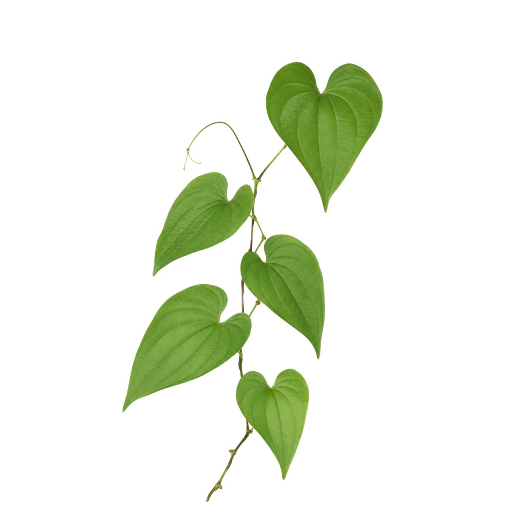 Principal source Wild Yam Dioscorea villosa Herb monograph →
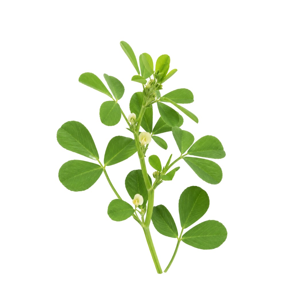 Source Fenugreek Trigonella foenum-graecum Herb monograph →
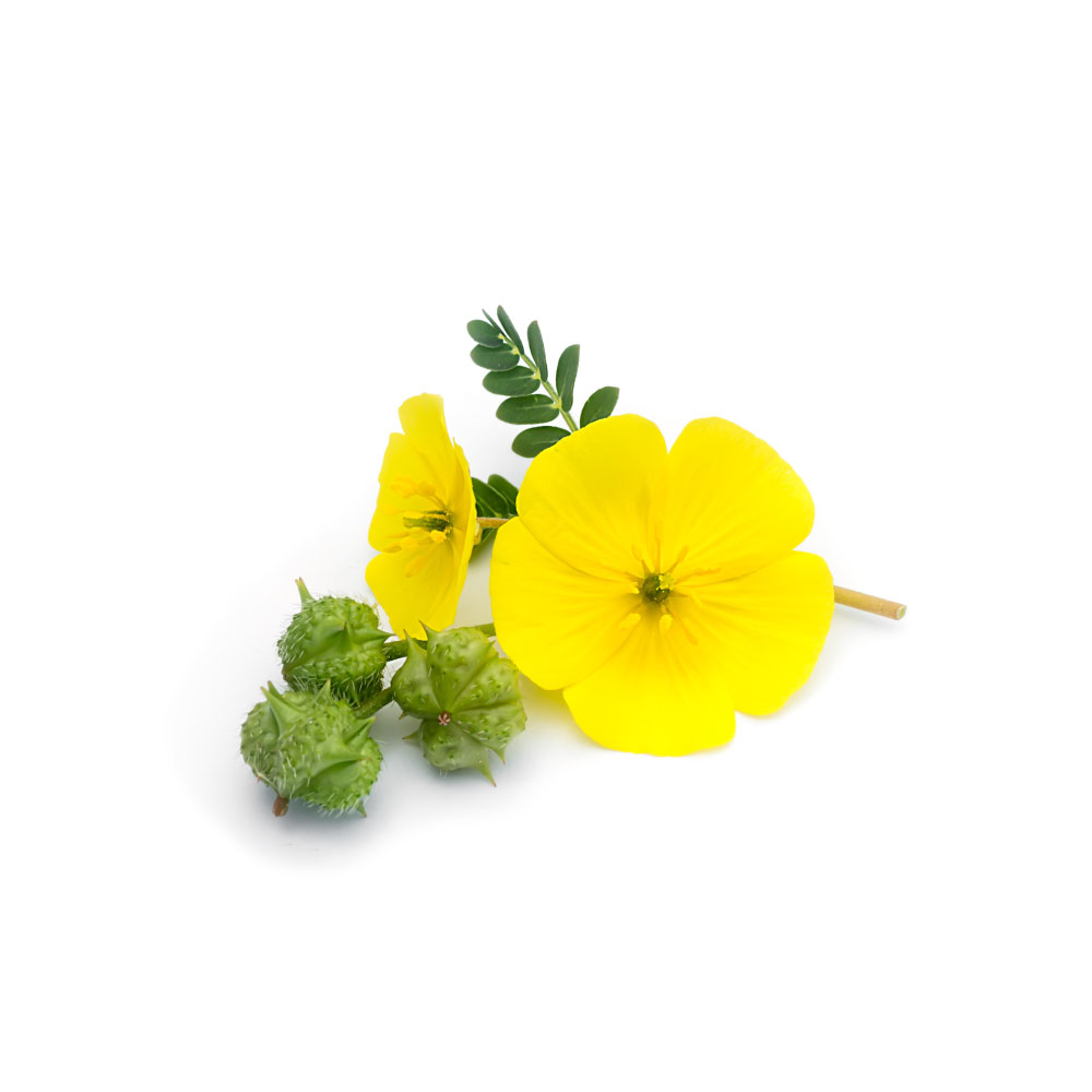 Source Tribulus Tribulus terrestris Herb monograph →
Concentration
Content by Source
Concentrations not reported. No percent-dry-weight or mg/g figures have been published for this compound in its source plants.
Dioscin at a Glance
Dioscin is a steroidal saponin — the glycoside form of diosgenin. Structurally it is one molecule doing two jobs at once: a rigid, fat-soluble spirostanol steroid at one end, and a branched three-sugar chacotriose chain (one glucose, two rhamnoses) at the other 2Reference 2AnimalCharacterization of the pharmacokinetics of dioscin in ratView study →. That amphiphilic shape is not incidental chemistry. It is a soap, and almost everything interesting or misleading about this molecule follows from that.
It is the reason Dioscorea rhizomes foam in water, the reason dioscin lyses red blood cells, and the reason a large fraction of the “dioscin kills cancer cells” literature has to be read with suspicion. It is also, more usefully, one of the better-characterised protective compounds in rodent models of chemically induced liver and kidney injury.
Two facts frame the whole page. First, dioscin is barely absorbed — absolute oral bioavailability in the rat is 0.2% 2Reference 2AnimalCharacterization of the pharmacokinetics of dioscin in ratView study →, and what does get in is partly hydrolysed to diosgenin on the way 1,
Dioscin & Diosgenin: The Glycoside–Aglycone Pair
Dioscin and diosgenin are the same steroid with and without its sugars. Strip the chacotriose off dioscin — by acid, by a fungal glycosidase, or by gut bacteria — and you have diosgenin. That relationship is the mechanism of exposure for this whole class, so it is worth getting right.
The counterintuitive finding is real, but smaller than it sounds. Glycosides are usually the less permeable member of a pair, so the reported result that dioscin crosses intestinal epithelium better than its own aglycone deserves checking. It holds. In the only side-by-side ADME study — and, unusually, one performed on material from D. villosa itself — dioscin showed higher apparent permeability across a Caco-2 monolayer with no significant efflux, while diosgenin was subject to P-glycoprotein-mediated efflux 1Reference 1In vitroCharacterization of in vitro ADME properties of diosgenin and dioscin from Dioscorea villosaView study →.
But read what that actually measured. The comparison is relative, and it is relative to an aglycone whose net absorption is being actively pumped back by a transporter. It is not evidence that dioscin is well absorbed. In a living rat, dioscin’s absolute oral bioavailability is 0.2% 2Reference 2AnimalCharacterization of the pharmacokinetics of dioscin in ratView study →; in rats given a Paris polyphylla extract, dioscin and the eight other steroidal saponins measured alongside it all came in below 1% bioavailability 3Reference 3AnimalThe pharmacokinetics and tissue distributions of nine steroidal saponins from Paris polyphylla in ratsView study →. Both numbers are consistent with the general saponin picture: molecular mass over 500 Da, high hydrogen-bonding capacity and high flexibility make membrane permeation poor, and saponins are additionally hydrolysed by colonic microflora before the host ever sees them 4Reference 4ReviewAbsorption, disposition, and pharmacokinetics of saponins from Chinese medicinal herbs: what do we know and what do we need to know more? Current Drug Metabolism (review). https://pubmed.ncbi.nlm.nih.gov/22292787/View study →. Dioscin is a permeability outlier only within a category defined by not being absorbed.
The rest of the same ADME study fills in the chain:
| Property | Dioscin | Diosgenin |
|---|---|---|
| Stability in simulated gastric fluid (pH 1.2) | degraded 28.3%, with 24.2% appearing as diosgenin | depleted <10% |
| Stability in simulated intestinal fluid (pH 6.8) | degraded 12.4%, with 2.4% appearing as diosgenin | depleted <10% |
| Caco-2 transport | higher permeability, no significant efflux | P-glycoprotein efflux substrate |
| Human liver microsomes (phase I) | stable | stable |
| S9 fraction (phase II) | stable | disappeared, t½ 11.3 min |
| CYP3A4 inhibition | IC50 ~33 µM | IC50 ~17 µM |
All figures from Manda et al. 2013 1Reference 1In vitroCharacterization of in vitro ADME properties of diosgenin and dioscin from Dioscorea villosaView study →; no other CYP isoform (2D6, 2C9, 1A2) was affected by either compound.
So the two molecules trade weaknesses. Dioscin survives metabolism but is a huge polar glycoside that mostly stays in the gut; diosgenin passes membranes but is effluxed by P-gp and rapidly conjugated in phase II. Neither reaches plasma in quantity. Whichever of them is doing the work in a rodent study, the concentration at the target is far below the micromolar figures the in-vitro literature reports — except in the gut lumen and, per tissue-distribution work, in the liver, where saponins concentrate preferentially 3,
What Dioscin Does Not Do
Diosgenin — dioscin’s aglycone — was the industrial feedstock from which progesterone and cortisone were semi-synthesised in the mid-twentieth century, by the Marker degradation. That is a sequence of laboratory reactions run with acid, heat and reagents. The human body cannot perform it. There is no human enzymatic pathway from diosgenin or dioscin to progesterone, DHEA or testosterone, and the persistent claim that a wild yam cream or capsule delivers “natural progesterone” is false. Where such a cream demonstrably works, progesterone was added to it.
Dioscin is one hydrolysis step upstream of diosgenin, so the claim gets attached to it too. It should not be softened into “supports hormonal balance.” Dioscin has no established steroid-receptor pharmacology in humans, and the one prominent paper reporting a receptor mechanism — dioscin activating estrogen receptor-β in prostate cancer cells — was retracted in 2021 28Reference 28Dioscin induces prostate cancer cell apoptosis through activation of estrogen receptor-βView study →. A regulatory safety assessment of D. villosa root extract standardised to ≤3.5% diosgenin found no estrogenic activity in a juvenile rat uterotrophic assay at up to 500 mg/kg/day 12Reference 12ReviewFinal report of the amended safety assessment of Dioscorea villosa (wild yam) root extractView study →.
The Detergent Problem
Dioscin is a surfactant, and it is one of the more haemolytic saponins known. Molecular-dynamics work describes the mechanism concretely: dioscin inserts into a lipid bilayer, accumulates in cholesterol-rich raft microdomains, and binds cholesterol more favourably than cholesterol binds itself — destabilising the raft, inducing membrane curvature, and ultimately rupturing the cell 5Reference 5Hemolytic mechanism of dioscin proposed by molecular dynamics simulationsView study →. Structure–activity work supports the same picture from the other direction: chemically capping the 6′-hydroxyl of the chacotriose chain sharply reduced cytotoxicity while only partly reducing haemolysis, showing that both properties are carried by the sugar chain 6Reference 6Synthesis, cytotoxicity, and hemolytic activity of 6′-O-substituted dioscin derivativesView study →.
This is the single most important caveat here, because it means a saponin can register as “cytotoxic to cancer cells at low micromolar” for reasons that have nothing to do with cancer biology. The authors of a whole-transcriptome study of wild yam extract in triple-negative breast cancer cells said so explicitly, calling the extract’s detergent saponins “an interfering variable” and deliberately restricting their analysis to sub-lethal concentrations to get past it 8Reference 8Effects of wild yam root (Dioscorea villosa) extract on the gene expression profile of triple-negative breast cancer cellsView study →.
Does the dioscin oncology literature control for this? Largely, no. A PubMed search pairing dioscin with normal cells or a selectivity index returns 31 records against roughly 560 for dioscin overall and 73 for the cancer-apoptosis subset — and the isolate studies cited here do not report a selectivity index. The flagship D. villosa study, for instance, compared dioscin’s effect across MCF-7 and MDA-MB-231 cells, both of which are tumour lines, with no normal-cell comparator 10Reference 10In vitroThe anticancer potential of steroidal saponin, dioscin, isolated from wild yam (Dioscorea villosa) root extract in invasive human breast cancer cell line MDA-MB-231 in vitroView study →. The one place a selectivity claim is made is not oncology at all: an antifungal study reported that dioscin inhibited Candida albicans virulence factors “with low cytotoxicity against mammalian cells” 30Reference 30Dioscin inhibits virulence factors of Candida albicansView study → — which makes sense, since a fungal membrane’s ergosterol is a plausible selective target in a way that a tumour cell’s plasma membrane is not.
There is one finely observed demonstration of the problem, and it comes from wild yam. When nine steroidal glycosides isolated from D. villosa were screened on HepG2 liver cells at 10–50 µM, the spirostanol saponins — dioscin, deltonin, progenin III and zingiberensis saponin I — were cytotoxic, while the furostanol saponins (protodioscin, pseudoprotodioscin, protodeltonin, methyl protobioside, huangjiangsu A) were not. Only the non-cytotoxic furostanols were carried forward into the hepatoprotection experiment, where they raised glutathione and reduced ROS 9Reference 9Hepatoprotective effect of steroidal glycosides from Dioscorea villosa on hydrogen peroxide-induced hepatotoxicity in HepG2 cellsView study →. In the one study that tested wild yam’s own saponins for liver protection, dioscin was excluded because it killed the cells. The furostanol/spirostanol distinction is not a taxonomic nicety; it changes the sign of the result.
Where the Evidence Comes From
Two structural features of dioscin’s literature should shape how you read the pharmacology below.
It is not a wild yam molecule in practice. Attribution to Dioscorea villosa is sound — dioscin has been directly isolated from D. villosa rhizome by countercurrent chromatography 13,dioscin AND "Dioscorea villosa" returns 7 records; dioscin AND Dioscorea returns 129, and dioscin overall 560. The research plants are D. nipponica (32 records) and D. zingiberensis (19). Chemotypes differ enough to be forensically useful: dioscoreavilloside A and B and parvifloside occur only in D. villosa, while asperin and gracillin occur only in D. cayenensis, and these markers are used to detect substitution in commercial “Dioscorea” products 11Reference 11Characterization of steroidal saponins from Dioscorea villosa and D. cayenensis using ultrahigh performance liquid chromatography/electrospray ionization quadrupole time-of-flight mass spectrometryView study →. A finding obtained on D. nipponica rhizome is a finding about that herb, not about wild yam.
A large share of the in-vivo work comes from one laboratory, and much of it has been corrected. The rodent hepatoprotective, nephroprotective and metabolic programme that constitutes dioscin’s strongest evidence is dominated by a single group at Dalian Medical University — a PubMed author search returns 82 dioscin papers for its principal investigator. Between 2017 and 2021, corrigenda were issued for a striking number of them, including the alcohol-liver-injury study 18Reference 18Protective effects of dioscin against alcohol-induced liver injuryView study →, the obesity study 21Reference 21AnimalPotent effects of dioscin against obesity in miceView study →, the 90-day toxicology study 7Reference 7AnimalA 90-day subchronic toxicological assessment of dioscin, a natural steroid saponin, in Sprague-Dawley ratsView study →, and further papers on hepatic fibrosis, doxorubicin cardiotoxicity, intestinal ischaemia–reperfusion, ovariectomy-induced bone loss and hepatic stellate cell migration; in total, PubMed indexes 28 published errata for dioscin. One dioscin paper — the estrogen-receptor-β prostate cancer study — has been retracted 28Reference 28Dioscin induces prostate cancer cell apoptosis through activation of estrogen receptor-βView study →.
Corrigenda are corrections, not retractions, and the corrected claims mostly stand. But a body of work this concentrated in one lab, with this density of post-publication correction, does not have the independent-replication depth its raw paper count suggests. The most reassuring findings here are the ones from other groups: the independent cisplatin kidney-injury study 24Reference 24Dioscin protects against cisplatin-induced acute kidney injury by reducing ferroptosis and apoptosis through activating Nrf2/HO-1 signalingView study →, the hyperuricaemia work 25Reference 25Protective effects of dioscin and diosgenin on plateau hyperuricemia by attenuating renal inflammation via EPHX2View study →, and the sub-micromolar anti-inflammatory result obtained during quality-control analysis of D. nipponica 26Reference 26Identification of anti-inflammatory components in Dioscorea nipponica Makino based on HPLC-MS/MS, quantitative analysis of multiple components by single marker and chemometric methodsView study →.
What It's Used For
Dioscin’s pharmacology has a clear shape: a broad, mechanistically coherent cytoprotective signal in rodent models of chemically induced organ injury, a plausible anti-inflammatory action at genuinely low concentrations, and a very large in-vitro oncology literature whose value is limited by the detergent problem above. Nothing has been tested in a human.
Support is an experimental score — weighted by study type (human > animal > in vitro) and volume. A beta, not a validated metric. No row here is supported by human data; the ceiling for the whole table is “replicated rodent model.”
The largest and most internally consistent body of work, all rodent, all on the isolated molecule. Dioscin protected against acute liver damage from carbon tetrachloride — at 100 mg/kg producing an effect equal to silymarin at 200 mg/kg, with proteomic validation of six differentially expressed proteins 15Reference 15AnimalApplication of proteomic and bioinformatic techniques for studying the hepatoprotective effect of dioscin against CCl4-induced liver damage in miceView study → — and separately through suppression of lipid peroxidation, TNF-α and IL-6, Fas/FasL downregulation and an improved Bcl-2/Bax ratio 16Reference 16AnimalMechanism investigation of dioscin against CCl4-induced acute liver damage in miceView study →.
Against paracetamol it prevented mitochondrial dysfunction and inhibited CYP2E1 induction in both HepG2 cells and mice 17Reference 17In vitroDioscin, a natural steroid saponin, shows remarkable protective effect against acetaminophen-induced liver damage in vitro and in vivoView study →.
Against ethanol it inhibited CYP2E1, suppressed NF-κB and ER-stress markers, and shifted lipid handling toward PPARα-driven β-oxidation 18Reference 18Protective effects of dioscin against alcohol-induced liver injuryView study →.
Against endotoxin it acted through TLR4/MyD88 19Reference 19Dioscin reduces lipopolysaccharide-induced inflammatory liver injury via regulating TLR4/MyD88 signal pathwayView study →.
The recurring targets — CYP2E1, NF-κB, mitochondrial apoptosis, antioxidant enzymes — are the same in every model, which is a genuine coherence argument.
Gap: No human data, no clinical trial, and no dose-ranging that establishes a human-relevant exposure. The programme is concentrated in one laboratory and several of the constituent papers carry published corrigenda. Note also the contrary cell-level result: in D. villosa’s own saponin set, dioscin was cytotoxic to HepG2 cells at 10–50 µM and was excluded from the hepatoprotection assay 9Reference 9Hepatoprotective effect of steroidal glycosides from Dioscorea villosa on hydrogen peroxide-induced hepatotoxicity in HepG2 cellsView study → — protection in a whole animal and toxicity in a dish are not obviously reconcilable, and no one has reconciled them.
The cleanest isolate result on the page, and it comes from an independent group doing quality-control chemistry rather than pharmacology. Screening the saponins of Dioscorea nipponica rhizome against LPS-stimulated RAW264.7 macrophages, dioscin inhibited nitric-oxide production with an IC50 of 0.469 µM — more potent than protodioscin (0.712 µM) or gracillin (0.815 µM) — and significantly reduced TNF-α, IL-1β and IL-6 26Reference 26Identification of anti-inflammatory components in Dioscorea nipponica Makino based on HPLC-MS/MS, quantitative analysis of multiple components by single marker and chemometric methodsView study →.
The concentration matters: 0.47 µM is far below the range where saponin membrane lysis begins, so this is unlikely to be a detergent artefact. In vivo, the endotoxin liver-injury model attributes dioscin’s effect to TLR4/MyD88 signalling 19Reference 19Dioscin reduces lipopolysaccharide-induced inflammatory liver injury via regulating TLR4/MyD88 signal pathwayView study →, which is the pathway a macrophage NO result would predict.
Gap: Isolate-only, and the source herb is D. nipponica, not wild yam. There is no anti-inflammatory trial of dioscin in an animal disease model outside the liver, and no human data. Whether 0.47 µM is reachable at any tissue other than gut epithelium, given 0.2% oral bioavailability 2Reference 2AnimalCharacterization of the pharmacokinetics of dioscin in ratView study →, is unaddressed.
Rodent, isolate, mechanistically specific. In high-fat-diet mice and ob/ob mice, dioscin reduced body weight and hepatic lipid accumulation, increased oxygen consumption and energy expenditure, promoted fatty-acid β-oxidation and induced autophagy 21Reference 21AnimalPotent effects of dioscin against obesity in miceView study →.
A follow-up in palmitate-treated hepatocytes and HFD rodents traced the effect to SIRT1/AMPK signalling regulating SREBP-1c, CPT, FAS, SCD, FoxO1 and ATGL — with the effect abolished by SIRT1 inhibition (nicotinamide) or AMPK blockade (compound C), which is a real mechanistic test rather than a correlation 20Reference 20Dioscin alleviates non-alcoholic fatty liver disease through adjusting lipid metabolism via SIRT1/AMPK signaling pathwayView study →.
The alcohol-liver work independently found PPARα upregulation without any change to the fatty-acid synthesis machinery 18Reference 18Protective effects of dioscin against alcohol-induced liver injuryView study →.
Gap: Both principal papers carry published author corrections, and both come from the same laboratory. Doses are in the tens of mg/kg in mice with no human-equivalent modelling, and dioscin’s aglycone diosgenin has its own, better-replicated hypolipidemic mechanism through intestinal NPC1L1 — so how much of a whole-plant metabolic effect belongs to dioscin rather than to the diosgenin liberated from it is not separable from these data.
The line with the most independent replication. An independent Chinese group showed dioscin protects against cisplatin-induced acute kidney injury by reducing ferroptosis and apoptosis through Nrf2/HO-1 activation 24Reference 24Dioscin protects against cisplatin-induced acute kidney injury by reducing ferroptosis and apoptosis through activating Nrf2/HO-1 signalingView study →.
Separately, dioscin protected against fructose-induced renal damage via Sirt3-mediated control of oxidative stress, fibrosis, lipid metabolism and inflammation 22Reference 22Protective effects of dioscin against fructose-induced renal damage via adjusting Sirt3-mediated oxidative stress, fibrosis, lipid metabolism and inflammationView study →, and against methotrexate-induced liver and kidney damage through a miR-145-5p-mediated oxidative-stress axis 23Reference 23Dioscin ameliorates methotrexate-induced liver and kidney damages via adjusting miRNA-145-5p-mediated oxidative stressView study →.
In a high-altitude hyperuricaemia model, dioscin and diosgenin together attenuated renal inflammation via the soluble epoxide hydrolase gene EPHX2 25Reference 25Protective effects of dioscin and diosgenin on plateau hyperuricemia by attenuating renal inflammation via EPHX2View study → — one of the few studies to test the glycoside and aglycone side by side.
Gap: All rodent, all isolate, no human data, and the mechanisms proposed (Nrf2/HO-1, Sirt3, a specific miRNA, EPHX2) do not yet converge on one story the way the liver work does. Note the tension with safety: dioscin is renally as well as faecally excreted and is haemolytic at higher doses 7Reference 7AnimalA 90-day subchronic toxicological assessment of dioscin, a natural steroid saponin, in Sprague-Dawley ratsView study →, so a nephroprotective claim needs a dose window that has not been defined.
The application where dioscin’s membrane activity is a feature rather than a confound. Against Candida albicans, dioscin inhibited biofilm formation and development, the yeast-to-hypha morphological transition, adhesion and secreted phospholipase — the organism’s principal virulence factors — and the authors report low cytotoxicity against mammalian cells at the active concentrations 30Reference 30Dioscin inhibits virulence factors of Candida albicansView study →.
That selectivity is mechanistically plausible: fungal membranes are built on ergosterol rather than cholesterol, and dioscin’s lytic action is a sterol-binding one 5Reference 5Hemolytic mechanism of dioscin proposed by molecular dynamics simulationsView study →.
A dioscin-related steroidal saponin from Allium paradoxum bulbs showed leishmanicidal activity 34Reference 34Isolation of dioscin-related steroidal saponin from the bulbs of Allium paradoxum L. with leishmanicidal activityView study →, and dioscin appears among Tribulus and Smilax saponins screened as multidrug-resistance modulators 39Reference 39AnimalScreening of some saponins and phenolic components of Tribulus terrestris and Smilax excelsa as MDR modulatorsView study →.
Gap: Single-organism, in vitro, and no animal infection model. The comparison against a standard antifungal is not established, and no formulation work exists for a compound with 0.2% oral bioavailability 2Reference 2AnimalCharacterization of the pharmacokinetics of dioscin in ratView study →. Read as a topical or luminal lead at most.
Large in volume, thin in interpretable content. Dioscin isolated from wild yam root extract reduced viability of MCF-7 and MDA-MB-231 breast cancer cells, upregulated GATA3, shifted MDA-MB-231 cells toward an epithelial morphology and cut cellular invasion by 65% at 5.76 µM 10Reference 10In vitroThe anticancer potential of steroidal saponin, dioscin, isolated from wild yam (Dioscorea villosa) root extract in invasive human breast cancer cell line MDA-MB-231 in vitroView study →.
Other work reports caspase-independent apoptosis via apoptosis-inducing factor in breast cancer cells, p38/JNK activation in HL-60 leukaemia cells, and mapped inhibitory targets in HepG2 27Reference 27Molecular mechanism and inhibitory targets of dioscin in HepG2 cellsView study →.
A 2022 review catalogues activity against a long list of malignancies 29Reference 29ReviewDioscin: a review on pharmacological properties and therapeutic valuesView study →.
The problem is that none of this distinguishes targeted cytotoxicity from membrane lysis. No selectivity index against normal cells is reported in the isolate studies cited here; the D. villosa study used two tumour lines and no normal comparator 10Reference 10In vitroThe anticancer potential of steroidal saponin, dioscin, isolated from wild yam (Dioscorea villosa) root extract in invasive human breast cancer cell line MDA-MB-231 in vitroView study →.
Structure–activity work shows cytotoxicity tracks the chacotriose sugar chain — the same feature that causes haemolysis 6Reference 6Synthesis, cytotoxicity, and hemolytic activity of 6′-O-substituted dioscin derivativesView study →.
The authors of the whole-extract transcriptomic study named the detergent saponins as an interfering variable and worked below lytic concentrations to avoid them 8Reference 8Effects of wild yam root (Dioscorea villosa) extract on the gene expression profile of triple-negative breast cancer cellsView study →.
And the most mechanistically specific paper in the set, claiming estrogen-receptor-β activation in prostate cancer, was retracted in 2021 28Reference 28Dioscin induces prostate cancer cell apoptosis through activation of estrogen receptor-βView study →.
Gap: Until a study reports dioscin’s IC50 against matched normal cells alongside the tumour figure, and shows the effect survives at concentrations below the haemolytic threshold, this application should be treated as unproven at the molecule level. There is no in-vivo isolate oncology result here that survives that filter.
Early and single-source, but mechanistically detailed. Dioscin purified from Smilax china rhizome raised cAMP and cGMP in human platelets, phosphorylating the IP3 receptor to blunt calcium mobilisation and phosphorylating VASP to suppress integrin αIIbβ3 activation and fibrinogen binding; it also modulated PI3K/Akt, MAPK and cPLA2 signalling associated with granule release. Feeding S. china rhizome reduced thrombus formation in an FeCl3 mouse model 31Reference 31Dioscin from Smilax china rhizomes inhibits platelet activation and thrombus formation via up-regulating cyclic nucleotidesView study →.
Gap: One study, one group, 2025. The in-vitro work is on isolated dioscin but the in-vivo confirmation used whole rhizome, so the animal result is a plant result. No dose–response against an established antiplatelet agent, and the bleeding-risk implication has not been assessed — which matters more here than for most applications, since dioscin is separately haemolytic 5,
Mechanisms
| Target / pathway | Effect | Relevant to |
|---|---|---|
| Membrane cholesterol / lipid rafts | inserts into bilayer, binds cholesterol, destabilises rafts → curvature → lysis | haemolysisantifungal activityin-vitro cytotoxicity artefacts |
| CYP2E1 | induction and activity suppressed in ethanol and paracetamol liver injury | hepatoprotection |
| NF-κB / TLR4–MyD88 | suppressed; ↓TNF-α, ↓IL-6, ↓IL-1β | anti-inflammatoryhepatoprotection |
| Inducible nitric-oxide synthase output | NO production inhibited in LPS macrophages, IC50 0.469 µM | anti-inflammatory |
| Nrf2 / HO-1 | activated; ↓ferroptosis and apoptosis in cisplatin kidney injury | nephroprotection |
| SIRT1 / AMPK | activated; regulates SREBP-1c, FAS, SCD, CPT, FoxO1, ATGL | NAFLDobesity |
| Sirt3 | mediates antioxidant, antifibrotic and lipid effects in renal injury | nephroprotection |
| PPARα | upregulated → fatty-acid β-oxidation (no effect on lipogenic enzymes) | alcoholic liver injurylipid handling |
| Mitochondrial apoptosis (Bcl-2/Bax, cytochrome c, caspase-3/-8/-9) | modulated in both directions depending on tissue — protective in hepatocytes, pro-apoptotic in tumour lines | hepatoprotectionin-vitro anticancer |
| CYP3A4 | inhibited, IC50 ~33 µM (2D6, 2C9, 1A2 unaffected) | drug interaction risk |
| P-glycoprotein | dioscin is not an efflux substrate; its aglycone diosgenin is | the permeability difference between the pair |
| Platelet cAMP/cGMP → IP3R, VASP | phosphorylation → ↓Ca²⁺ mobilisation, ↓αIIbβ3 activation | antiplatelet |
The first row is the one to keep in mind while reading the rest. A molecule that dissolves membranes will produce a positive result in almost any cell-based assay if the concentration is high enough; the rows that matter are the ones demonstrated well below that threshold or in a whole animal.
Pharmacokinetics
The defining fact is 0.2%. In the only dedicated rat pharmacokinetic study, dioscin’s absolute oral bioavailability was 0.2% after intravenous and oral dosing 2Reference 2AnimalCharacterization of the pharmacokinetics of dioscin in ratView study →. Clearance decreased with increasing intravenous dose (4.67 mL/min/kg at 0.064 mg/kg falling to 3.49 mL/min/kg at 1.0 mg/kg), indicating dose-dependent, saturable kinetics. Oral activated charcoal did not change the parameters, so enterohepatic recycling is not important in the rat; only a small amount appeared in bile, and the parent compound was excreted mainly in the faeces 2Reference 2AnimalCharacterization of the pharmacokinetics of dioscin in ratView study →. Absorption from the intestine was prolonged and elimination from organs slow — consistent with the observation that dioscin is one of the few saponins excreted slowly enough into bile to give a long elimination half-life 4Reference 4ReviewAbsorption, disposition, and pharmacokinetics of saponins from Chinese medicinal herbs: what do we know and what do we need to know more? Current Drug Metabolism (review). https://pubmed.ncbi.nlm.nih.gov/22292787/View study →.
The same picture recurs elsewhere. In rats given a nine-saponin Paris polyphylla extract, dioscin reached a Cmax of 16.17 µg/L, every component had bioavailability under 1%, all nine were hydrolysed by intestinal flora in anaerobic incubation, and tissue distribution was predominantly liver and lung 3Reference 3AnimalThe pharmacokinetics and tissue distributions of nine steroidal saponins from Paris polyphylla in ratsView study →. That last detail is the most useful thing in dioscin’s PK: the organ where the compound concentrates is the organ where its pharmacology is strongest.
Three further points shape interpretation:
- Hydrolysis begins before absorption. In simulated gastric fluid dioscin degraded 28.3%, with 24.2% recovered as diosgenin; in simulated intestinal fluid, 12.4% degraded with 2.4% appearing as diosgenin 1Reference 1In vitroCharacterization of in vitro ADME properties of diosgenin and dioscin from Dioscorea villosaView study →. Colonic microflora complete the job 3,
4Reference 3AnimalThe pharmacokinetics and tissue distributions of nine steroidal saponins from Paris polyphylla in ratsView study →Reference 4ReviewAbsorption, disposition, and pharmacokinetics of saponins from Chinese medicinal herbs: what do we know and what do we need to know more? Current Drug Metabolism (review). https://pubmed.ncbi.nlm.nih.gov/22292787/View study → . A meaningful share of what an oral dose “does” is therefore attributable to diosgenin, not dioscin. - Dioscin resists host metabolism. It was stable in human liver microsomes and S9 fraction, meaning neither phase I nor phase II metabolism removes it — unlike diosgenin, which vanished from S9 with a half-life of 11.3 minutes 1Reference 1In vitroCharacterization of in vitro ADME properties of diosgenin and dioscin from Dioscorea villosaView study →.
- It inhibits CYP3A4 at IC50 ~33 µM in vitro 1Reference 1In vitroCharacterization of in vitro ADME properties of diosgenin and dioscin from Dioscorea villosaView study →. Whether that is achievable systemically at 0.2% bioavailability is doubtful, but gut-wall CYP3A4 sees luminal concentrations, not plasma concentrations, and that is the interaction worth flagging.
Clinical Trials
No clinical trial of dioscin has been conducted. A PubMed search pairing dioscin with clinical trials returns two records, neither of which is a trial of the compound. Human exposure to dioscin occurs only through whole-plant preparations — wild yam, Dioscorea nipponica rhizome, Smilax — none of which is standardised to dioscin content in the Western supplement market. One analytical study did detect dioscin in the plasma of a volunteer who drank an aqueous D. nipponica rhizome extract, confirming that some crosses the human gut, but it was a method-development paper, not a pharmacology study.
| Completed | Planned | Terminated | Preclinical |
|---|---|---|---|
| None | None registered | — | Hepatoprotection, nephroprotection, metabolic, anti-inflammatory, antifungal, oncology |
Last checked: August 2026.
Isolate vs. Plant Studies
Dioscin sits unusually well on this axis, and it is the page’s main strength.
Almost all of the pharmacology is isolate work. The liver, kidney and metabolic rodent studies dose purified dioscin at defined mg/kg 15,
But isolate work is not the same as relevant work. Three qualifications:
- The plant behind the isolate is usually not the plant on the label. Most of the material is D. nipponica or D. zingiberensis; the herb Western readers buy is D. villosa, whose saponin profile is demonstrably different 11Reference 11Characterization of steroidal saponins from Dioscorea villosa and D. cayenensis using ultrahigh performance liquid chromatography/electrospray ionization quadrupole time-of-flight mass spectrometryView study →.
- The isolate does not survive contact with a gut. A quarter of an oral dose converts to diosgenin in the stomach 1Reference 1In vitroCharacterization of in vitro ADME properties of diosgenin and dioscin from Dioscorea villosaView study →, the rest is largely hydrolysed by colonic flora 3,
4Reference 3AnimalThe pharmacokinetics and tissue distributions of nine steroidal saponins from Paris polyphylla in ratsView study →Reference 4ReviewAbsorption, disposition, and pharmacokinetics of saponins from Chinese medicinal herbs: what do we know and what do we need to know more? Current Drug Metabolism (review). https://pubmed.ncbi.nlm.nih.gov/22292787/View study → , and 0.2% reaches the circulation intact 2Reference 2AnimalCharacterization of the pharmacokinetics of dioscin in ratView study →. An isolate result at 10 µM describes an exposure that oral dosing does not produce. - The isolate is a detergent. Purity does not fix the artefact problem; if anything it concentrates it. A pure saponin at a lytic concentration is a cleaner way to lyse cells than a crude extract, not a more meaningful one 5,
6, .8Reference 5Hemolytic mechanism of dioscin proposed by molecular dynamics simulationsView study →Reference 6Synthesis, cytotoxicity, and hemolytic activity of 6′-O-substituted dioscin derivativesView study →Reference 8Effects of wild yam root (Dioscorea villosa) extract on the gene expression profile of triple-negative breast cancer cellsView study →
Where whole-plant data exist, they cut against the enthusiasm. The only human trial of any D. villosa preparation — a randomised, placebo-controlled crossover of a wild yam cream — found no change in symptoms, lipids or any sex hormone, and the wild yam monograph carries that result in full.
Its Role in Plants
Dioscin is a defence saponin. Its detergent action against membranes is not a pharmacological curiosity but the point of the molecule: steroidal saponins in Dioscorea, Paris and Smilax rhizomes deter fungi, insects and grazing animals by attacking sterol-containing membranes, exactly as characterised in the Candida virulence work 30Reference 30Dioscin inhibits virulence factors of Candida albicansView study → and the cholesterol-binding simulations 5Reference 5Hemolytic mechanism of dioscin proposed by molecular dynamics simulationsView study →. Its concentration in underground storage organs — rhizomes and tubers, the tissue the plant most needs to protect through a dormant season — follows the same logic.
The plant stores the compound in its glycosylated, water-soluble form, which is the standard saponin strategy: the sugar chain keeps the molecule compartmentalised and comparatively inert until a glycosidase releases the membrane-active aglycone. That the enzymatic step is real, and efficient, is demonstrated industrially — a Penicillium strain isolated for this purpose converts dioscin in D. zingiberensis rhizome directly to diosgenin at over 90% yield 37Reference 37Direct biotransformation of dioscin into diosgenin in rhizome of Dioscorea zingiberensis by Penicillium dioscinView study →.
Prevalence in Nature
Dioscin’s documented range centres on the monocot order Liliales and its relatives — Dioscoreaceae, Smilacaceae, Melanthiaceae, Asparagaceae — and extends into one legume.
Major Natural Sources
| Source | Part | Note |
|---|---|---|
| Dioscorea nipponica | rhizome | The principal research plant. A pharmacopoeial Chinese herb in which dioscin is used as the quantitative reference marker for the other saponins 26Reference 26Identification of anti-inflammatory components in Dioscorea nipponica Makino based on HPLC-MS/MS, quantitative analysis of multiple components by single marker and chemometric methodsView study → |
| Dioscorea zingiberensis | rhizome | The industrial diosgenin feedstock; dioscin is the precursor converted on scale 37, |
| Wild yam (D. villosa) | rhizome | Confirmed by direct isolation 13, |
| Dioscorea tokoro | rhizome | The plant dioscin was first isolated from, in 1936 36Reference 36Untersuchung der Glykoside von Dioscorea Tokoro Makino (IView study → |
| China root (Smilax china) | rhizome | Isolated as a discrete compound; also the source material for the antiplatelet work 31, |
| Paris polyphylla | rhizome | One of nine steroidal saponins quantified in a rat PK study of the extract 3Reference 3AnimalThe pharmacokinetics and tissue distributions of nine steroidal saponins from Paris polyphylla in ratsView study → |
| Polygonatum zanlanscianense | root | Isolated alongside methyl protodioscin 35Reference 35Effects of two saponins extracted from the Polygonatum zanlanscianense Pamp on the human leukemia (HL-60) cellsView study → |
| Fenugreek (Trigonella foenum-graecum) | seed | Among the steroidal saponins isolated and assayed for α-glucosidase inhibition 33Reference 33Steroidal saponins and sapogenins from fenugreek and their inhibitory activity against α-glucosidaseView study →; also measurable in the plasma of fenugreek-fed mice |
| Allium paradoxum | bulb | A dioscin-related steroidal saponin with leishmanicidal activity 34Reference 34Isolation of dioscin-related steroidal saponin from the bulbs of Allium paradoxum L. with leishmanicidal activityView study → |
| Tribulus (T. terrestris) | aerial parts | Reported among the saponins screened as multidrug-resistance modulators 39Reference 39AnimalScreening of some saponins and phenolic components of Tribulus terrestris and Smilax excelsa as MDR modulatorsView study → |
No dioscin-specific concentration figure could be verified for any of these sources. The available numbers are either for the whole saponin fraction (≤0.4% of a D. villosa root extract 12Reference 12ReviewFinal report of the amended safety assessment of Dioscorea villosa (wild yam) root extractView study →) or for the aglycone (diosgenin ≤3.5% of the same extract 12Reference 12ReviewFinal report of the amended safety assessment of Dioscorea villosa (wild yam) root extractView study →). Dioscin content in Dioscorea rhizome is known to vary substantially with producing area, growing conditions, harvest time and processing, and commercial yam foods differ enormously — in one survey only three of ten commercial products contained significant saponin at all. That variability, rather than any single number, is the practical point.
Note also that dioscin is a member of a tightly related family that co-occurs with it and is easily confused for it: gracillin (the same aglycone with a different sugar), protodioscin and protogracillin (the corresponding furostanol saponins, which convert to their spirostanol counterparts on drying and processing), deltonin, progenin III, and the D. villosa-specific dioscoreavillosides. Furostanols behave differently — in the wild yam HepG2 screen they were the non-cytotoxic ones 9Reference 9Hepatoprotective effect of steroidal glycosides from Dioscorea villosa on hydrogen peroxide-induced hepatotoxicity in HepG2 cellsView study → — so a result for protodioscin is not a result for dioscin.
Discovery & Synthesis
Dioscin was isolated and named in 1936 by Tsukamoto and Ueno, working on the glycosides of Dioscorea tokoro Makino, in the first paper of a series in Yakugaku Zasshi 36Reference 36Untersuchung der Glykoside von Dioscorea Tokoro Makino (IView study →. The aglycone was named diosgenin in the same programme — both names derive from the genus. The molecule’s structure, 25(R)-spirost-5-en-3β-yl 2,4-di-O-α-L-rhamnopyranosyl-β-D-glucopyranoside, was refined over subsequent decades; the branched trisaccharide is now conventionally called chacotriose.
Its historical significance is downstream. Diosgenin, liberated from dioscin and its relatives by acid hydrolysis of Dioscorea rhizome, became the feedstock for the Marker degradation and hence for the industrial synthesis of progesterone, cortisone and the first oral contraceptives — one of the twentieth century’s largest natural-product-to-drug programmes. Dioscin’s practical role in that story was as the thing you hydrolysed.
Manufacture today is extractive, and total synthesis is not commercially relevant. Research-grade dioscin is purified from Dioscorea rhizome, typically by countercurrent chromatography or preparative HPLC with evaporative light-scattering detection 13,
Patents: not yet researched.
Commercial Production
There is no meaningful market for dioscin itself. There is an enormous one for what it becomes.
The industrial route has historically been acid hydrolysis of Dioscorea zingiberensis rhizome to strip dioscin’s sugars and precipitate diosgenin. That process is notoriously dirty — it generates large volumes of acidic, saponin-laden wastewater and has been a significant pollution problem in the Chinese producing regions. The alternative under development is direct biotransformation: a Penicillium strain selected for glycosidase activity converts dioscin in the rhizome to diosgenin in solid fermentation at over 90% yield, without acid 37Reference 37Direct biotransformation of dioscin into diosgenin in rhizome of Dioscorea zingiberensis by Penicillium dioscinView study →. Whether that displaces acid hydrolysis at scale is an economic question rather than a chemical one.
For the herbal market, the relevant fact is that no wild yam or Dioscorea supplement sold in the West is standardised to dioscin. Products are standardised, if at all, to diosgenin — and the two are not interchangeable measures, since processing conditions determine how much of the glycoside survives as glycoside. A D. villosa extract prepared under the process described in the CIR safety assessment carries ≤3.5% diosgenin and ≤0.4% total steroidal saponins 12Reference 12ReviewFinal report of the amended safety assessment of Dioscorea villosa (wild yam) root extractView study →; a differently prepared extract has a different profile, and the analytical markers that distinguish D. villosa from substituted species are specific saponins, not diosgenin 11Reference 11Characterization of steroidal saponins from Dioscorea villosa and D. cayenensis using ultrahigh performance liquid chromatography/electrospray ionization quadrupole time-of-flight mass spectrometryView study →.
Toxicity & Safety
Dioscin carries a moderate flag. It is not acutely dangerous at dietary or traditional-preparation exposures, but it is a haemolytic surfactant with a documented adverse effect in a formal repeat-dose study, and that is more than a “low” badge should cover.
The Repeat-Dose Data
A 90-day subchronic oral study in Sprague-Dawley rats at 0, 75, 150 and 300 mg/kg/day found no subchronic toxicity in females and slight toxicity in males. Male rats at 300 mg/kg/day showed gastrointestinal tract distension and haemolytic anaemia on haematology, with significantly reduced body-weight gain. The authors set the NOAEL at 300 mg/kg/day for females and the LOAEL at 300 mg/kg/day for males 7Reference 7AnimalA 90-day subchronic toxicological assessment of dioscin, a natural steroid saponin, in Sprague-Dawley ratsView study →. (An erratum to this paper was published in 2019.) That is a wide margin above the 20–100 mg/kg doses used in the efficacy studies, but the adverse effect that appears is precisely the one the molecule’s chemistry predicts.
Haemolysis
Dioscin is among the more haemolytic saponins characterised. The mechanism is direct: it partitions into the lipid bilayer, concentrates in cholesterol-rich domains, and binds cholesterol preferentially, destabilising the membrane 5Reference 5Hemolytic mechanism of dioscin proposed by molecular dynamics simulationsView study →. Structure–activity work confirms the sugar chain carries the activity — modifying the chacotriose 6′-hydroxyl reduced haemolysis only partially 6Reference 6Synthesis, cytotoxicity, and hemolytic activity of 6′-O-substituted dioscin derivativesView study →. In practice, oral exposure is protected by the 0.2% bioavailability 2Reference 2AnimalCharacterization of the pharmacokinetics of dioscin in ratView study →; the finding matters for parenteral formulations, for concentrated extracts, and as the explanation for the gastrointestinal irritation saponins generally cause.
Interactions
Dioscin inhibited CYP3A4 with an IC50 of ~33 µM in vitro, without affecting CYP2D6, 2C9 or 1A2 1Reference 1In vitroCharacterization of in vitro ADME properties of diosgenin and dioscin from Dioscorea villosaView study →. Its aglycone diosgenin is the more potent inhibitor of the two (IC50 ~17 µM) and is additionally a P-glycoprotein efflux substrate 1Reference 1In vitroCharacterization of in vitro ADME properties of diosgenin and dioscin from Dioscorea villosaView study →. Systemic CYP3A4 inhibition at 0.2% oral bioavailability is improbable, but intestinal CYP3A4 is exposed to luminal rather than plasma concentrations, so a first-pass interaction with CYP3A4-metabolised drugs is theoretically plausible and has not been tested clinically. No transporter-induction or protein-binding data exist.
Reproductive & Developmental
No reproductive or developmental toxicity study has been performed on isolated dioscin. What exists is at the extract level: a D. villosa root extract standardised to ≤3.5% diosgenin showed no estrogenic activity in a juvenile rat uterotrophic assay up to 500 mg/kg/day, and its genotoxicity battery was negative except for one Ames strain at a single dose level by one method 12Reference 12ReviewFinal report of the amended safety assessment of Dioscorea villosa (wild yam) root extractView study →. Decisions about intake in pregnancy or lactation belong on the herb or product page where intake is actually decided, not on a molecule page.
- Dietary and traditional exposures are low. Dioscin is one saponin among many in a rhizome preparation, and 0.2% of an oral dose reaches the circulation intact.
- Haemolytic anaemia is a documented repeat-dose finding. Male rats at 300 mg/kg/day for 90 days developed haemolytic anaemia and gut distension; dioscin is intrinsically a cholesterol-binding membrane lytic.
- CYP3A4 inhibition in vitro (IC50 ~33 µM). Gut-wall CYP3A4 sees luminal concentrations; a first-pass interaction with CYP3A4 substrates is untested rather than excluded.
- Saponin gastrointestinal irritation. Surfactant activity on gut epithelium is the likeliest source of the nausea and gastric discomfort associated with concentrated Dioscorea preparations.
- No human safety data of any kind. No clinical trial, no human pharmacokinetics beyond a single analytical detection, no interaction study. Silence is not clearance.
Dosage
There is no established human dose of dioscin, and no product supplies the isolated molecule. The figures below are what research has used, for orientation only.
| Setting | Form | Dose |
|---|---|---|
| CCl4 liver injury (mouse) | isolated dioscin, oral | 100 mg/kg — equivalent in effect to silymarin at 200 mg/kg 15Reference 15AnimalApplication of proteomic and bioinformatic techniques for studying the hepatoprotective effect of dioscin against CCl4-induced liver damage in miceView study → |
| Chemical liver injury, general (rodent) | isolated dioscin, oral | typically 20–80 mg/kg/day for 5–14 days 16, |
| NAFLD / obesity (mouse, rat) | isolated dioscin, oral | tens of mg/kg/day over weeks 20, |
| Renal injury models (rodent) | isolated dioscin, oral | comparable range 22, |
| 90-day subchronic toxicity (rat) | isolated dioscin, oral | NOAEL 300 mg/kg/day (female); LOAEL 300 mg/kg/day (male) 7Reference 7AnimalA 90-day subchronic toxicological assessment of dioscin, a natural steroid saponin, in Sprague-Dawley ratsView study → |
| Anti-inflammatory (cell) | isolated dioscin | NO inhibition IC50 0.469 µM 26Reference 26Identification of anti-inflammatory components in Dioscorea nipponica Makino based on HPLC-MS/MS, quantitative analysis of multiple components by single marker and chemometric methodsView study → |
| Anticancer (cell) | isolated dioscin | active around 5–6 µM in breast cancer lines — with no normal-cell comparator 10Reference 10In vitroThe anticancer potential of steroidal saponin, dioscin, isolated from wild yam (Dioscorea villosa) root extract in invasive human breast cancer cell line MDA-MB-231 in vitroView study → |
| Human | — | no trial; exposure only via whole Dioscorea or Smilax preparations |
Scaling any of these to a person is not meaningful. Rodent efficacy doses of 20–100 mg/kg imply gram-scale human intake of a purified saponin with 0.2% oral bioavailability and documented haemolytic activity — a combination no one has attempted and no one should. These are doses studied in research and are not a personal recommendation.
References
- Manda VK, Avula B, Ali Z, Wong YH, Smillie TJ, Khan IA, Khan SI (2013). Characterization of in vitro ADME properties of diosgenin and dioscin from Dioscorea villosa. Planta Medica. https://pubmed.ncbi.nlm.nih.gov/23970424/
- Li K, Tang Y, Fawcett JP, Gu J, Zhong D (2005). Characterization of the pharmacokinetics of dioscin in rat. Steroids. https://pubmed.ncbi.nlm.nih.gov/15894036/
- Lu W, Pan M, Zhang P, Zheng T, Huang L, Ye F, Lei P (2020). The pharmacokinetics and tissue distributions of nine steroidal saponins from Paris polyphylla in rats. European Journal of Drug Metabolism and Pharmacokinetics. https://pubmed.ncbi.nlm.nih.gov/32661907/
- Yu K, Chen F, Li C (2012). Absorption, disposition, and pharmacokinetics of saponins from Chinese medicinal herbs: what do we know and what do we need to know more? Current Drug Metabolism (review). https://pubmed.ncbi.nlm.nih.gov/22292787/
- Lin F, Wang R (2010). Hemolytic mechanism of dioscin proposed by molecular dynamics simulations. Journal of Molecular Modeling. https://pubmed.ncbi.nlm.nih.gov/19513766/
- Li W, Qiu Z, Wang Y, Zhang Y, Li M, Yu J, Zhang L, Zhu Z, Yu B (2007). Synthesis, cytotoxicity, and hemolytic activity of 6′-O-substituted dioscin derivatives. Carbohydrate Research. https://pubmed.ncbi.nlm.nih.gov/17945208/
- Xu T, Zhang S, Zheng L, Yin L, Xu L, Peng J (2012). A 90-day subchronic toxicological assessment of dioscin, a natural steroid saponin, in Sprague-Dawley rats. Food and Chemical Toxicology. Erratum published 2019. https://pubmed.ncbi.nlm.nih.gov/22386816/
- Mazzio E, Almalki A, Darling-Reed SF, Soliman KFA (2021). Effects of wild yam root (Dioscorea villosa) extract on the gene expression profile of triple-negative breast cancer cells. Cancer Genomics & Proteomics. https://pubmed.ncbi.nlm.nih.gov/34697066/
- Siddiqui MA, Ali Z, Chittiboyina AG, Khan IA (2018). Hepatoprotective effect of steroidal glycosides from Dioscorea villosa on hydrogen peroxide-induced hepatotoxicity in HepG2 cells. Frontiers in Pharmacology. https://pubmed.ncbi.nlm.nih.gov/30083104/
- Aumsuwan P, Khan SI, Khan IA, Ali Z, Avula B, Walker LA, Shariat-Madar Z, Helferich WG, Katzenellenbogen BS, Dasmahapatra AK (2016). The anticancer potential of steroidal saponin, dioscin, isolated from wild yam (Dioscorea villosa) root extract in invasive human breast cancer cell line MDA-MB-231 in vitro. Archives of Biochemistry and Biophysics. https://pubmed.ncbi.nlm.nih.gov/26682631/
- Avula B, Wang YH, Wang M, Ali Z, Smillie TJ, Zweigenbaum J, Khan IA (2014). Characterization of steroidal saponins from Dioscorea villosa and D. cayenensis using ultrahigh performance liquid chromatography/electrospray ionization quadrupole time-of-flight mass spectrometry. Planta Medica. https://pubmed.ncbi.nlm.nih.gov/24510365/
- Cosmetic Ingredient Review Expert Panel (2004). Final report of the amended safety assessment of Dioscorea villosa (wild yam) root extract. International Journal of Toxicology. https://pubmed.ncbi.nlm.nih.gov/15513824/
- Yoon KD, Kim J (2008). Preparative separation of dioscin derivatives from Dioscorea villosa by centrifugal partition chromatography coupled with evaporative light scattering detection. Journal of Separation Science. https://pubmed.ncbi.nlm.nih.gov/18646278/
- Yoon KD, Chin YW, Yang MH, Choi J, Kim J (2012). Application of high-speed countercurrent chromatography–evaporative light scattering detection for the separation of seven steroidal saponins from Dioscorea villosa. Phytochemical Analysis. https://pubmed.ncbi.nlm.nih.gov/22407490/
- Lu B, Yin L, Xu L, Peng J (2011). Application of proteomic and bioinformatic techniques for studying the hepatoprotective effect of dioscin against CCl4-induced liver damage in mice. Planta Medica. https://pubmed.ncbi.nlm.nih.gov/20979020/
- Lu B, Xu Y, Xu L, Cong X, Yin L, Li H, Peng J (2012). Mechanism investigation of dioscin against CCl4-induced acute liver damage in mice. Environmental Toxicology and Pharmacology. https://pubmed.ncbi.nlm.nih.gov/22516057/
- Zhao X, Cong X, Zheng L, Xu L, Yin L, Peng J (2012). Dioscin, a natural steroid saponin, shows remarkable protective effect against acetaminophen-induced liver damage in vitro and in vivo. Toxicology Letters. https://pubmed.ncbi.nlm.nih.gov/22939915/
- Xu T, Zheng L, Xu L, Yin L, Qi Y, Xu Y, Han X, Peng J (2014). Protective effects of dioscin against alcohol-induced liver injury. Archives of Toxicology. Correction published 2019. https://pubmed.ncbi.nlm.nih.gov/24146112/
- Yao H, Hu C, Yin L, Tao X, Xu L, Qi Y, Han X, Xu Y, Zhao Y, Wang C, Peng J (2016). Dioscin reduces lipopolysaccharide-induced inflammatory liver injury via regulating TLR4/MyD88 signal pathway. International Immunopharmacology. https://pubmed.ncbi.nlm.nih.gov/27135544/
- Yao H, Tao X, Xu L, Qi Y, Yin L, Han X, Xu Y, Zheng L, Peng J (2018). Dioscin alleviates non-alcoholic fatty liver disease through adjusting lipid metabolism via SIRT1/AMPK signaling pathway. Pharmacological Research. https://pubmed.ncbi.nlm.nih.gov/29574225/
- Liu M, Xu L, Yin L, Qi Y, Xu Y, Han X, Zhao Y, Sun H, Yao J, Lin Y, Liu K, Peng J (2015). Potent effects of dioscin against obesity in mice. Scientific Reports. Author correction published 2020. https://pubmed.ncbi.nlm.nih.gov/25609476/
- Qiao Y, Xu L, Tao X, Yin L, Qi Y, Xu Y, Han X, Tang Z, Ma X, Liu K, Peng J (2018). Protective effects of dioscin against fructose-induced renal damage via adjusting Sirt3-mediated oxidative stress, fibrosis, lipid metabolism and inflammation. Toxicology Letters. https://pubmed.ncbi.nlm.nih.gov/29197622/
- Li Y, Gao M, Yin LH, Xu LN, Qi Y, Sun P, Peng JY (2021). Dioscin ameliorates methotrexate-induced liver and kidney damages via adjusting miRNA-145-5p-mediated oxidative stress. Free Radical Biology and Medicine. https://pubmed.ncbi.nlm.nih.gov/33836263/
- Wang S, Zheng Y, Jin S, Fu Y, Liu Y (2022). Dioscin protects against cisplatin-induced acute kidney injury by reducing ferroptosis and apoptosis through activating Nrf2/HO-1 signaling. Antioxidants. https://pubmed.ncbi.nlm.nih.gov/36552651/
- Dang W, Li F, Gao R, Zhang C, Cheng H, Wu Z, Yang T, Pan J, Tang X, Gao Y (2024). Protective effects of dioscin and diosgenin on plateau hyperuricemia by attenuating renal inflammation via EPHX2. International Journal of Molecular Sciences. https://pubmed.ncbi.nlm.nih.gov/39769164/
- Yang G, Liu P, Shi H, Fan W, Feng X, Chen J, Jing S, Wang L, Zheng Y, Zhang D, Guo L (2022). Identification of anti-inflammatory components in Dioscorea nipponica Makino based on HPLC-MS/MS, quantitative analysis of multiple components by single marker and chemometric methods. Journal of Chromatography B. https://pubmed.ncbi.nlm.nih.gov/36371964/
- Zhang YS, Ma YL, Thakur K, Hussain SS, Wang J, Zhang Q, Zhang JG, Wei ZJ (2018). Molecular mechanism and inhibitory targets of dioscin in HepG2 cells. Food and Chemical Toxicology. https://pubmed.ncbi.nlm.nih.gov/29990575/
- Tao X, Xu L, Yin L, Han X, Qi Y, Xu Y, Song S, Zhao Y, Peng J (2017). Dioscin induces prostate cancer cell apoptosis through activation of estrogen receptor-β. Cell Death & Disease. RETRACTED — retraction notice Cell Death & Disease 2021;12:800, https://doi.org/10.1038/s41419-021-04083-y. https://pubmed.ncbi.nlm.nih.gov/28796245/
- Bandopadhyay S, Anand U, Gadekar VS, Jha NK, Gupta PK, Behl T, Kumar M, Radha, Shekhawat MS, Dey A (2022). Dioscin: a review on pharmacological properties and therapeutic values. BioFactors (review). https://pubmed.ncbi.nlm.nih.gov/34919768/
- Yang L, Liu X, Zhong L, Sui Y, Quan G, Huang Y, Wang F, Ma T (2018). Dioscin inhibits virulence factors of Candida albicans. BioMed Research International. https://pubmed.ncbi.nlm.nih.gov/30598996/
- Lee GH, Lee JP, Heo NY, Lee CD, Kim G, Wahab AA, Rhee MH, Lee S, Lee DH (2025). Dioscin from Smilax china rhizomes inhibits platelet activation and thrombus formation via up-regulating cyclic nucleotides. Scientific Reports. https://pubmed.ncbi.nlm.nih.gov/40664973/
- Liang C, Lim JH, Kim SH, Kim DS (2012). Dioscin: a synergistic tyrosinase inhibitor from the roots of Smilax china. Food Chemistry. https://pubmed.ncbi.nlm.nih.gov/23107741/
- Zhang H, Xu J, Wang M, Xia X, Dai R, Zhao Y (2020). Steroidal saponins and sapogenins from fenugreek and their inhibitory activity against α-glucosidase. Steroids. https://pubmed.ncbi.nlm.nih.gov/32598954/
- Rezaee F, Zolfaghari B, Dinani MS (2018). Isolation of dioscin-related steroidal saponin from the bulbs of Allium paradoxum L. with leishmanicidal activity. Research in Pharmaceutical Sciences. https://pubmed.ncbi.nlm.nih.gov/30271449/
- Wang Z, Zhou J, Ju Y, Zhang H, Liu M, Li X (2001). Effects of two saponins extracted from the Polygonatum zanlanscianense Pamp on the human leukemia (HL-60) cells. Biological & Pharmaceutical Bulletin. https://pubmed.ncbi.nlm.nih.gov/11217084/
- Tsukamoto T, Ueno Y (1936). Untersuchung der Glykoside von Dioscorea Tokoro Makino (I. Mitteilung). Yakugaku Zasshi 56:802–807. https://doi.org/10.1248/yakushi1881.56.10_802
- Dong J, Lei C, Lu D, Wang Y (2015). Direct biotransformation of dioscin into diosgenin in rhizome of Dioscorea zingiberensis by Penicillium dioscin. Indian Journal of Microbiology. https://pubmed.ncbi.nlm.nih.gov/25805907/
- Zhang X, Li J, Ito Y, Sun W (2015). Simultaneous quantification of five steroid saponins from Dioscorea zingiberensis C.H. Wright in rat plasma by HPLC-MS/MS and its application to the pharmacokinetic studies. Steroids. https://pubmed.ncbi.nlm.nih.gov/25201262/
- Ivanova A, Serly J, Dinchev D, Ocsovszki I, Kostova I, Molnár J (2009). Screening of some saponins and phenolic components of Tribulus terrestris and Smilax excelsa as MDR modulators. In Vivo. https://pubmed.ncbi.nlm.nih.gov/19567388/