Compound Monograph
Eleutheroside E
Eleutheroside E is a furofuran lignan diglucoside — the syringaresinol di-O-glucoside earlier named acanthoside D — and, with eleutheroside B, one of the two marker compounds used to standardise eleuthero (Siberian ginseng). Its own isolate literature is entirely preclinical: anti-inflammatory NF-κB suppression, ischaemic cardio- and cerebroprotection, and metabolic effects. Eleuthero's human anti-fatigue reputation belongs to the whole standardised extract, not this single glycoside.

Sources · 3
Where Does It Come From?
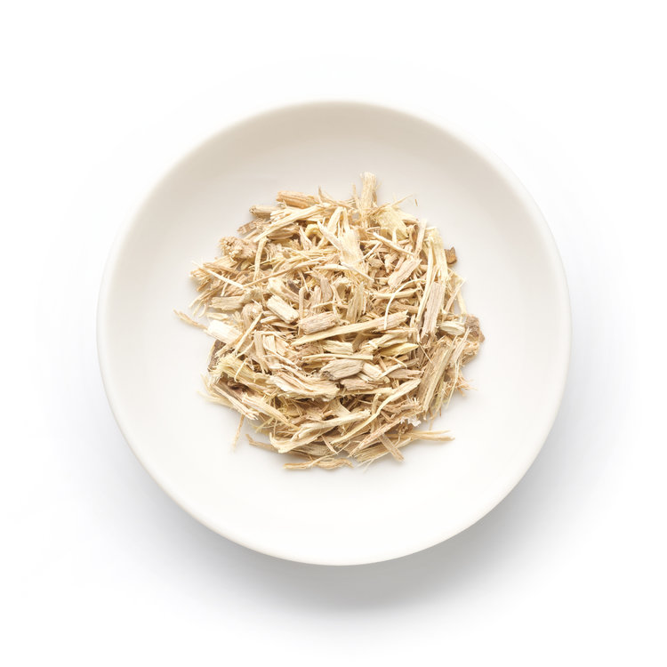
Eleutheroside E is a naturally occurring furofuran lignan diglucoside (syringaresinol di-o-glucoside), found in Eleuthero Root and 2 other sources. It is well tolerated orally (low toxicity).
Concentration
Content by Source
Concentrations not reported. No percent-dry-weight or mg/g figures have been published for this compound in its source plants.
Eleutheroside E at a Glance
Eleutheroside E is a furofuran lignan diglucoside — the molecule that results when the aglycone syringaresinol carries a glucose on each of its two phenolic oxygens. It is chemically identical to the compound earlier named acanthoside D. With eleutheroside B (syringin), it is one of the two marker compounds used to standardise eleuthero (Siberian ginseng, Eleutherococcus senticosus), the classic adaptogen.
That marker status is the source of most confusion about it. Eleuthero’s human reputation — anti-fatigue, endurance, stress resistance — rests on the whole standardised root extract, a mixture of eleutherosides, coumarins (isofraxidin), caffeoylquinic acids, phytosterols and immune-active polysaccharides 11,
What the isolate does have is a coherent, entirely preclinical signature. It suppresses NF-κB-driven inflammation in arthritis and immune-cell models 1,
Eleutheroside E & Syringaresinol
The single most useful thing to understand about this molecule is its relationship to syringaresinol, its aglycone. Eleutheroside E is (−)-syringaresinol-4,4′-O-β-D-diglucoside — the rigid furofuran (bis-epoxylignan) core of syringaresinol, built from two syringyl (4-hydroxy-3,5-dimethoxyphenyl) units, carrying a glucose on each phenolic oxygen. Remove those two sugars and you are left with the aglycone; add them back and you have the diglucoside.
Two naming points are worth fixing in place, because the older literature is scattered across synonyms:
- Acanthoside D = eleutheroside E. The compound isolated and named acanthoside D from Acanthopanax (an older genus name for Eleutherococcus) was later shown to be the same (−)-syringaresinol diglucoside now catalogued as eleutheroside E. Much of the pharmacology appears under the synonym Acanthopanax senticosus; keep those on the same line.
- Diglucoside, not aglycone. Findings on eleutheroside E (the glycoside) and on syringaresinol (the aglycone) are not interchangeable. They differ in polarity, in oral absorption, and in the exact assays run. Where a result is the glycoside’s, it stays the glycoside’s — only eleutheroside-E-specific studies are reported here and cross-refers to the aglycone page for the syringaresinol isolate work.
Structurally it belongs to the same furofuran (bis-epoxylignan) family as sesamin, pinoresinol and lariciresinol — the group whose two aryl rings are locked through a fused bicyclic diether — but as a glycoside rather than a free lignan, which is what governs how it behaves in the gut (see Pharmacokinetics). A closely related eleuthero/lignan glycoside, liriodendrin, is a symmetrical syringaresinol diglucoside variant that co-occurs with it in some plants; it is a distinct molecule and does not yet have its own page here.
The Eleuthero Adaptogen Marker
In eleuthero, the two pharmacopoeial markers are eleutheroside B (syringin, a phenylpropanoid glycoside) and eleutheroside E (this syringaresinol diglucoside). European Pharmacopoeia dried root is standardised to a minimum 0.08% for the sum of eleutheroside B + E, and Russian- and Korean-grown root generally runs several-fold higher in E than Chinese material 12Reference 12ReviewEleutherococcus root: a comprehensive review of its phytochemistry and pharmacological potential in the context of its adaptogenic effectView study →. The B+E pair is thus a quality-control fingerprint for the herb — how you verify you have genuine eleuthero root and not a substitute species.
Two cautions follow, and they are the point of this section:
- Marker ≠ active agent. Standardising an extract to eleutheroside E does not make eleutheroside E the thing responsible for eleuthero’s effect. The best-evidenced human outcomes for eleuthero — improved endurance and mental performance in mild fatigue and weakness — are documented for the whole extract 11Reference 11Evidence-based efficacy of adaptogens in fatigue, and molecular mechanisms related to their stress-protective activityView study →, and a comprehensive 2025 review concludes the adaptogenic action is a combination of anti-inflammatory, immunomodulatory, antioxidant and neuroprotective activity from the unfractionated raw material, not any single eleutheroside 12Reference 12ReviewEleutherococcus root: a comprehensive review of its phytochemistry and pharmacological potential in the context of its adaptogenic effectView study →. The marker is how you confirm the herb; it is not a claim that the marker carries the pharmacology.
- The anti-fatigue reputation is the extract’s, not the isolate’s. Where eleutheroside E has been singled out for anti-fatigue, the evidence is bioactivity-guided fractionation of the plant — the eleutheroside-containing fraction extended weight-loaded swimming time in mice, and the authors inferred eleutheroside E as the responsible constituent 2Reference 2Bioactivity-guided fractionation for anti-fatigue property of Acanthopanax senticosusView study →. That is a fraction-level attribution in rodents, not a trial of the purified molecule in people. Even the 2025 review flags the lack of robust clinical evidence for eleuthero in asthenia 12Reference 12ReviewEleutherococcus root: a comprehensive review of its phytochemistry and pharmacological potential in the context of its adaptogenic effectView study →.
What It's Used For
The isolate literature is preclinical throughout — cell and animal work, no human trials of the pure compound. It clusters into an anti-inflammatory core, an ischaemia-protection thread that recurs across heart and brain, and scattered metabolic, neurological and skeletal signals.
Support is an experimental score — weighted by study type (human > animal > in vitro) and volume. A beta, not a validated metric. Every row here is isolate preclinical (no human data), so scores stay low by design. Each application links to its write-up.
The most developed thread, and it is the isolated molecule. Purified eleutheroside E reduced the severity of collagen-induced arthritis in mice — lowering arthritis score and incidence, and cutting inflammatory-cell infiltration, pannus, cartilage damage and bone erosion — with a marked fall in TNF-α and IL-6 both in vivo and in vitro, attributed to NF-κB inhibition 1Reference 1AnimalEleutheroside E ameliorates arthritis severity in collagen-induced arthritis mice model by suppressing inflammatory cytokine releaseView study →.
The same NF-κB axis recurs in its cardiac and pulmonary hypoxia work: it suppressed IκBα phosphorylation and NF-κB p65 nuclear translocation in cardiomyocytes 5Reference 5Eleutheroside E decreases oxidative stress and NF-κB activation and reprograms the metabolic response against hypoxia-reoxygenation injury in H9c2 cellsView study → and downregulated NLRP3-inflammasome/pyroptosis signalling in high-altitude heart injury 6Reference 6Eleutheroside E from pre-treatment of Acanthopanax senticosus ameliorates high-altitude-induced heart injury by regulating NLRP3 inflammasome-mediated pyroptosis via the NLRP3/caspase-1 pathwayView study →.
Gap: entirely isolate cell/animal work — no human anti-inflammatory trial — and the mechanistic anchor (NF-κB) is shared with the aglycone syringaresinol, so the effect is consistent but clinically unproven.
A genuinely recurring isolate signal across organs. In cardiomyocytes (H9c2), eleutheroside E pre-treatment attenuated hypoxia-reoxygenation apoptosis, inhibiting mitochondrial oxidative stress and NF-κB activation and reprogramming the metabolic response to injury 5Reference 5Eleutheroside E decreases oxidative stress and NF-κB activation and reprograms the metabolic response against hypoxia-reoxygenation injury in H9c2 cellsView study →.
In rats exposed to simulated high altitude it dose-dependently reduced heart injury by suppressing NLRP3-inflammasome-mediated pyroptosis via the NLRP3/caspase-1 pathway 6Reference 6Eleutheroside E from pre-treatment of Acanthopanax senticosus ameliorates high-altitude-induced heart injury by regulating NLRP3 inflammasome-mediated pyroptosis via the NLRP3/caspase-1 pathwayView study →.
And in a rat cerebral ischaemia-reperfusion model it reduced hippocampal-neuron apoptosis in a 5-HT receptor 2C (Htr2c)-dependent manner, upregulating Htr2c and inhibiting caspase-3/6/7 — silencing Htr2c abolished the protection 7Reference 7AnimalEleutheroside E alleviates cerebral ischemia-reperfusion injury in a 5-hydroxytryptamine receptor 2C (Htr2c)-dependent manner in ratsView study →.
Gap: three separate but each single isolate models (cell + rodent), all on surrogate injury/apoptosis endpoints; no clinical ischaemia outcome, and the mechanisms proposed (metabolic, pyroptotic, serotonergic) do not yet cohere into one pathway 5,
The reputational headline — and the weakest link between molecule and claim. In bioactivity-guided fractionation of Acanthopanax senticosus, the eleutheroside-containing n-butanol/ethanol fraction significantly extended weight-loaded swimming time in mice, and the authors concluded “eleutheroside E may be responsible” for the anti-fatigue effect, via lower triglycerides (increased fat utilisation), delayed blood-urea-nitrogen accumulation and raised LDH to limit lactic-acid buildup 2Reference 2Bioactivity-guided fractionation for anti-fatigue property of Acanthopanax senticosusView study →.
This is a fraction-level inference in rodents, not a purified-isolate trial.
Gap: the anti-fatigue and endurance evidence in humans belongs to the whole standardised eleuthero extract 11Reference 11Evidence-based efficacy of adaptogens in fatigue, and molecular mechanisms related to their stress-protective activityView study →, not to isolated eleutheroside E; the isolate attribution here is indirect, and a 2025 review still flags the lack of robust clinical adaptogen evidence 12Reference 12ReviewEleutherococcus root: a comprehensive review of its phytochemistry and pharmacological potential in the context of its adaptogenic effectView study →.
A single but reasonably complete isolate study. In type-2-diabetic db/db mice, eleutheroside E lowered blood glucose and serum insulin, improved the lipid profile, attenuated HOMA-IR and increased insulin sensitivity on glucose- and insulin-tolerance testing; mechanistically it raised insulin-stimulated glucose uptake in C2C12 myotubes, reversed TNF-α-suppressed uptake in 3T3-L1 adipocytes, and improved hepatic glucose handling (up glycolysis, down gluconeogenesis) 4Reference 4AnimalEleutheroside E, an active component of Eleutherococcus senticosus, ameliorates insulin resistance in type 2 diabetic db/db miceView study →.
Both the whole extract and the isolate were active.
Gap: one animal/cell study, no human metabolic data, and no dose-ranging or long-term safety for the isolate 4Reference 4AnimalEleutheroside E, an active component of Eleutherococcus senticosus, ameliorates insulin resistance in type 2 diabetic db/db miceView study →.
Two isolate rodent studies, on different endpoints. In aged rats, eleutheroside E (50 mg/kg i.p.) reversed isoflurane/anaesthesia-induced cognitive dysfunction on novel-object-recognition and Morris-water-maze testing, working through the α7-nAChR–NMDAR pathway and restored acetylcholine signalling 8Reference 8Eleutheroside E attenuates isoflurane-induced cognitive dysfunction by regulating the α7-nAChR-NMDAR pathwayView study →.
In a mouse 72-hour sleep-deprivation model, repeated eleutheroside E (10 or 50 mg/kg) normalised sleep-deprivation-induced behavioural and biochemical alterations (passive avoidance, locomotion, Y-maze) 3Reference 3AnimalThe effect of eleutheroside E on behavioral alterations in murine sleep deprivation stress modelView study → — the closest thing to the “circadian isolate” work, though it tests sleep-stress recovery rather than clock genes directly.
Gap: two small, unreplicated rodent behavioural studies with different models and doses; no human cognitive or sleep data, and the cerebral-ischaemia result 7Reference 7AnimalEleutheroside E alleviates cerebral ischemia-reperfusion injury in a 5-hydroxytryptamine receptor 2C (Htr2c)-dependent manner in ratsView study → sits alongside these as a separate neuro-thread rather than a unified mechanism 3,
The thinnest thread. A combined network-pharmacology, molecular-docking and ovariectomy (OVX) mouse study reported that eleutheroside E prevented ovariectomy-induced bone loss by regulating gut microbiota (raising Lactobacillus, lowering Clostridiaceae) and the associated inflammatory/osteoclast response, framing it as a candidate osteoporosis agent 9Reference 9Decoding the mechanism of eleutheroside E in treating osteoporosis via network pharmacological analysis and molecular docking of osteoclast-related genes and gut microbiotaView study →.
Gap: a single hypothesis-generating study leaning heavily on in-silico target prediction plus one animal model — no dedicated bone-density pharmacology, no human data 9Reference 9Decoding the mechanism of eleutheroside E in treating osteoporosis via network pharmacological analysis and molecular docking of osteoclast-related genes and gut microbiotaView study →.
Mechanisms
| Target / pathway | Effect | Relevant to |
|---|---|---|
| NF-κB (IκBα phosphorylation, p65 translocation) | suppressed | anti-inflammatorycardioprotection |
| TNF-α, IL-6 (and other cytokines) | downregulated | arthritissystemic inflammation |
| NLRP3 inflammasome / caspase-1 (pyroptosis) | inhibited | high-altitude heart injury |
| Mitochondrial oxidative stress + metabolite reprogramming | reduced / re-modulated | cardiomyocyte hypoxia-reoxygenation |
| 5-HT receptor 2C (Htr2c) → caspase-3/6/7 | Htr2c ↑, caspases ↓ → less apoptosis | cerebral ischaemia-reperfusion |
| Insulin signalling / glucose uptake (myotubes, adipocytes); hepatic glycolysis vs gluconeogenesis | insulin sensitivity ↑; HOMA-IR ↓ | insulin resistance (db/db) |
| α7-nAChR–NMDAR / acetylcholine | restored | anaesthesia-induced cognitive dysfunction |
| Gut microbiota (Lactobacillus ↑, Clostridiaceae ↓) | remodelled | ovariectomy bone loss |
| CYP2C9, CYP2E1 | weak inhibition (high μM) | drug-interaction potential |
The load-bearing observation is that the anti-inflammatory (NF-κB) and ischaemia-protection threads converge on inflammation and cell-survival signalling — a plausible rationale for why the diglucoside is an adaptogen marker, but one demonstrated only in cells and rodents, and never for the isolate in people.
Pharmacokinetics
Human pharmacokinetics of eleutheroside E are uncharacterised, and the compound’s structure predicts poor oral bioavailability — the key caveat behind every in-vitro potency claim. As a furofuran lignan diglucoside it is large and polar; like other lignan glycosides it is expected to be deglycosylated by gut microflora before the lipophilic aglycone (syringaresinol) can be absorbed, and syringaresinol itself is not efficiently converted to the mammalian lignans enterolactone/enterodiol and shows P-glycoprotein-limited intestinal absorption. Rodent studies dose eleutheroside E parenterally (e.g. 10–50 mg/kg intraperitoneally) 3,
Clinical Trials
There are no clinical trials of isolated eleutheroside E for any indication. The human evidence often invoked alongside it belongs to whole eleuthero extract standardised to the eleutheroside B+E marker pair — a different, multi-component product — where it has been studied for fatigue, endurance and stress 11,
| Completed | Planned | Terminated | Preclinical |
|---|---|---|---|
| None(isolate); whole-eleuthero-extract adaptogen/fatigue trials are a separate entity | — | — | Anti-inflammatory, ischaemic cardio-/cerebroprotection, insulin resistance, cognition/sleep, bone |
Last checked: July 2026.
Isolate vs. Plant Studies
This molecule shows a clean split. The mechanistic work is genuine isolate science — purified eleutheroside E in defined arthritis, cardiomyocyte, ischaemia, diabetic-mouse and cognition models 1,
Prevalence in Nature
Eleutheroside E is a plant lignan glycoside whose signature concentrated form is in eleuthero (Eleutherococcus senticosus / Acanthopanax senticosus) root, where it and eleutheroside B (syringin) are the pharmacopoeial marker pair. Its content is strongly provenance-dependent: Russian- and Korean-grown root generally carries several-fold more eleutheroside E than Chinese material, and the two markers together must reach ≥0.08% of dried root under the European Pharmacopoeia standard 12Reference 12ReviewEleutherococcus root: a comprehensive review of its phytochemistry and pharmacological potential in the context of its adaptogenic effectView study →. As the diglucoside of the widespread lignan syringaresinol, it (or close variants such as liriodendrin) also occurs across various lignan-bearing plants, but eleuthero root is its defining source.
Major Natural Sources
| Source (Botanical) | Part | Form | Notes |
|---|---|---|---|
| Eleuthero / Siberian ginseng (Eleutherococcus senticosus) | Root / underground organs | Eleutheroside E (diglucoside) | Pharmacopoeial marker with eleutheroside B; ≥0.08% B+E; higher in Russian/Korean root 12Reference 12ReviewEleutherococcus root: a comprehensive review of its phytochemistry and pharmacological potential in the context of its adaptogenic effectView study → |
| Other Eleutherococcus / Acanthopanax spp. | Root / bark | Eleutheroside E / acanthoside D | Historical acanthoside D isolations; watch for substitute-species chemotypes |
| Various lignan-bearing plants | Various | Syringaresinol glycosides | Background occurrence as the syringaresinol diglucoside / related liriodendrin |
Quantified % dry-weight figures per source (beyond the combined B+E pharmacopoeial minimum) were not pinned — the E-specific concentration data live in the gaps tracker to chase on a later pass.
Discovery & Synthesis
Eleutheroside E entered the literature partly under a different name. The compound isolated from Acanthopanax and named acanthoside D was later identified as (−)-syringaresinol-4,4′-O-β-D-diglucoside — the same molecule catalogued as eleutheroside E once Eleutherococcus pharmacology consolidated around a numbered “eleutheroside” marker series. Because it is simply the diglucoside of syringaresinol, its biosynthesis follows the general lignan route — oxidative (peroxidase/laccase-type) coupling of sinapyl alcohol to the symmetrical furofuran core, followed by glucosylation at both phenolic oxygens. In practice, research-grade eleutheroside E is obtained by extraction and isolation from eleuthero root (and used as an analytical standard for herb standardisation) rather than by total synthesis.
Patents: not yet researched.
Toxicity & Safety
Eleutheroside E carries a low toxicity flag. It is a dietary lignan glycoside consumed at marker-level quantities within a herb (eleuthero) that has a long traditional-use record, and dedicated isolate testing has surfaced no cytotoxicity or genotoxicity signal for it or its aglycone. The one measured pharmacological caution is a weak drug-metabolism interaction, and the broader picture is defined by absence of dedicated human data rather than by any established harm.
- Marker-level dietary exposure. Human intake is small and indirect, chiefly as one of two standardisation markers in eleuthero preparations (≥0.08% combined B+E) 12Reference 12ReviewEleutherococcus root: a comprehensive review of its phytochemistry and pharmacological potential in the context of its adaptogenic effectView study →.
- No isolate cyto-/genotoxicity signal. The aglycone syringaresinol tested negative for cytotoxicity and DNA damage in liver and colon cells; no adverse isolate finding is reported for eleutheroside E itself.
- Weak CYP2C9 / CYP2E1 inhibition. In rat liver microsomes, eleutherosides B and E weakly inhibited CYP2C9 and CYP2E1 (IC₅₀ in the high-μM range) with no effect on CYP2D6 or CYP3A4 — a theoretical interaction for co-administered CYP2C9/2E1 substrates 10Reference 10AnimalEffects of eleutheroside B and eleutheroside E on activity of cytochrome P450 in rat liver microsomesView study →.
- No human safety, PK or interaction data for the isolate. Absence of evidence is not evidence of safety; poor oral bioavailability further limits what the in-vitro data imply.
Full Safety & Interactions Detail
The dedicated interaction data are the informative part. In rat liver microsomes, eleutheroside E (with eleutheroside B) weakly inhibited CYP2C9 and CYP2E1 — IC₅₀ ≈ 262 μM (CYP2C9) and ≈ 188 μM (CYP2E1), a mixed-type inhibition of CYP2E1 (Ki ≈ 172 μM) — while leaving CYP2D6 and CYP3A4 unaffected 10Reference 10AnimalEffects of eleutheroside B and eleutheroside E on activity of cytochrome P450 in rat liver microsomesView study →.
Those concentrations are high relative to plausible exposure from a standardised herbal dose, so the practical interaction risk is modest, but it flags CYP2C9/2E1 substrates as the theoretical concern rather than the CYP3A4/P-gp axis. Separately, whole-eleuthero-root extracts are known to contain digoxin-like immunoreactive substances that distort digoxin immunoassays — an assay interference, not a pharmacokinetic effect, and a property of the extract rather than of purified eleutheroside E, but worth flagging on any eleuthero-related page. Beyond this, the safety picture is defined by missing data: there are no human safety studies, no repeat-dose or reproductive-toxicity package, and no characterised half-life for the isolate. Pregnancy and lactation decisions belong to the eleuthero herb page and any supplement product — where intake is actually decided — not to this molecule page.
Dosage
There is no established human dose for isolated eleutheroside E, and nothing here is a recommendation — the figures are the preclinical research doses behind the sections above:
| Application | Model | Exposure studied |
|---|---|---|
| Neuroprotection / cognition | Aged rats | 50 mg/kg intraperitoneally 8Reference 8Eleutheroside E attenuates isoflurane-induced cognitive dysfunction by regulating the α7-nAChR-NMDAR pathwayView study → |
| Sleep-deprivation behaviour | Mice | 10 or 50 mg/kg, repeated 3Reference 3AnimalThe effect of eleutheroside E on behavioral alterations in murine sleep deprivation stress modelView study → |
| Anti-inflammatory (arthritis) | CIA mice / cells | in-vivo dosing + μM in vitro 1Reference 1AnimalEleutheroside E ameliorates arthritis severity in collagen-induced arthritis mice model by suppressing inflammatory cytokine releaseView study → |
| Insulin resistance | db/db mice; C2C12 / 3T3-L1 cells | dietary/oral in mice; μM in cells 4Reference 4AnimalEleutheroside E, an active component of Eleutherococcus senticosus, ameliorates insulin resistance in type 2 diabetic db/db miceView study → |
| Ischaemia protection | H9c2 cells; rats | μM in vitro; dose-dependent in vivo 5, |
Human exposure in practice is indirect and small — as one of two marker eleutherosides within eleuthero preparations standardised to the combined eleutheroside B + E fraction (≥0.08% of dried root), not as the free isolate 12Reference 12ReviewEleutherococcus root: a comprehensive review of its phytochemistry and pharmacological potential in the context of its adaptogenic effectView study →. Note that the rodent studies frequently dose it by injection, bypassing the glycoside’s poor oral absorption; an oral human equivalent cannot be inferred from them. These are doses studied in research and are not a personal recommendation.
References
- He C, Chen X, Zhao C, et al. (2014). Eleutheroside E ameliorates arthritis severity in collagen-induced arthritis mice model by suppressing inflammatory cytokine release. Inflammation. https://pubmed.ncbi.nlm.nih.gov/24917466/
- Huang LZ, Huang BK, Ye Q, Qin LP (2011). Bioactivity-guided fractionation for anti-fatigue property of Acanthopanax senticosus. Journal of Ethnopharmacology. https://pubmed.ncbi.nlm.nih.gov/20920564/
- Huang LZ, Wei L, Zhao HF, Huang BK, Rahman K, Qin LP (2011). The effect of eleutheroside E on behavioral alterations in murine sleep deprivation stress model. European Journal of Pharmacology. https://pubmed.ncbi.nlm.nih.gov/21376030/
- Ahn J, Um MY, Lee H, et al. (2013). Eleutheroside E, an active component of Eleutherococcus senticosus, ameliorates insulin resistance in type 2 diabetic db/db mice. Evidence-Based Complementary and Alternative Medicine. https://pubmed.ncbi.nlm.nih.gov/23690865/
- Wang S, Yang X (2020). Eleutheroside E decreases oxidative stress and NF-κB activation and reprograms the metabolic response against hypoxia-reoxygenation injury in H9c2 cells. International Immunopharmacology. https://pubmed.ncbi.nlm.nih.gov/32330867/
- Jia N, Shen Z, Zhao S, et al. (2023). Eleutheroside E from pre-treatment of Acanthopanax senticosus ameliorates high-altitude-induced heart injury by regulating NLRP3 inflammasome-mediated pyroptosis via the NLRP3/caspase-1 pathway. International Immunopharmacology. https://pubmed.ncbi.nlm.nih.gov/37331291/
- Liu Z, Gao W, Xu Y (2022). Eleutheroside E alleviates cerebral ischemia-reperfusion injury in a 5-hydroxytryptamine receptor 2C (Htr2c)-dependent manner in rats. Bioengineered. https://pubmed.ncbi.nlm.nih.gov/35502892/
- Lu X, Chai XQ (2019). Eleutheroside E attenuates isoflurane-induced cognitive dysfunction by regulating the α7-nAChR-NMDAR pathway. Neuroreport. https://pubmed.ncbi.nlm.nih.gov/30585907/
- Zhou T, Zhou Y, Ge D, et al. (2023). Decoding the mechanism of eleutheroside E in treating osteoporosis via network pharmacological analysis and molecular docking of osteoclast-related genes and gut microbiota. Frontiers in Endocrinology. https://pubmed.ncbi.nlm.nih.gov/38027135/
- Guo S, Liu Y, Lin Z, et al. (2014). Effects of eleutheroside B and eleutheroside E on activity of cytochrome P450 in rat liver microsomes. BMC Complementary and Alternative Medicine. https://pubmed.ncbi.nlm.nih.gov/24383621/
- Panossian A, Wikman G (2009). Evidence-based efficacy of adaptogens in fatigue, and molecular mechanisms related to their stress-protective activity. Current Clinical Pharmacology. https://pubmed.ncbi.nlm.nih.gov/19500070/
- Patyra A, Kołtun-Jasion M, Kupniewska K, Parzonko A, Kiss AK (2025). Eleutherococcus root: a comprehensive review of its phytochemistry and pharmacological potential in the context of its adaptogenic effect. Frontiers in Pharmacology. https://pubmed.ncbi.nlm.nih.gov/41235111/