Compound Monograph
Rugosin D
Rugosin D is a large dimeric ellagitannin from rose petals and meadowsweet — two tellimagrandin II units bridged by a valoneoyl group. It is the most potent natural inhibitor yet found of histidine decarboxylase, the enzyme that makes histamine, which is the leading mechanistic explanation for meadowsweet's odd reputation as a salicylate herb that protects the stomach. That explanation is enzyme-level only: at nearly 1,900 daltons the molecule is almost certainly not absorbed intact.

Sources · 6
Where Does It Come From?
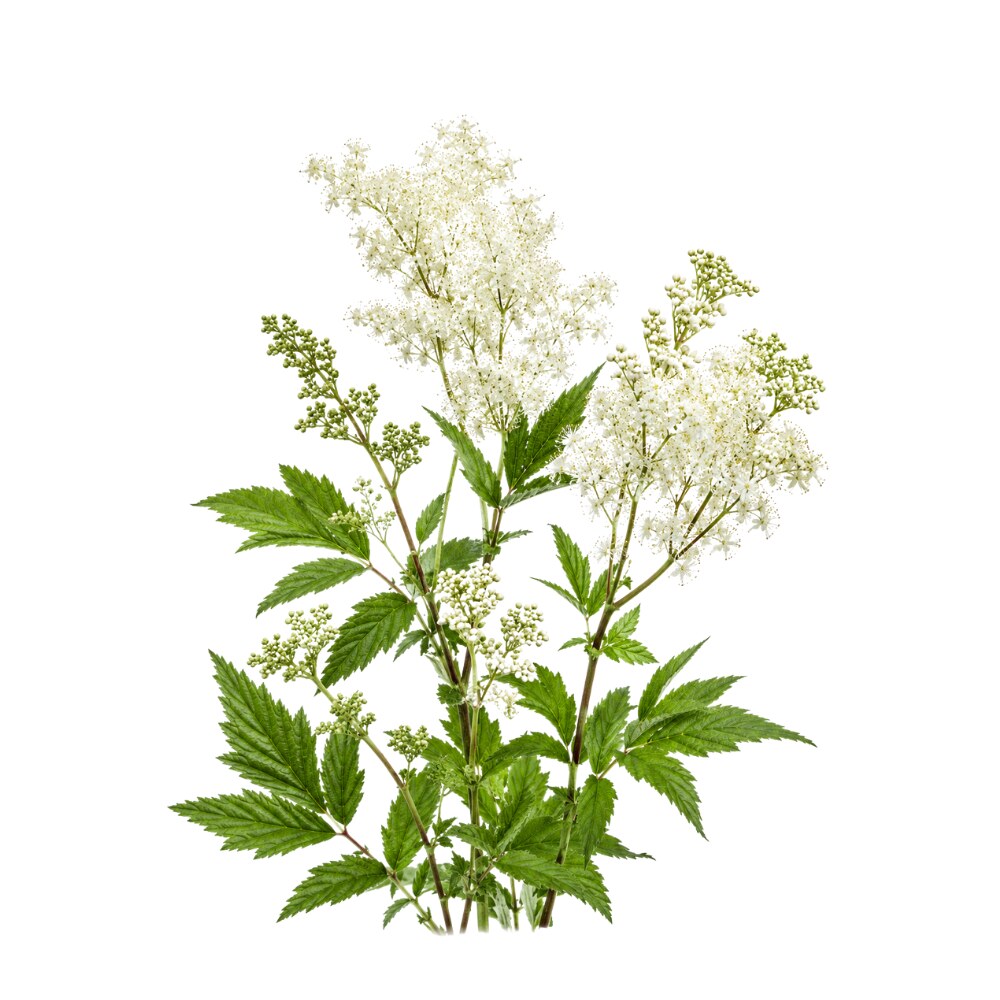
Rugosin D is a naturally occurring ellagitannin (dimeric hydrolysable tannin; valoneoyl-linked), found in Meadowsweet, Rugosa rose, Prickly rose and 3 other sources. It is well tolerated orally (low toxicity).
Concentration
Content by Source
Reported concentrations across the plants that contain rugosin D — the bar marks the typical level, the line shows the reported range.
Literature figures for varying plant parts and preparations. Read them as a comparative guide, not exact assays.
Rugosin D at a Glance
Rugosin D is a dimeric ellagitannin — a hydrolysable tannin built from two smaller tannin units joined together, with a molecular weight near 1,875 daltons. It was isolated in 1982 from the petals of the rugosa rose, Rosa rugosa, which is where the whole rugosin family gets its name 5Reference 5Rugosin D, E, F and G, dimeric and trimeric hydrolyzable tanninsView study →. Chemically it is two tellimagrandin II units bridged by a valoneoyl group, and that dimeric architecture is the reason the molecule matters pharmacologically.
Its claim to attention is narrow but genuinely interesting. Rugosin D is the most potent natural inhibitor of histidine decarboxylase (HDC) yet reported — the enzyme that decarboxylates the amino acid histidine into histamine, including the histamine that drives gastric-acid secretion 1,
The load-bearing caveat is size. At nearly 1,900 daltons with more than fifty oxygen atoms, rugosin D is far outside the range of molecules that cross the gut wall intact. Like other hydrolysable tannins it is expected to be hydrolysed in the gut and converted by colonic bacteria toward ellagic acid and then urolithins, with large person-to-person variation 14,
The Salicylate Paradox
This is the reason rugosin D has a page at all, so it is worth stating precisely — including where the evidence runs out.
The paradox. Meadowsweet (Filipendula ulmaria) is the plant salicin and salicylic acid were named after — Spiraea, its old genus, is the “spir” in aspirin. Yet the herb’s traditional and experimental reputation is gastroprotective. In classic rat work a flower decoction reduced ulceration from several ulcerogenic procedures and, pointedly, prevented acetylsalicylic-acid-induced gastric lesions 11Reference 11Anti-ulcer action of a decoction of the flowers of the dropwort, Filipendula ulmariaView study →; a modern study confirmed protection against ethanol-induced acute gastric injury with lyophilised flower infusions at 100–300 mg/kg orally 12Reference 12Antioxidant, anti-inflammatory and gastroprotective activity of Filipendula ulmaria (L.) Maxim. and Filipendula vulgaris MoenchView study →. A salicylate-bearing plant that guards the stomach is the opposite of what the pharmacology of aspirin would predict.
The proposed resolution. Gastric acid secretion is driven substantially by histamine acting on parietal-cell H2 receptors, and that histamine is made on demand by histidine decarboxylase. Inhibit HDC and you reduce the histamine supply upstream of the receptor — a different point of attack from the H2 blockers and proton-pump inhibitors used clinically. Nitta and colleagues fractionated the ethyl-acetate extract of meadowsweet petals and found the activity concentrated in four ellagitannins: rugosin D, rugosin A, rugosin A methyl ester (a compound not previously described) and tellimagrandin II. All four inhibited recombinant human HDC non-competitively, with inhibition constants of roughly 0.35–1 µM — comparable to purpose-designed substrate-analogue inhibitors, and a big step up from the best dietary HDC inhibitor previously known, epicatechin gallate, which is competitive with a Ki near 10 µM 1,
Where it stops being demonstrated. Three gaps, in order of importance:
- It is an enzyme assay. Nitta 2013 used recombinant human HDC in vitro — not cells, not stomach tissue, not an animal 1Reference 1Inhibitory activity of Filipendula ulmaria constituents on recombinant human histidine decarboxylaseView study →. No study has shown rugosin D lowering gastric histamine, gastric acid or ulcer scores in a living animal.
- A µM constant on a 1,875-dalton molecule is not a systemic effect. Hydrolysable tannins of this size are not meaningfully absorbed intact; a low-micromolar Ki is achievable in the stomach and gut lumen but not in plasma 14,
15, . If the mechanism works at all in a person, it works locally — which happens to be where gastric HDC is, so the argument is not absurd, only unproven.16, 19Reference 14Ellagic acid metabolism by human gut microbiota — consistent observation of three urolithin phenotypes in intervention trials, independent of food source, age and health statusView study →Reference 15Biological significance of urolithins, the gut microbial ellagic-acid-derived metabolites — the evidence so farView study →Reference 16Bioavailability of ellagic acid in human plasma after consumption of ellagitannins from pomegranate (Punica granatum L.) juiceView study →Reference 19Partition coefficients (logP) of hydrolysable tanninsView study → - The closest in-vivo evidence is for the monomer, not for rugosin D. In the ethanol-injury rat model, tellimagrandin II at 40 mg/kg orally showed ulcer-preventive ability 12Reference 12Antioxidant, anti-inflammatory and gastroprotective activity of Filipendula ulmaria (L.) Maxim. and Filipendula vulgaris MoenchView study → — tellimagrandin II being precisely the unit rugosin D is built from. That is the best bridge in the literature, and it is still one step to the side: the study tested a different (smaller) molecule and did not test the HDC mechanism.
No animal or human gastroprotection study of isolated rugosin D, or of a rugosin-standardised extract, exists. The honest summary is that HDC inhibition is a well-measured enzyme-level property of the right molecule from the right plant, plausibly acting at the right anatomical site, that has never been tested in a stomach.
A Dimer of Tellimagrandin II
Rugosin D is not a dimer of rugosin A, despite the shared name. The rugosin series was named for its source plant, not for a shared build.
Rugosin D consists of two tellimagrandin II units linked by a single valoneoyl (m-depside, m-DOG) bond 5,
Dimerisation is the structure–activity story. Against bacterial HDC from Morganella morganii, the four meadowsweet ellagitannins line up in a clear order — rugosin D (dimer) IC50 1.5 µM > rugosin A methyl ester 4.4 µM > tellimagrandin II 6.1 µM > rugosin A 6.8 µM 2Reference 2Inhibition of Morganella morganii histidine decarboxylase activity and histamine accumulation in mackerel muscle derived from Filipendula ulmaria extractsView study →. The dimer is roughly four times more potent than either monomer. Extending the chain extends the trend: the trimer rugosin G inhibits recombinant human HDC more strongly than the monomers and more strongly than oenothein B, a dimer with a macrocyclic rather than linear structure — from which Nitta’s group concluded that potency tracks a linear oligomeric conformation, not merely molecular size 3Reference 3The ellagitannin trimer rugosin G inhibits recombinant human histidine decarboxylaseView study →. A separate metabolomic study reached a compatible conclusion from a different direction, finding rugosin D the most growth-inhibitory of the hydrolysable tannins tested against E. coli and S. aureus and arguing that effective tannins need both hydrophobic and hydrophilic regions — a balance a linear dimer supplies and a small monomer does not 8Reference 8NMR metabolomics and DNA sequencing of Escherichia coli and Staphylococcus aureus cultures treated with hydrolyzable tanninsView study →.
The catch is that the same property cuts both ways: the extended, multivalent surface that makes rugosin D a better enzyme inhibitor is exactly what makes it a worse drug candidate. Bigger, more polar, more protein-binding, less absorbable.
What It's Used For
The literature on rugosin D is small — a PubMed search for the molecule by name returns single digits — and it is entirely in vitro apart from a whole-extract food-safety experiment. What exists is unusually clean, though: several papers test the purified molecule rather than a crude tannin fraction, which is rare in this field.
Support is an experimental score — weighted by study type (human > animal > in vitro) and volume. A beta, not a validated metric. Every row here is capped low because no application has been tested in an animal or a person using the isolated molecule.
The marquee finding, and the reason this molecule appears in the meadowsweet monograph. Screening the ethyl-acetate fraction of Filipendula ulmaria petals against recombinant human HDC, Nitta and colleagues identified four ellagitannins as the active principles — rugosin D, rugosin A, rugosin A methyl ester and tellimagrandin II — all inhibiting non-competitively with Ki ≈ 0.35–1 µM, values the authors describe as comparable to designed substrate-analogue inhibitors 1Reference 1Inhibitory activity of Filipendula ulmaria constituents on recombinant human histidine decarboxylaseView study →.
For context, the same group’s earlier survey of dietary phenolics found epicatechin gallate the best of the food compounds at Ki = 10 µM, and competitive 4Reference 4Food components inhibiting recombinant human histidine decarboxylase activityView study →; the ellagitannins are both more potent and mechanistically different. A follow-up against the bacterial HDC of Morganella morganii — the enzyme behind scombroid histamine poisoning — ranked the same four compounds and put rugosin D first at IC50 1.5 µM; a 2% meadowsweet extract applied to mackerel muscle significantly reduced histamine accumulation 2Reference 2Inhibition of Morganella morganii histidine decarboxylase activity and histamine accumulation in mackerel muscle derived from Filipendula ulmaria extractsView study →.
Because parietal-cell histamine drives gastric acid secretion, HDC inhibition is a coherent upstream explanation for meadowsweet’s anti-ulcer behaviour 11,
Gap: enzyme assays only. Nothing has been measured in a stomach — no gastric histamine, acid-output or ulcer-score data for rugosin D in any animal, and no human data of any kind. The nearest in-vivo evidence is that its monomer tellimagrandin II at 40 mg/kg p.o. prevented ethanol-induced gastric ulceration in rats, in a study that tested outcome and not mechanism 12Reference 12Antioxidant, anti-inflammatory and gastroprotective activity of Filipendula ulmaria (L.) Maxim. and Filipendula vulgaris MoenchView study →. The mackerel result is a food-preservation finding using a whole extract, not a gastric one.
The best-quantified application, and the one where the “gut lumen, not plasma” framing is a feature rather than a limitation — digestive enzymes are in the lumen. Working with Rosa acicularis, in which rugosin D is the dominant ellagitannin, Olennikov and colleagues measured the purified molecule against three α-amylases: human pancreatic IC50 30.84 µg/mL, porcine pancreatic 32.09 µg/mL, human salivary 67.59 µg/mL (roughly 16–36 µM at MW 1,875) 6Reference 6Metabolites of prickly rose — chemodiversity and digestive-enzyme-inhibiting potential of Rosa acicularis and the main ellagitannin rugosin DView study →.
In a simulated gastrointestinal digestion model rugosin D produced dose-dependent suppression of amylase activity across 1–1,000 µg/mL, in the same range as the drug acarbose. Earlier work had already flagged rugosins A and D from R. gallica petals as principal inhibitors of a bacterial α-amylase 6Reference 6Metabolites of prickly rose — chemodiversity and digestive-enzyme-inhibiting potential of Rosa acicularis and the main ellagitannin rugosin DView study →.
Separately, rugosin D inhibited diacylglycerol acyltransferase (DGAT) — the final step of triglyceride assembly — by 82% at 10 µM, in a screen of Rosa centifolia petal ellagitannins where rugosin B (96%) and eusupinin A (84%) scored similarly 7Reference 7Identification of diacylglycerol acyltransferase inhibitors from Rosa centifolia petalsView study →.
Gap: all cell-free or cell-based; no glycaemic, lipid or body-weight outcome has been measured in an animal or a person for rugosin D, and no dose–response has been established for a rugosin-containing preparation. The DGAT result is a single screen at a single concentration.
A study combining NMR metabolomics with sequencing exposed Escherichia coli and Staphylococcus aureus — and faecal cultures — to a panel of hydrolysable tannins. Rugosin D was the most effective at inhibiting growth and at shifting bacterial metabolic profiles, with E. coli more perturbed than S. aureus; the authors concluded that a tannin needs both hydrophobic and hydrophilic regions to do this, a structural argument that favours linear dimers 8Reference 8NMR metabolomics and DNA sequencing of Escherichia coli and Staphylococcus aureus cultures treated with hydrolyzable tanninsView study →.
The selectivity question has been probed at the extract level: powdered Rosa rugosa petals, whose actives were identified as hydrolysable tannins, inhibited E. coli and S. aureus while leaving bifidobacteria and lactobacilli unaffected — a prebiotic-like pattern 18Reference 18Effects of Rosa rugosa petals on intestinal bacteriaView study →.
Gap: the selectivity result is for whole rose-petal tannins, not for rugosin D specifically, so the flattering “kills pathogens, spares commensals” line cannot be assigned to this molecule. Neither study is an infection model, and no minimum inhibitory concentration in a clinically relevant range has been established.
Included because the ellagitannin literature makes a structural claim that rugosin D fits, and because that is easy to misread as evidence about rugosin D. Miyamoto and colleagues tested fifteen tannins in mice (10 mg/kg intraperitoneally, four days before S-180 inoculation) and found significant antitumour activity in those built from tellimagrandin I or II units — tellimagrandin II itself, rugosin A, hirtellin B, oenothein A and oenothein B — with the oligomers also the strongest inducers of IL-1β from human peripheral macrophages 9Reference 9Antitumor activity and interleukin-1 induction by tanninsView study →. Rugosin D is a tellimagrandin II dimer, so the class principle predicts it should behave the same way.
Gap: rugosin D was not one of the fifteen tannins tested 9Reference 9Antitumor activity and interleukin-1 induction by tanninsView study →. This is a prediction from structure, not a result. The route is also intraperitoneal, which bypasses exactly the absorption problem that governs oral tannins. Note as well that the cytokine in this literature is IL-1β, not IL-2 — the two are frequently conflated in secondary sources. Related antiproliferative/NF-κB work on Rosa rugosa tannins concerns rugosin E, a different dimer, and does not transfer.
Mechanisms
| Target / pathway | Effect | Relevant to |
|---|---|---|
| Histidine decarboxylase (human, recombinant) | non-competitive inhibition, Ki ≈ 0.35–1 µM → less histidine converted to histamine | gastroprotection; histamine-mediated symptoms |
| Histidine decarboxylase (Morganella morganii) | IC50 1.5 µM — most potent of the four meadowsweet ellagitannins | food-borne histamine (scombroid)SAR ranking |
| α-Amylase (human pancreatic / salivary; porcine) | IC50 ≈ 31 / 68 / 32 µg/mL — starch digestion slowed in the lumen | carbohydrate digestion |
| Diacylglycerol acyltransferase (DGAT) | 82% inhibition at 10 µM | triglyceride synthesis (screen-level) |
| Bacterial growth / metabolome (E. coli, S. aureus) | most inhibitory tannin of its panel; requires balanced hydrophobic + hydrophilic surface | antibacterial |
| Non-specific protein binding (astringency) | multivalent polyphenol–protein complexation — the general tannin action | astringent effectiron chelationmouthfeel |
The pattern is consistent: rugosin D is a promiscuous, high-affinity protein binder whose large linear surface lets it inhibit structurally unrelated enzymes. That explains both the impressive potency numbers and why they should not be over-read — an inhibitor this indiscriminate at micromolar concentrations in a test tube tells you less about physiology than a weaker, more selective one would.
Pharmacokinetics
There is no pharmacokinetic study of rugosin D — no absorption, distribution or metabolite data for the molecule in any species. What can be said is inferential, and it is the most important thing on the page.
At MW ≈ 1,875 with 52 oxygens, rugosin D sits at the far end of the hydrolysable-tannin size range and well outside the physicochemical window for passive intestinal absorption; measured partition coefficients for hydrolysable tannins confirm the class is highly hydrophilic and size-penalised 19Reference 19Partition coefficients (logP) of hydrolysable tanninsView study →. The established fate of dietary ellagitannins is hydrolysis in the gut releasing HHDP units that lactonise to ellagic acid, followed by colonic microbial conversion to urolithins (A, B, C, D) — and even free ellagic acid barely reaches plasma, peaking near 0.06 µmol/L after pomegranate juice 16Reference 16Bioavailability of ellagic acid in human plasma after consumption of ellagitannins from pomegranate (Punica granatum L.) juiceView study →. Urolithin production is strongly person-dependent: people fall into reproducible metabotypes A, B and 0, so two people drinking the same infusion get very different internal exposures 14,
The practical consequence: a 0.35–1 µM Ki is a luminal concentration, not a systemic one. Any real effect of rugosin D is most likely exerted in the gastrointestinal tract itself — on gastric HDC, on digestive enzymes, on gut bacteria — with anything downstream belonging to ellagic acid and urolithins rather than to this molecule. Notably, the two studies that have modelled meadowsweet’s gut biotransformation both tracked the flavonoid side of the extract (rutin, spiraeoside and isoquercitrin losing their sugars to quercetin and kaempferol) and did not follow the ellagitannins at all 13,
Clinical Trials
None. There are no clinical trials of rugosin D, no trials of a rugosin-standardised extract, and no human data of any kind for the isolated molecule. Human-adjacent evidence exists only at the level of the whole herb, where the EU herbal monograph for meadowsweet rests on traditional use rather than trial efficacy.
| Completed | Planned | Terminated | Preclinical |
|---|---|---|---|
| — | — | — | Enzyme kinetics (HDC, α-amylase, DGAT), antibacterial in vitro; animal data only for whole meadowsweet infusion or the monomer tellimagrandin II |
Last checked: August 2026.
Isolate vs. Plant Studies
Unusually for a tannin, the enzyme work here is genuine isolate work — Nitta’s group purified four individual ellagitannins from the petal fraction and ran kinetics on each, and Olennikov’s α-amylase IC50s are for purified rugosin D 1,
The weakness is on the other side of the ledger: every outcome-level result belongs to something else. The anti-ulcer evidence is for a meadowsweet flower decoction and lyophilised flower infusions 11,
Prevalence in Nature
Rugosin D is a Rosaceae molecule. It was first found in, and named after, the petals of Rosa rugosa 5Reference 5Rugosin D, E, F and G, dimeric and trimeric hydrolyzable tanninsView study →, and it recurs across the ornamental and medicinal roses — R. gallica (French rose), R. centifolia (cabbage rose) and R. acicularis (prickly rose) — and in Filipendula ulmaria, meadowsweet, the same family 1,
Its distribution within a plant is strongly tissue- and season-dependent. In prickly rose it is overwhelmingly a leaf compound, peaking during flowering: 41.15 mg/g dry weight in July leaf, falling to 32.67 mg/g in September and 18.35 mg/g in May, against only 4.06 mg/g in the flowers — and undetectable in roots and fruits 6Reference 6Metabolites of prickly rose — chemodiversity and digestive-enzyme-inhibiting potential of Rosa acicularis and the main ellagitannin rugosin DView study →. That roughly ten-fold leaf-to-flower gradient is worth holding onto, because the herbal preparations people actually use are made from flowers (meadowsweet flowering tops, rose petals), which in the one plant where this has been mapped is the tissue with the least.
A caution about the name: several unrelated things are called “rugosin”. The rugosins A, B and C of the frog Rana rugosa are antimicrobial peptides with no relation to these tannins, and isorugosin D from Liquidambar formosana is a distinct compound differing in the orientation of the valoneoyl group. Attribution to Eucalyptus and Rubus also does not hold up for this molecule specifically: those genera carry their own valoneoyl-type oligomeric ellagitannins (eucarpanins, rubusuaviins) rather than rugosin D itself.
Major Natural Sources
| Source | Part | Status of the figure |
|---|---|---|
| Meadowsweet (Filipendula ulmaria) | Flower petal | Identified as one of four principal ellagitannins of the ethyl-acetate petal fraction; not quantified — and the comprehensive LC-MS profile of the herb does not supply a figure either 1, |
| Prickly rose (Rosa acicularis) | Leaf (and flower) | The best-quantified source: 18–41 mg/g dry wt leaf, 4 mg/g flower 6Reference 6Metabolites of prickly rose — chemodiversity and digestive-enzyme-inhibiting potential of Rosa acicularis and the main ellagitannin rugosin DView study → |
| Rugosa rose (Rosa rugosa) | Flower petal | Type source of the 1982 isolation; qualitative 5Reference 5Rugosin D, E, F and G, dimeric and trimeric hydrolyzable tanninsView study → |
| French rose (Rosa gallica) | Petal / bud | Reported among the petal ellagitannins; qualitative 6Reference 6Metabolites of prickly rose — chemodiversity and digestive-enzyme-inhibiting potential of Rosa acicularis and the main ellagitannin rugosin DView study → |
| Cabbage rose (Rosa centifolia) | Petal | Identified in the DGAT-inhibitor screen; qualitative 7Reference 7Identification of diacylglycerol acyltransferase inhibitors from Rosa centifolia petalsView study → |
Discovery & Synthesis
Rugosin D was described in 1982 by Takuo Okuda, Tsutomu Hatano and Noboru Ogawa at Okayama University, in a Chemical and Pharmaceutical Bulletin paper reporting four new hydrolysable tannins from Rosa rugosa flower petals: the dimers rugosin D, E and F, each carrying a valoneoyl group, and the trimer rugosin G carrying two — with the orientation of the valoneoyl groups established as part of the structural work 5Reference 5Rugosin D, E, F and G, dimeric and trimeric hydrolyzable tanninsView study →. The paper sits inside Okuda’s long programme mapping the oligomeric ellagitannins, which produced much of the vocabulary this field still uses.
There is no total synthesis of rugosin D, and no commercial production. Molecules of this class are obtained by extraction and chromatographic purification from plant material — which is why the analytical papers cited here start from rose leaf or petal rather than from a bottle. Biosynthetically it arises the way ellagitannins generally do: galloyl esters of glucose (via pentagalloylglucose) undergo oxidative C–C coupling to form HHDP groups, and a further intermolecular oxidative coupling joins two tellimagrandin II units through the valoneoyl bridge that defines the dimer.
Patents: not yet researched.
Toxicity & Safety
Rugosin D carries a low flag. It is a dietary hydrolysable tannin from edible flowers and a long-used herbal tea plant, with no reported specific organ toxicity; its cautions are the generic tannin-class ones, and its very poor absorption caps systemic risk. No dedicated toxicology study of the isolated molecule exists.
- Long dietary and herbal exposure. Consumed for centuries in meadowsweet infusion and rose-petal tea as part of the tannin fraction, with no specific toxicity attributed to it.
- Very poorly absorbed. At ~1,875 daltons it is essentially confined to the gut lumen, which limits systemic exposure.
- Tannin-class cautions. Astringent polyphenols bind dietary protein and reduce non-heme iron absorption; concentrated tannin intake can cause gastric irritation, nausea or constipation, and matters most for people managing iron-deficiency anaemia.
- No isolate safety data. There is no toxicology, genotoxicity or reproductive dataset for purified rugosin D — the low rating is inferred from dietary use and class behaviour, not measured.
Full Safety & Interactions Detail
Class behaviour, not molecule data. Everything known about rugosin D’s safety is inherited from hydrolysable tannins generally: reversible protein precipitation (the astringency you taste), chelation of non-heme iron and other divalent cations, and — at high concentrated intakes rather than tea-level ones — gastrointestinal irritation. Because tannins bind proteins non-specifically, they can in principle reduce the absorption of co-ingested drugs and of dietary minerals; separating a tannin-rich infusion from iron supplements or iron-rich meals is the standard, conservative precaution.
Herb-level context. Rugosin D’s practical exposure route is meadowsweet, whose own safety profile carries the salicylate cautions (aspirin/salicylate allergy, salicylate sensitivity, the class Reye’s-syndrome caution in children with viral illness) and a reported heparin-like anticoagulant fraction. Those belong to the herb and to its salicylic acid/salicin content, not to this tannin — see the meadowsweet monograph, where the decision about intake is actually made.
A theoretical mechanism-side caution. If HDC inhibition did operate in vivo, the consequence would be less histamine, which is the direction of benefit for gastric acid and for histamine-mediated symptoms rather than a hazard. Histamine also has physiological roles in neurotransmission and immune signalling, so a systemically active HDC inhibitor would need a proper safety evaluation — but nothing suggests a poorly absorbed tannin achieves systemic HDC inhibition, and this remains hypothetical.
Reproductive note (prose only). No reproductive-toxicity data exist for isolated rugosin D. Pregnancy and lactation decisions belong to the herb or product page where intake is decided, not to this molecule page.
Dosage
There is no dose for rugosin D. It is not sold as an isolate, no preparation is standardised to it, and no human study has administered it. Every quantity in the literature is one of three things:
| Kind of number | Typical values | What it is |
|---|---|---|
| Enzyme inhibition constants | Ki ≈ 0.35–1 µM (human HDC); IC50 1.5 µM (bacterial HDC) | Test-tube kinetics 1, |
| In-vitro concentrations | ~31–68 µg/mL (α-amylase); 10 µM (DGAT) | Cell-free assay concentrations 6, |
| Plant content | 4–41 mg/g dry weight (prickly rose flower → leaf) | How much is in the plant, not how much reaches you 6Reference 6Metabolites of prickly rose — chemodiversity and digestive-enzyme-inhibiting potential of Rosa acicularis and the main ellagitannin rugosin DView study → |
Real-world exposure comes from meadowsweet infusion and rose-petal preparations, where rugosin D is one member of the tannin fraction and where the dose that matters is the herb’s, not the molecule’s. Because it is large and poorly absorbed, whatever it does it almost certainly does in the gut. Treat the numbers above as research figures, not recommendations.
References
- Nitta Y, Kikuzaki H, Azuma T, Ye Y, Sakaue M, Higuchi Y, Komori H, Ueno H. (2013). Inhibitory activity of Filipendula ulmaria constituents on recombinant human histidine decarboxylase. Food Chemistry, 138(2–3), 1551–1556. https://pubmed.ncbi.nlm.nih.gov/23411280/
- Nitta Y, Yasukata F, Kitamoto N, Ito M, Sakaue M, Kikuzaki H, Ueno H. (2016). Inhibition of Morganella morganii histidine decarboxylase activity and histamine accumulation in mackerel muscle derived from Filipendula ulmaria extracts. Journal of Food Protection, 79(3), 463–467. https://pubmed.ncbi.nlm.nih.gov/26939657/
- Nitta Y, Ito H, Komori H, Ueno H, Takeshima D, Ito M, Sakaue M, Kikuzaki H. (2019). The ellagitannin trimer rugosin G inhibits recombinant human histidine decarboxylase. Bioscience, Biotechnology, and Biochemistry, 83(8), 1428–1435. https://pubmed.ncbi.nlm.nih.gov/30995171/
- Nitta Y, Kikuzaki H, Ueno H. (2007). Food components inhibiting recombinant human histidine decarboxylase activity. Journal of Agricultural and Food Chemistry, 55(2), 299–304. https://pubmed.ncbi.nlm.nih.gov/17227057/
- Okuda T, Hatano T, Ogawa N. (1982). Rugosin D, E, F and G, dimeric and trimeric hydrolyzable tannins. Chemical and Pharmaceutical Bulletin, 30(11), 4234–4237. https://doi.org/10.1248/cpb.30.4234
- Olennikov DN, Chemposov VV, Chirikova NK. (2021). Metabolites of prickly rose — chemodiversity and digestive-enzyme-inhibiting potential of Rosa acicularis and the main ellagitannin rugosin D. Plants, 10(11), 2525. https://pubmed.ncbi.nlm.nih.gov/34834888/
- Kondo H, Hashizume K, Shibuya Y, Hase T, Murase T. (2011). Identification of diacylglycerol acyltransferase inhibitors from Rosa centifolia petals. Lipids, 46(8), 691–700. https://pubmed.ncbi.nlm.nih.gov/21538210/
- Virtanen V, Puljula E, Walton G, Woodward MJ, Karonen M. (2023). NMR metabolomics and DNA sequencing of Escherichia coli and Staphylococcus aureus cultures treated with hydrolyzable tannins. Metabolites, 13(4), 551. https://pubmed.ncbi.nlm.nih.gov/36984760/
- Miyamoto K, Murayama T, Nomura M, Hatano T, Yoshida T, Furukawa T, Koshiura R, Okuda T. (1993). Antitumor activity and interleukin-1 induction by tannins. Anticancer Research, 13(1), 37–42. https://pubmed.ncbi.nlm.nih.gov/8476228/
- Bijttebier S, Van der Auwera A, Voorspoels S, et al. (2016). A first step in the quest for the active constituents in Filipendula ulmaria (meadowsweet) — comprehensive phytochemical identification by liquid chromatography coupled to quadrupole-orbitrap mass spectrometry. Planta Medica, 82(6), 559–572. https://pubmed.ncbi.nlm.nih.gov/26845709/
- Barnaulov OD, Denisenko PP. (1980). Anti-ulcer action of a decoction of the flowers of the dropwort, Filipendula ulmaria. Farmakologiya i Toksikologiya, 43(6), 700–705. https://pubmed.ncbi.nlm.nih.gov/7450008/
- Samardžić S, Arsenijević J, Božić D, et al. (2018). Antioxidant, anti-inflammatory and gastroprotective activity of Filipendula ulmaria (L.) Maxim. and Filipendula vulgaris Moench. Journal of Ethnopharmacology, 213, 132–137. https://pubmed.ncbi.nlm.nih.gov/29132911/
- Van der Auwera A, Peeters L, Foubert K, et al. (2023). In vitro biotransformation and anti-inflammatory activity of constituents and metabolites of Filipendula ulmaria. Pharmaceutics, 15(4), 1291. https://pubmed.ncbi.nlm.nih.gov/37111776/
- Tomás-Barberán FA, García-Villalba R, González-Sarrías A, et al. (2014). Ellagic acid metabolism by human gut microbiota — consistent observation of three urolithin phenotypes in intervention trials, independent of food source, age and health status. Journal of Agricultural and Food Chemistry, 62(28), 6535–6538. https://pubmed.ncbi.nlm.nih.gov/24976365/
- Espín JC, Larrosa M, García-Conesa MT, Tomás-Barberán F. (2013). Biological significance of urolithins, the gut microbial ellagic-acid-derived metabolites — the evidence so far. Evidence-Based Complementary and Alternative Medicine, 2013, 270418. https://pubmed.ncbi.nlm.nih.gov/23781257/
- Seeram NP, Lee R, Heber D. (2004). Bioavailability of ellagic acid in human plasma after consumption of ellagitannins from pomegranate (Punica granatum L.) juice. Clinica Chimica Acta, 348(1–2), 63–68. https://pubmed.ncbi.nlm.nih.gov/15369737/
- Popowski D, Zentek J, Piwowarski JP, Granica S. (2022). Gut microbiota of pigs metabolizes extracts of Filipendula ulmaria and Orthosiphon aristatus — herbal remedies used in urinary tract disorders. Planta Medica, 88(11), 1050–1062. https://pubmed.ncbi.nlm.nih.gov/34624904/
- Kamijo M, Kanazawa T, Funaki M, Nishizawa M, Yamagishi T. (2008). Effects of Rosa rugosa petals on intestinal bacteria. Bioscience, Biotechnology, and Biochemistry, 72(3), 773–777. https://pubmed.ncbi.nlm.nih.gov/18323655/
- Virtanen V, Karonen M. (2020). Partition coefficients (logP) of hydrolysable tannins. Molecules, 25(16), 3691. https://pubmed.ncbi.nlm.nih.gov/32823639/