Compound Monograph
Schisandrin A
Schisandrin A (deoxyschisandrin / wuweizisu A) is a dibenzocyclooctadiene lignan of schisandra berries with a broad but almost entirely preclinical profile — antioxidant, anti-photoaging, anti-inflammatory and neuroprotective — and a genuine, well-documented drug-interaction angle: it inhibits P-glycoprotein and is used as an in-vivo CYP3A probe substrate.

Sources · 3
Where Does It Come From?
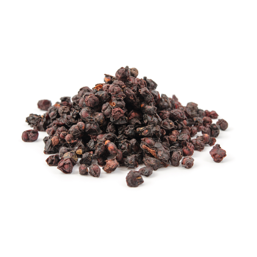
Schisandrin A is a naturally occurring lignan (dibenzocyclooctadiene), found in Schisandra / five-flavor berry and 2 other sources. It is well tolerated orally (low toxicity).
Concentration
Content by Source
Concentrations not reported. No percent-dry-weight or mg/g figures have been published for this compound in its source plants.
Schisandrin A at a Glance
Schisandrin A is a dibenzocyclooctadiene lignan of the schisandra berry (Schisandra chinensis, wuweizi), one of the family of lignans that give this “five-flavour berry” its reputation as an adaptogen and liver tonic in traditional Chinese medicine. It is more often met under its older names — deoxyschisandrin, deoxyschizandrin or wuweizisu A — and the same molecule sits behind all of them 1Reference 1ReviewA review: Pharmacology and pharmacokinetics of Schisandrin AView study →.
Almost everything known about the isolated compound is preclinical. Cell and rodent studies report antioxidant and anti-photoaging effects (through AMPK/Nrf2 signalling), anti-inflammatory activity (largely by suppressing NF-κB), and a scatter of organ-protective findings in the liver, kidney, heart, bone and brain 1,
The most concrete, real-world point about this molecule is not an efficacy claim at all — it is a drug interaction. Deoxyschisandrin is a potent inhibitor of the P-glycoprotein efflux pump and a substrate/modulator of the CYP3A drug-metabolising enzymes; it is reliable enough at CYP3A that pharmacologists have proposed it as an in-vivo CYP3A probe 6,
Schisandrin A vs. Schisandrin & Schisandrin B
The single most common confusion about this molecule is a naming one, so it is worth settling first.
- Schisandrin A (this one) = deoxyschisandrin = deoxyschizandrin = wuweizisu A. These are all the same dibenzocyclooctadiene lignan; the “deoxy” name reflects that it lacks a hydroxyl group carried by its close relative.
- Schisandrin (unqualified) = schisandrol A, the most abundant single lignan of the berry and a different, hydroxylated molecule. Despite the near-identical name it is not schisandrin A.
- Schisandrin B (wuweizisu B) is another distinct lignan — the one most studied for hepatoprotection and neuroprotection.
Head-to-head work shows the A and B forms are not interchangeable: in LPS-stimulated macrophages both are anti-inflammatory, but schisandrin A acts more by depleting reduced glutathione and driving glutathione-S-transferase, while schisandrin B is the one that activates Nrf2 and induces thioredoxin — different biochemistry behind a superficially similar effect 3Reference 3Differential action between schisandrin A and schisandrin B in eliciting an anti-inflammatory action: the depletion of reduced glutathione and the induction of an antioxidant responseView study →. When reading the literature, the exact lignan named matters; results do not transfer freely between them. (Other berry lignans with their own pages: schisandrol B (a.k.a. gomisin A), schisantherin A.)
What It's Used For
The evidence for schisandrin A is broad in scope but shallow in depth: many mechanisms across many organ systems, all in cells or rodents, none in humans. Read the per-row basis before weighing any single claim.
Support is an experimental score — weighted by study type (human > animal > in vitro) and volume. A beta, not a validated metric. Every row here is preclinical, so scores stay modest by design; they rank the relative weight of evidence within an all-preclinical set.
The most fully worked-out line. In UVB-irradiated HaCaT keratinocytes and ICR mice, isolated schisandrin A improved cell viability, reduced senescence and apoptosis, raised superoxide-dismutase activity and glutathione, lowered malondialdehyde, preserved dermal collagen and epidermal thickness, and inhibited matrix-metalloproteinase expression — traced to activation of AMPK and Nrf2 with enhanced autophagy 2Reference 2Schisandrin A alleviates UVB-induced photoaging via AMPK/Nrf2-mediated antioxidant and autophagy pathwaysView study →. This is consistent with the broader antioxidant signature the compound shows in other oxidative-stress models 1,
Gap: entirely a cell-line + mouse-skin story with no human (or even topical human-skin) data; the anti-photoaging framing is a mechanistic hypothesis, not a demonstrated cosmetic or clinical outcome 2Reference 2Schisandrin A alleviates UVB-induced photoaging via AMPK/Nrf2-mediated antioxidant and autophagy pathwaysView study →.
Schisandrin A suppresses inflammatory signalling across several isolated-compound models: it inhibits the JNK/p38/NF-κB pathway and pro-inflammatory cytokine release in LPS-stimulated RAW264.7 macrophages (more strongly than schisandrin B in that assay) 3Reference 3Differential action between schisandrin A and schisandrin B in eliciting an anti-inflammatory action: the depletion of reduced glutathione and the induction of an antioxidant responseView study →, protects human intestinal epithelial (HCT116) cells from H₂O₂-induced apoptosis by blocking IκBα degradation and NF-κB translocation 4Reference 4Deoxyschisandrin inhibits H2O2-induced apoptotic cell death in intestinal epithelial cells through nuclear factor-kappaBView study →, and — in a mouse asthma model — attenuates the Th2 response and airway inflammation with NF-κB implicated by network analysis 5Reference 5Schisandrin A ameliorates airway inflammation in model of asthma by attenuating Th2 responseView study →.
Gap: the mechanism is well sampled but the models are heterogeneous single studies; there is no human anti-inflammatory data, and the whole-berry vehicle is never separated from the isolate in the traditional-use context 3,
Two threads. Electrophysiologically, deoxyschisandrin dampens neuronal excitability — in cultured hippocampal neurons it reduced synchronised intracellular Ca²⁺ oscillations (IC₅₀ ≈ 3.8 mg/L) and inhibitory postsynaptic currents, depressing high-voltage-gated Ca²⁺ and voltage-gated Na⁺ channel currents 9Reference 9Deoxyschisandrin modulates synchronized Ca2+ oscillations and spontaneous synaptic transmission of cultured hippocampal neuronsView study →.
Behaviourally, in diabetic rats it improved insulin sensitivity and fear-memory, raised synaptic proteins, lowered Aβ42 and microglial activation, and reduced ferroptosis markers in the prefrontal cortex 10Reference 10Schisandrin A ameliorates the diabetes-associated memory impairment by alleviating inflammation and ferroptosisView study →.
Gap: mechanistically interesting but disparate (ion-channel modulation in vitro vs. a diabetes-memory rodent model); no cognitive-outcome data in humans, and doses/routes are not human-translatable 9,
A cluster of single-organ rodent findings, each from one study group. In renal fibrosis, schisandrin A inhibited PKCβ and oxidative stress to slow fibrotic progression 11Reference 11Schisandrin A alleviates renal fibrosis by inhibiting PKCβ and oxidative stressView study →. In isoproterenol-induced chronic heart failure, it curbed myocardial hypertrophy via miR-155 12Reference 12Schisandrin A protects against isoproterenol-induced chronic heart failure via miR-155View study →. In glucocorticoid-induced osteoporosis, it promoted osteoblast differentiation by activating Wnt signalling 13Reference 13Schisandrin A induces osteoblast differentiation to treat glucocorticoid-induced osteoporosis through activating Wnt pathwayView study →.
Gap: each organ rests on a lone rodent model with no replication or human follow-up; these are hypothesis-generating, not established uses 11,
Mechanisms
| Target / pathway | Effect | Relevant to |
|---|---|---|
| AMPK / Nrf2 | activation → antioxidant enzyme induction (SOD, GSH), autophagy, ↓MMP | antioxidant / anti-photoaging |
| NF-κB (via JNK/p38, IκBα) | suppression of inflammatory transcription and cytokine release | anti-inflammatory |
| Glutathione / glutathione-S-transferase | schisandrin A depletes reduced GSH and raises GST (contrast: schisandrin B induces Nrf2/thioredoxin) | anti-inflammatory (the A-vs-B distinction) |
| P-glycoprotein (ABCB1) | inhibition of efflux — most potent of 12 Schisandra lignans in Caco-2; ↑absorption of P-gp substrates (digoxin, rhodamine-123) | drug interaction |
| CYP3A | substrate/modulator; used as an in-vivo CYP3A probe; contributes to raised tacrolimus exposure | drug interaction / metabolism |
| PKCβ / oxidative stress | inhibition → reduced renal fibrosis | organ-protective (kidney) |
| miR-155 | modulation → reduced myocardial hypertrophy | organ-protective (heart) |
| Wnt | activation → osteoblast differentiation | organ-protective (bone) |
The load-bearing rows are the P-gp and CYP3A lines: they are the one part of this molecule’s pharmacology with direct, documented human relevance — see the interaction section below.
Pharmacokinetics
A dedicated review notes that schisandrin A has “good” pharmacokinetic characteristics, but that these are altered by co-administered drugs and by disease state (e.g. hepatic fibrosis changes its disposition) 1Reference 1ReviewA review: Pharmacology and pharmacokinetics of Schisandrin AView study →. As a lipophilic dibenzocyclooctadiene lignan it is subject to intestinal first-pass processing by P-glycoprotein and CYP3A — the same machinery it in turn inhibits, which is why it self-interacts and interacts with other substrates 6,
Clinical Trials
There are no human clinical trials of isolated schisandrin A. All efficacy evidence is in-vitro or rodent; the only human-adjacent data are pharmacokinetic-interaction studies of schisandra lignans (Wuzhi tablet) affecting tacrolimus, where schisandrin A is one of several lignans tested 8Reference 8In vitroIn vivo to in vitro effects of six bioactive lignans of Wuzhi tablet (Schisandra sphenanthera extract) on the CYP3A/P-glycoprotein-mediated absorption and metabolism of tacrolimusView study →.
| Completed | Planned | Terminated | Preclinical |
|---|---|---|---|
| None(isolate) | — | — | Antioxidant/anti-photoaging, anti-inflammatory, neuroprotective, organ-protective, PK-interaction mechanistic |
Last checked: July 2026.
Deoxyschisandrin, CYP3A & P-Glycoprotein
This is the part of the molecule’s profile with the clearest human relevance — and it is a caution, not a benefit.
In human intestinal Caco-2 cells, deoxyschisandrin was the most potent P-glycoprotein inhibitor of twelve schisandra dibenzocyclooctadiene lignans tested: it increased the cellular accumulation and absorptive (apical-to-basal) transport of P-gp substrates such as digoxin and rhodamine-123, collapsing their efflux ratios 7Reference 7Effects of Schisandra lignans on P-glycoprotein-mediated drug efflux in human intestinal Caco-2View study →. Because P-gp guards the absorption of many drugs, inhibiting it can raise their blood levels.
The same logic extends to metabolism. Schisandrin A is one of six major lignans in Wuzhi tablet (a Schisandra sphenanthera extract) shown to inhibit both P-gp-mediated efflux and CYP3A-mediated metabolism of tacrolimus (FK506), increasing the immunosuppressant’s exposure in rats — though schisandrol B was the strongest of the six 8Reference 8In vitroIn vivo to in vitro effects of six bioactive lignans of Wuzhi tablet (Schisandra sphenanthera extract) on the CYP3A/P-glycoprotein-mediated absorption and metabolism of tacrolimusView study →. And in 2026, deoxyschizandrin was formalised as an in-vivo CYP3A probe substrate, precisely because its handling tracks CYP3A activity reliably 6Reference 6AnimalNew strategy to quantify hepatic cytochrome P450 3A activity with deoxyschizandrin as an in vivo probe: a Vmax,app approachView study →.
The practical reading: a molecule that alters CYP3A and P-gp is a molecule that can change the pharmacokinetics of co-administered medicines — many of which (immunosuppressants, some statins, certain anticoagulants and others) are CYP3A/P-gp substrates with narrow margins. This is a whole-berry consideration too, since schisandra preparations deliver this lignan alongside its relatives.
Isolate vs. Plant Studies
The evidence base here is unusually isolate-heavy — which, unlike most herbs, is a mixed picture rather than a strength. The antioxidant, anti-inflammatory, neuroprotective and organ-protective studies almost all use purified schisandrin A in cell lines or rodents 2,
Prevalence in Nature
Schisandrin A is one of the principal dibenzocyclooctadiene lignans of the schisandra berry (Schisandra chinensis, wuweizi), sitting within the total-lignan fraction that makes up a few percent of the dried fruit 1Reference 1ReviewA review: Pharmacology and pharmacokinetics of Schisandrin AView study →. It also occurs in the related Schisandra sphenanthera (nan-wuweizi / the source of Wuzhi tablet), and species discipline matters: several CYP3A/P-gp interaction findings come specifically from S. sphenanthera and should not be silently attributed to S. chinensis 8Reference 8In vitroIn vivo to in vitro effects of six bioactive lignans of Wuzhi tablet (Schisandra sphenanthera extract) on the CYP3A/P-glycoprotein-mediated absorption and metabolism of tacrolimusView study →. Reported lignan content varies with species, cultivation region and chemotype (northern varieties generally carry more lignan than southern), so any single concentration figure is provenance-dependent. A precisely-cited percentage for schisandrin A specifically is not established in the sources reviewed here (tracked in the gaps note).
Discovery & Synthesis
Schisandrin A was isolated from Schisandra chinensis fruit as one of the classic dibenzocyclooctadiene lignans characterised during the mid-twentieth-century study of the schizandrin series; its “deoxy” name (deoxyschisandrin) marks it as lacking a hydroxyl group present on the parent schisandrin (schisandrol A). The dibenzocyclooctadiene skeleton has since been the target of documented total-synthesis work, though a firm first-synthesis date is not established in the sources reviewed here. In practice, schisandrin A is obtained by extraction and chromatographic isolation from schisandra berries rather than manufactured synthetically — it reaches people as a natural constituent of whole-berry preparations, not as a made single-molecule ingredient 1Reference 1ReviewA review: Pharmacology and pharmacokinetics of Schisandrin AView study →.
Patents: not yet researched.
Toxicity & Safety
Schisandrin A carries a low intrinsic-toxicity flag. The schisandra berry has a long history of food and medicinal use, and the isolated lignan shows no notable toxicity signal at the doses used in the preclinical literature — indeed it is repeatedly protective in oxidative-stress and organ-injury models 1,
- Low intrinsic toxicity. From a long-used food/medicinal berry; the isolate is protective, not injurious, in preclinical oxidative-stress models.
- Drug-interaction potential (the main concern). Schisandrin A inhibits P-glycoprotein and modulates CYP3A, so it can raise blood levels of co-administered CYP3A/P-gp substrates (e.g. tacrolimus, digoxin, some statins). Whole-berry schisandra shares this caution.
- No human efficacy or safety trials of the isolate. Do not read the preclinical protective findings as established human benefits or as a safety clearance.
- Species & chemotype matter. Interaction data partly derive from Schisandra sphenanthera (Wuzhi); don’t assume identical behaviour across species or provenance.
Because reproductive-toxicity data for the isolate are absent, no pregnancy/lactation verdict is made at the molecule level — that decision belongs to the schisandra herb page where intake is actually decided.
Dosage
There is no established human dose for isolated schisandrin A, and it is not sold as a stand-alone supplement. The figures in the research are preclinical: rodent oral doses in the injury/protection models and micromolar in-vitro concentrations in the cell work (e.g. up to ~50 µM deoxyschisandrin in the Caco-2 P-gp assay) 7Reference 7Effects of Schisandra lignans on P-glycoprotein-mediated drug efflux in human intestinal Caco-2View study →. Human exposure occurs only as one lignan within whole schisandra-berry preparations, whose dosing is set at the berry/extract level, not the single molecule. These are doses studied in research and are not a personal recommendation.
References
- Fu K, Zhou H, Wang C, et al. (2022). A review: Pharmacology and pharmacokinetics of Schisandrin A. Phytotherapy Research. https://pubmed.ncbi.nlm.nih.gov/35384105/
- Wang S, Liu K, Zhang M, et al. (2026). Schisandrin A alleviates UVB-induced photoaging via AMPK/Nrf2-mediated antioxidant and autophagy pathways. Photochemical & Photobiological Sciences. https://pubmed.ncbi.nlm.nih.gov/41801672/
- Leong PK, Wong HS, Chen J, et al. (2016). Differential action between schisandrin A and schisandrin B in eliciting an anti-inflammatory action: the depletion of reduced glutathione and the induction of an antioxidant response. PLoS One. https://pubmed.ncbi.nlm.nih.gov/27195753/
- Gu BH, Minh NV, Lee SH, et al. (2010). Deoxyschisandrin inhibits H2O2-induced apoptotic cell death in intestinal epithelial cells through nuclear factor-kappaB. International Journal of Molecular Medicine. https://pubmed.ncbi.nlm.nih.gov/20664957/
- Qiu Q, Zhang W, Liu K, et al. (2023). Schisandrin A ameliorates airway inflammation in model of asthma by attenuating Th2 response. European Journal of Pharmacology. https://pubmed.ncbi.nlm.nih.gov/37329976/
- Li X, Hu C, Wei G, et al. (2026). New strategy to quantify hepatic cytochrome P450 3A activity with deoxyschizandrin as an in vivo probe: a Vmax,app approach. Drug Metabolism and Disposition. https://pubmed.ncbi.nlm.nih.gov/41780174/
- Yoo HH, Lee M, Lee MW, et al. (2007). Effects of Schisandra lignans on P-glycoprotein-mediated drug efflux in human intestinal Caco-2. Planta Medica. https://pubmed.ncbi.nlm.nih.gov/17566147/
- Qin XL, Chen X, Wang Y, et al. (2014). In vivo to in vitro effects of six bioactive lignans of Wuzhi tablet (Schisandra sphenanthera extract) on the CYP3A/P-glycoprotein-mediated absorption and metabolism of tacrolimus. Drug Metabolism and Disposition. https://pubmed.ncbi.nlm.nih.gov/24195812/
- Fu M, Sun ZH, Zong M, et al. (2008). Deoxyschisandrin modulates synchronized Ca2+ oscillations and spontaneous synaptic transmission of cultured hippocampal neurons. Acta Pharmacologica Sinica. https://pubmed.ncbi.nlm.nih.gov/18664321/
- Ma G, Lei M, Guo S, et al. (2025). Schisandrin A ameliorates the diabetes-associated memory impairment by alleviating inflammation and ferroptosis. Acta Biochimica et Biophysica Sinica. https://pubmed.ncbi.nlm.nih.gov/40589410/
- Liu HL, Huang Z, Li QZ, et al. (2024). Schisandrin A alleviates renal fibrosis by inhibiting PKCβ and oxidative stress. Phytomedicine. https://pubmed.ncbi.nlm.nih.gov/38382281/
- Gao L, Li T, Li S, et al. (2022). Schisandrin A protects against isoproterenol-induced chronic heart failure via miR-155. Molecular Medicine Reports. https://pubmed.ncbi.nlm.nih.gov/34812475/
- Ai Y, Xie X, Guo Y, et al. (2025). Schisandrin A induces osteoblast differentiation to treat glucocorticoid-induced osteoporosis through activating Wnt pathway. Biochemical and Biophysical Research Communications. https://pubmed.ncbi.nlm.nih.gov/40555002/