
Condition
Estrogen Dominance
Estrogen dominance is a hormonal imbalance involving a high ratio of estrogen to progesterone.

Overview
Aetiology
Estrogen Excess
Estrogen excess causes symptoms such as fluid retention (through changes to fluid osmolarity threshold through mechanisms involving ADH and ACTH), extended menstrual cycle and spotting in the luteal phase.
Estrogen excess is a condition involving a high estrogen to progesterone ratio. It can occur for many reasons, and can change depending on the timing in the menstrual cycle. Estrogen dominance can involve high or normal estrogen and low or normal progesterone.
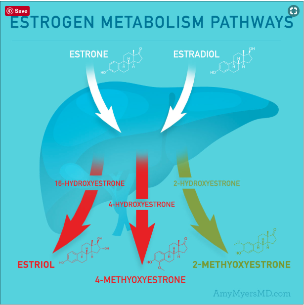
Estrogen is converted from C19 steroids to estrogen via aromatase. This enzyme is encoded in the liver by the CYP19 gene 9Reference 9Aromatase cytochrome P450, the enzyme responsible for estrogen biosynthesis. E1 and E2 are metabolized by the phase 1 liver detoxification enzyme CYP1A1 into 2-hydroxy catechol metabolites with weak binding capacity and estrogenic activity (potentially even antiestrogenic) 10Reference 10Soy Protein Isolate Increases Urinary Estrogens and the Ratio of 2: 16 α-Hydroxyestrone in Men at High Risk of Prostate Cancer.
Another set of enzymes encoded by the CYP3A4 and CYP3A7 convert E1 and E2 into 16-hydroxy catechols that are reported to have strong estrogenic activity and binding capacity and are associated with increased proliferation of endometrial tissue 11Reference 11Estrogen metabolite ratio: Is the 2-hydroxyestrone to 16α-hydroxyestrone ratio predictive for breast cancer?. CYP1B1 converts E1 and E2 into 4-hydroxy catechol estrogen 12Reference 12Cytochrome P450 1B1 (CYP1B1) pharmacogenetics: association of polymorphisms with functional differences in estrogen hydroxylation activity, which is also an active metabolite.
Therefore, shifting the ratio between 2-hydroxy, 4-hydroxy, and 16-hydroxy catechol estrogens is an important therapeutic mechanism when addressing concerns over estrogen dominance.
This is achieved through inhibition of the 16-hydroxy catechol producing CYP3A4, and 4-hydroxy catechol producing CYP3B1, and inducing the 2-hydroxy catechol estrogen producing CYP1A1.
High Xenestrogen Exposure
Exposure to xenestrogens in the environment may cause symptoms of estrogen excess. Removal of these factors is the first line of treatment if this is suspected.
Poor Liver Metabolism
Estrogen is broken down in the liver, and eliminated through the digestive tract. There are 3 main reasons why the liver may have difficulty eliminating estrogen, allowing it to gradually increase in concentration, or remain elevated for longer than it should.
- Nutrient deficiencies (vitamin B6, magnesium, copper, and zinc are all needed for metabolizing estrogen)
- Liver disease, such as NAFLD, AFLD, or hepatitis may interfere with liver function
- Increased demand on the liver, reducing clearance of estrogens
Progesterone Deficiency
Progesterone has a negative feedback effect on the hypothalamus to reduce the effects of the HPG, while estrogen has a (mostly) positive feedback effect on the HPG. This means that estrogen is more likely to become in excess than progesterone is, however, it’s crucial that this differentiation is made in order to provide effective treatment.
Low Dopamine
Low dopamine results in elevated prolactin levels, which can reduce progesterone levels. A common contributor is nutrient deficiency, including tyrosine, magnesium, and vitamin B6.
High Stress
Stress is thought to be a major cause for lowered progesterone levels through a theory called the “progesterone steal”. This is a theory, and has not yet been proven. It suggests that the manufacture of cortisol during stressed states takes priority over progesterone production by robbing a common precursor, progesterone, from the cycle in order to make cortisol.
Symptoms of High Estrogen
- Fluid retention
- Breast tenderness/swelling
Symptoms of Low Progesterone
- Poor concentration
- Fluid retention
- Clotting
- Spotting
- Depression/anxiety
- Burning throat
- Cysts in breasts
- Swollen breasts
- Dry skin
- Hair loss
- Menorrhagia
General Symptoms
General features include premenstrual breast tenderness and swelling, bloating and fluid retention, mood changes such as irritability and low mood, headaches, heavy or prolonged menstrual bleeding, and worsening of premenstrual syndrome.
Assessment
Diagnostic Considerations
Diagnosis is supported by serum, salivary, or urinary measurement of estrogen and progesterone, ideally timed to the luteal phase to assess their ratio. Thyroid function, prolactin, and pelvic imaging may help identify contributing or alternative causes.
Assessment
Differential Diagnosis
- Hypothyroidism
- Menopause
- Ovarian cysts
- Ovarian cancer
- PCOS
- PCOD
Context
Comorbidities
- Anemia
- Ovarian cancer
- Ovarian cysts
- Endometriosis
- Hypothyroidism
References:
- Stachenfeld, N. S. (2008). Sex hormone effects on body fluid regulation. Exercise and sport sciences reviews, 36(3), 152.
- Fitzgerald, P., & Dinan, T. G. (2008). Prolactin and dopamine: what is the connection? A review article. Journal of Psychopharmacology, 22(2_suppl), 12-19.
- Bone, K., & Mills, S. Y. (2013). Principles and Practice of Phytotherapy, Modern Herbal Medicine, 2: Principles and Practice of Phytotherapy. Elsevier Health Sciences.
- Hoffmann, D. (2003). Medical herbalism: the science and practice of herbal medicine. Simon and Schuster.
- Lessey, B. A., & Young, S. L. (2014, September). Homeostasis imbalance in the endometrium of women with implantation defects: the role of estrogen and progesterone. In Seminars in reproductive medicine (Vol. 32, No. 05, pp. 365-375). Thieme Medical Publishers.
- Cousins, L. M., Hobel, C. J., Chang, R. J., Okada, D. M., & Marshall, J. R. (1977). Serum progesterone and estradiol-17β levels in premature and term labor. American Journal of Obstetrics & Gynecology, 127(6), 612-615.
- Yu, L. L., Song, P. L., Guo, Z. L., Mu, Z. X., & CHEN, M. (2003). The Therapeutic Effect of Traditional Chinese Medicine Prescription Zhuyun III on Kidney-Deficiency and Luteal Phase Defect Aborting Rat Model [J]. Reproduction and Contraception, 1, 003.
- Shozu, M., Sebastian, S., Takayama, K., Hsu, W. T., Schultz, R. A., Neely, K., … & Bulun, S. E. (2003). Estrogen excess associated with novel gain-of-function mutations affecting the aromatase gene. New England Journal of Medicine, 348(19), 1855-1865.
- Simpson, E. R., Mahendroo, M. S., Means, G. D., Kilgore, M. W., Hinshelwood, M. M., Graham-Lorence, S., … & Mendelson, C. R. (1994). Aromatase cytochrome P450, the enzyme responsible for estrogen biosynthesis. Endocrine reviews, 15(3), 342-355.
- Hamilton-Reeves, J. M., Rebello, S. A., Thomas, W., Slaton, J. W., & Kurzer, M. S. (2007). Soy Protein Isolate Increases Urinary Estrogens and the Ratio of 2: 16 α-Hydroxyestrone in Men at High Risk of Prostate Cancer. The Journal of nutrition, 137(10), 2258-2263.
- Obi, N., Vrieling, A., Heinz, J., & Chang-Claude, J. (2011). Estrogen metabolite ratio: Is the 2-hydroxyestrone to 16α-hydroxyestrone ratio predictive for breast cancer?. International journal of women’s health, 3, 37.
- Hanna, I. H., Dawling, S., Roodi, N., Guengerich, F. P., & Parl, F. F. (2000). Cytochrome P450 1B1 (CYP1B1) pharmacogenetics: association of polymorphisms with functional differences in estrogen hydroxylation activity. Cancer Research, 60(13), 3440-3444.