
Condition
Systemic Lupus Erythematosus (SLE)
Systemic lupus erythematosus is a chronic autoimmune disease in which the body attacks its own tissues, causing inflammation across many organ systems.

Overview
Aetiology
Systemic lupus erythematosus is a chronic multisystem autoimmune disease characterized by antibodies directed against the cell nucleus. It is the prototype immune complex disease — the damage comes less from the antibodies binding tissue directly than from antibody-antigen complexes depositing in small vessels and activating complement.
The initiating defect is a failure to clear apoptotic cells. Nuclear material that should be silently removed is instead retained and presented to the immune system, breaking tolerance to self-antigens: double-stranded DNA, histones, and ribonucleoproteins. The resulting complexes lodge in glomeruli, skin, serosal membranes, joints and choroid plexus, where complement activation recruits neutrophils and drives inflammation. Consumption of complement in that process is why C3 and C4 fall during a flare — a useful and counterintuitive marker, since most inflammatory diseases raise rather than lower their acute-phase proteins.
Susceptibility is multifactorial:
- Genetic — HLA-DR2 and DR3 associations, and hereditary deficiency of early complement components (C1q, C2, C4), which impairs clearance of apoptotic debris. Concordance in identical twins is around 25%, so genetics is necessary but not sufficient.
- Hormonal — the female-to-male ratio is roughly 9:1 in the reproductive years, and disease often begins or flares with pregnancy or estrogen exposure.
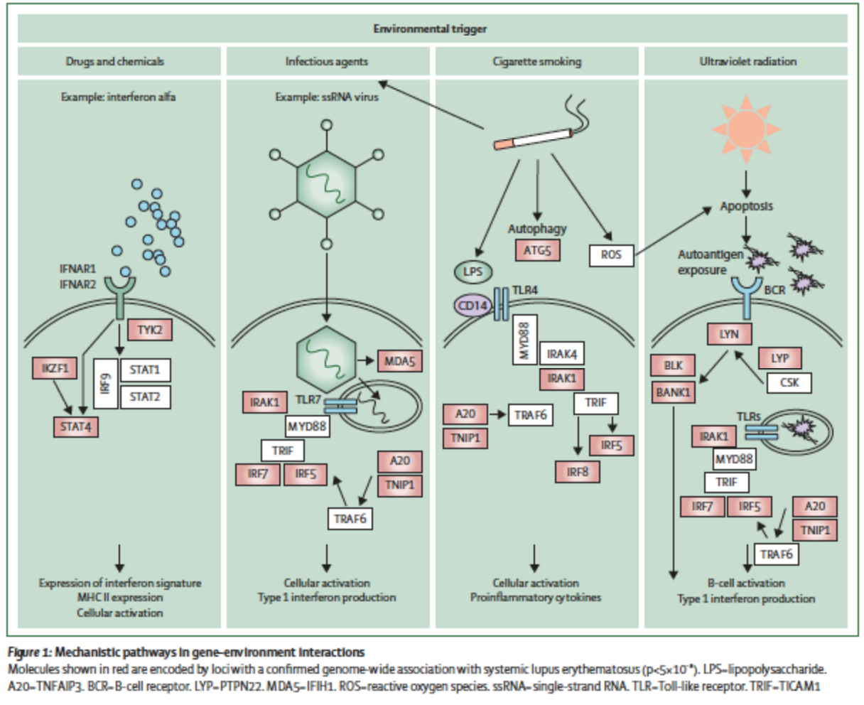
- Environmental — ultraviolet light is the best-established trigger, causing keratinocyte apoptosis and releasing nuclear antigen into the skin. Epstein-Barr virus infection, silica exposure and smoking are associated.
- Drug-induced lupus — hydralazine, procainamide, isoniazid, minocycline and TNF inhibitors. It is a distinct entity: anti-histone antibodies are typical, renal and CNS involvement is rare, and it resolves on withdrawal.
Prevalence is substantially higher, and disease typically more severe, in people of African, Hispanic and Asian ancestry.
Presentation
Symptoms
The characteristic pattern is a relapsing-remitting multisystem illness with constitutional symptoms, and its variability is the reason diagnosis is so often delayed.
Constitutional — fatigue, present in the great majority and often the most disabling symptom; fever, weight loss, and lymphadenopathy.
Musculoskeletal — arthralgia and arthritis affect around 90%. It is typically symmetrical and involves small joints, resembling rheumatoid arthritis but non-erosive; Jaccoud’s arthropathy produces reducible deformity without joint destruction.
Cutaneous — the malar “butterfly” rash over the cheeks and nasal bridge, sparing the nasolabial folds; discoid lesions, which scar; photosensitivity; oral and nasal ulcers, characteristically painless; and non-scarring alopecia.
Renal — lupus nephritis affects up to half and is a principal determinant of prognosis. It is frequently silent until advanced, which is why urinalysis at every review matters more than symptoms.
Serosal — pleurisy and pericarditis, often presenting as pleuritic chest pain.
Neuropsychiatric — headache, seizures, psychosis, cognitive dysfunction, mood disorder, and stroke.
Haematological — leukopenia, lymphopenia, thrombocytopenia and autoimmune haemolytic anemia.
Vascular — Raynaud’s phenomenon, vasculitis, and thrombosis where antiphospholipid antibodies are present.
Assessment
Diagnostic Considerations
Diagnosis rests on a compatible clinical picture together with supporting serology; no single test is sufficient.
ANA is the screening test and is positive in over 95% — a negative ANA makes SLE very unlikely. But it is not specific: it is positive in other connective tissue diseases, in chronic infection, and in a meaningful proportion of healthy people, particularly older women. A positive ANA in someone without compatible features is a common cause of misdiagnosis and unnecessary alarm.
Anti-double-stranded DNA is far more specific, and its titre often tracks disease activity, particularly renal activity. Anti-Smith is the most specific antibody of all, though present in only about 30%. Anti-Ro and anti-La are associated with sicca symptoms, subacute cutaneous lupus, and — importantly — with congenital heart block in the offspring of affected mothers. Antiphospholipid antibodies (lupus anticoagulant, anticardiolipin, anti-β2-glycoprotein I) should be checked in everyone, since they change management of thrombosis and pregnancy.
Monitoring uses complement C3 and C4, which fall in active disease, alongside anti-dsDNA titre. Urinalysis and urine protein:creatinine ratio at every visit are the standard of care for detecting silent nephritis, and renal biopsy classifies it and determines treatment. ESR is usually raised while CRP often remains normal — a rising CRP in a lupus patient should prompt a search for infection rather than being assumed to be a flare.
Assessment
Differential Diagnosis
- Rheumatoid arthritis — erosive, with anti-CCP antibodies
- Mixed connective tissue disease — high-titre anti-U1-RNP
- Sjögren’s syndrome — sicca symptoms dominate; frequently overlaps
- Systemic sclerosis — skin thickening, anti-centromere or anti-Scl-70
- Dermatomyositis — heliotrope rash, Gottron’s papules, proximal weakness
- Drug-induced lupus — anti-histone antibodies, resolves on withdrawal
- Adult-onset Still’s disease — quotidian fever, salmon rash, very high ferritin
- Viral infection — parvovirus B19, EBV, HIV, hepatitis C
- Fibromyalgia — widespread pain and fatigue with normal serology
- Vasculitis, and lymphoma where constitutional symptoms dominate
Context
Comorbidities
Accelerated atherosclerosis is a leading cause of late mortality, with cardiovascular risk several-fold higher than expected — chronic inflammation, corticosteroid exposure and renal disease compound one another, and conventional risk calculators underestimate it. Infection is the other major cause of death, driven by immunosuppression and by intrinsic complement and immune dysfunction.
Antiphospholipid syndrome coexists in around a third and causes arterial and venous thrombosis and recurrent pregnancy loss. Osteoporosis follows from corticosteroids and from disease activity. Chronic kidney disease and end-stage renal failure follow lupus nephritis. Depression and anxiety are common, and fatigue frequently persists even when serology is quiescent. Pregnancy requires planning — active disease and renal involvement raise the risk of pre-eclampsia, prematurity and fetal loss, and drug regimens need review before conception.