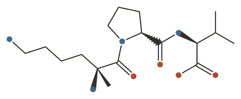
KPV
The anti-inflammatory C-terminal tripeptide of α-MSH, with a replicated rodent anti-colitis effect and no human data by any route.

KPV at a Glance
KPV is the C-terminal tripeptide of α-MSH — lysine-proline-valine — the smallest fragment that keeps the parent hormone’s anti-inflammatory activity while dropping its pigmentary and melanocortin effects. That separation is what makes it interesting: three amino acids carrying an immunomodulatory action without the rest of the melanocortin profile. It has a replicated anti-inflammatory effect in rodent colitis models, no human trial data, and no approval anywhere.
History & Development
KPV is three amino acids - the tail end of a hormone - and it carries a reproducible anti-inflammatory effect in animals that has never once been tested in a person.
| First described | Not established in the sources consulted for this section |
| Discovered / synthesised by | Not established in the sources consulted for this section |
| Institution | Not established in the sources consulted for this section |
| Origin | Lys-Pro-Val - the C-terminal tripeptide of alpha-MSH, residues 11-13 of the tridecapeptide alpha-MSH(1-13); MW 342.4 Da |
| Key patent | Not established in the sources consulted for this section |
| Assignee | Not established in the sources consulted for this section |
| Development status | Research chemical - no human data by any route; never approved |
Discovery
Alpha-MSH is best known for pigmentation, but it is also potently anti-inflammatory - and those two activities live in different parts of the molecule. The pigmentation depends on the melanocortin receptor binding region; the anti-inflammatory activity survives in the C-terminal tripeptide, Lys-Pro-Val.
KPV is therefore alpha-MSH’s anti-inflammatory effect without its pigmentary one, in three residues. That separation is elegant and it is the reason the fragment is studied at all.
Its most reproducible finding is a genuinely replicated rodent anti-colitis effect - better than most compounds on this hub can claim at the animal level.
Development & ownership
The human gap is total: there is no human data by any route - not oral, not topical, not injected. A federal regulator has formally noted that absence.
Two further gaps compound it. There is zero toxicology in any species. And substance identity is unresolved in a specific, documented way: one CAS number is used commercially for two chemically distinct substances - so the identifier on a certificate of analysis does not reliably establish what is in the vial.
US compounding records also show no outsourcing facility reporting it.
How it reached the market
KPV is sold for gut inflammation and healing, orally and by injection, on the strength of the rodent colitis work. It is an interesting endogenous fragment with a real animal effect, a complete absence of human evidence, no toxicology, and an ambiguous chemical identity in commerce.
What It's Used For
KPV is the C-terminal tripeptide of α-melanocyte-stimulating hormone — Lys-Pro-Val, residues 11–13 of the tridecapeptide α-MSH(1–13), molecular weight 342.4 Da. Three things have to be kept apart. α-MSH itself is a well-studied endogenous melanocortin with a large literature and real receptor pharmacology — it is not this compound. KPV is the minimal fragment claimed to keep α-MSH’s anti-inflammatory activity while shedding the pigment-inducing action of the parent hormone 25Reference 25In vitroBrzoska T, Luger TA, Maaser C, Abels C, Böhm M. (2008). α-Melanocyte-stimulating hormone and related tripeptides: biochemistry, antiinflammatory and protective effects in vitro and in vivo, and future perspectives — review. Endocrine Reviews. https://pubmed.ncbi.nlm.nih.gov/18612139/View study →.
And KdPT (Lys-D-Pro-Thr) is a closely related tripeptide studied alongside KPV and routinely conflated with it in this literature; where a study used KdPT, GKPV, or the [Ac-CKPV]₂ dimer rather than free KPV, it is labeled as such below.
That “activity without the side effect” claim has the same shape as AOD-9604 (growth hormone’s fat loss without GH’s growth effects) and it deserves the same two-part test — because on this compound, both halves survive scrutiny in a way they did not for AOD-9604, and yet the conclusion is bleaker. The “without pigmentation” half holds up unusually well: KPV does not displace radiolabeled α-MSH from MC1 receptor binding sites and does not raise cAMP the way α-MSH does 12,
That is not an inference drawn here. In May 2026 the FDA published a full evaluation of KPV for the section 503A Bulk Drug Substances List, and searched for exactly this. Its finding: “The nomination did not include, and FDA has not identified, any clinical studies or human exposure data for KPV via any route of administration.” FDA concluded that the balance of criteria weighs against allowing KPV to be compounded at all. See Clinical & regulatory status below — it is the single most decision-relevant fact here, and it is dated.
Route is load-bearing, and it cuts two ways. As a di/tripeptide, KPV is a substrate for the intestinal transporter PepT1, which is minimally expressed in healthy colon and strongly induced in inflamed gut — so oral KPV can act locally in inflamed intestine in rodents even though its systemic oral bioavailability is negligible 1,
Support is an experimental score I’m building — a composite weighted by study type (human > animal > in vitro > review) and study volume. It’s a beta: a fast way to rank strength of evidence at a glance, not a validated metric. Each application links down to its write-up.
Read the ceiling on this table, not just the ordering. Every row below is preclinical, and none rests on a human trial — so no row can rise far. This is the “never tested” kind of low score, not the AOD-9604 kind where a large human trial ran and failed. That distinction cuts both ways: nothing here has been disproven in people, and nothing here has been shown in people either.
KPV’s flagship application, and the only one with genuinely replicated animal data from more than one laboratory.
In mice, KPV is taken up into intestinal epithelial and immune cells via PepT1, a di/tripeptide transporter expressed at low levels in healthy colon and induced during inflammatory bowel disease. Nanomolar concentrations inhibited NF-κB and MAP-kinase signaling and cut pro-inflammatory cytokine output in human intestinal epithelial (Caco2-BBE, HT29-Cl.19A) and Jurkat T-cell lines, and KPV given in drinking water reduced both DSS- and TNBS-induced colitis 1Reference 1In vitroPepT1-mediated tripeptide KPV uptake reduces intestinal inflammation — animal (mouse) / in vitroView study →.
A second, independent group confirmed benefit in DSS and CD45RB-transfer colitis — and, notably, KPV rescued all treated animals from death during DSS colitis in mice expressing a non-functional MC1 receptor, indicating the effect is at least partly independent of melanocortin-receptor signaling 2Reference 2AnimalMelanocortin-derived tripeptide KPV has anti-inflammatory potential in murine models of inflammatory bowel disease — animal (mouse)View study →.
Later transgenic work tied the effect to the transporter directly: KPV prevented carcinogenesis in a mouse colitis-associated-cancer model, and produced no anti-tumorigenic effect at all in PepT1-knockout mice 3Reference 3AnimalCritical role of PepT1 in promoting colitis-associated cancer and therapeutic benefits of the anti-inflammatory PepT1-mediated tripeptide KPV in a murine model — animal (mouse) / human biopsyView study →.
Because free KPV is unstable, most subsequent efficacy work delivers it in an engineered vehicle, and the vehicle does a lot of the lifting. Colon-targeted nanoparticles in an alginate–chitosan hydrogel achieved comparable efficacy at a 12,000-fold lower KPV concentration than free peptide in DSS mice 4Reference 4In vitroDrug-loaded nanoparticles targeted to the colon with polysaccharide hydrogel reduce colitis in a mouse model — animal (mouse) / in vitroView study →.
Hyaluronic-acid-functionalized nanoparticles 5Reference 5AnimalOrally targeted delivery of tripeptide KPV via hyaluronic acid-functionalized nanoparticles efficiently alleviates ulcerative colitis — animal (mouse)View study →, a self-cross-linked hydrogel in TNBS rats 6Reference 6AnimalSelf-cross-linked hydrogel of cysteamine-grafted γ-polyglutamic acid stabilized tripeptide KPV for alleviating TNBS-induced ulcerative colitis in rats — animal (rat)View study →, a hybrid KPV-conjugated nanoparticle co-loaded with teduglutide 7Reference 7AnimalA nanoparticle platform for combined mucosal healing and immunomodulation in inflammatory bowel disease treatment (hyaluronate-KPV conjugated, teduglutide-loaded) — animal (mouse)View study →, and a PepT1-targeted co-assembled nanodrug 8Reference 8AnimalPepT1-targeted nanodrug based on co-assembly of anti-inflammatory peptide and immunosuppressant for combined treatment of acute and chronic DSS-induced colitis — animal (mouse)View study → all reduced murine or rat colitis. Most recently, a 2026 oral self-immolative prodrug (proKPV) reached inflamed colon at 3.8× the level of free KPV and was effective at a 20-fold lower dose in colitis mice 9Reference 9AnimalInflammation-triggered self-immolative conjugates enable oral peptide delivery by overcoming gastrointestinal barriers (proKPV) — animal (mouse)View study →.
Reviews frame the melanocortin system as a plausible IBD target generally 10,
Gap: every result is rodent or cell-line. There is no human clinical trial of KPV in IBD, or in anything else. The human melanocortin data in this space belong to different molecules in clinical development — not to KPV, and their progress does not transfer. Note also that several of the strongest results are properly results about a delivery system containing KPV, not about KPV as sold.
KPV’s anti-inflammatory identity was established in classic whole-animal models, and this is also where the mechanism question was settled against melanocortin receptors.
In mice, α-MSH(11–13) inhibited picryl-chloride-induced ear swelling dose-dependently, compared against saline and a large corticosteroid dose 14Reference 14AnimalAntiinflammatory activity of a COOH-terminal fragment of the neuropeptide α-MSH — animal (mouse)View study →.
In crystal-induced peritonitis, systemic KPV significantly reduced polymorphonuclear leukocyte accumulation — but the selective MC1R agonist did not, the MC3/4R antagonist SHU9119 did not block KPV’s effect, KPV failed to raise cAMP, and the anti-inflammatory effect persisted in IL-1β-induced peritonitis and in mice with a non-functional MC1R. The authors concluded KPV “is unlikely to mediate its effects through melanocortin receptors” and more likely acts by inhibiting IL-1β function 11Reference 11In vitroDissection of the anti-inflammatory effect of the core and C-terminal (KPV) α-melanocyte-stimulating hormone peptides — animal (mouse) / in vitroView study →.
Immobilized GKPV (α-MSH 10–13, an analogue — not KPV) inhibited TNF-α-driven NF-κB activation in vitro 16Reference 16In vitroImmobilized α-melanocyte stimulating hormone 10-13 (GKPV) inhibits tumor necrosis factor-α stimulated NF-κB activity — in vitro (GKPV analogue, not KPV)View study →.
The lineage traces to 1984, when Lys-Pro-Val was shown to carry α-MSH’s antipyretic activity in rabbits both centrally and peripherally, though at markedly lower potency than the parent peptide — the authors’ own reading being that other parts of α-MSH are needed for the full effect 15Reference 15Effect of α-MSH 11-13 (lysine-proline-valine) on fever in the rabbit — animal (rabbit)View study →.
Gap: multiple models, all animal or in vitro, and mostly decades old. Nothing here has been tested for any systemic anti-inflammatory indication in a human, and the lower-potency-than-parent finding is rarely mentioned in marketing that invokes α-MSH’s reputation.
The rationale is that KPV (and KdPT) retain α-MSH’s anti-inflammatory action without pigmentation, which is attractive for cutaneous repair 17Reference 17ReviewAre melanocortin peptides future therapeutics for cutaneous wound healing? — reviewView study →. The evidence is much thinner than the marketing.
The main in-vivo study is in diabetic mice: a three-layer “skin-adaptive” film dressing loaded with KPV in its bottom layer and epidermal growth factor in its upper layers improved full-thickness wound repair, with the mechanism attributed to inflammatory inhibition, angiogenesis and collagen deposition 18Reference 18AnimalSkin-adaptive film dressing with smart-release of growth factors accelerated diabetic wound healing (KPV co-loaded with EGF) — animal (mouse)View study →.
Read the design carefully — the benefit belongs to a dressing that released KPV plus EGF in sequence, so the peptide’s independent contribution is not isolable. Mechanistic work in human keratinocytes found that α-MSH, KPV and KP-D-V all raised intracellular calcium while producing no rise in cyclic AMP, i.e. not the cAMP route classically ascribed to α-MSH 19Reference 19In vitroElliott RJ, Szabo M, Wagner MJ, Kemp EH, MacNeil S, Haycock JW. (2004). α-Melanocyte-stimulating hormone, MSH 11-13 KPV and adrenocorticotropic hormone signalling in human keratinocyte cells — in vitro (human cells). Journal of Investigative Dermatology. https://pubmed.ncbi.nlm.nih.gov/15102092/View study → — though in MC1R-transfected CHO cells the KPV peptides did elevate calcium, so a receptor contribution is not fully excluded. A 2025 review notes KPV-loaded hydrogels reduce inflammation and show activity in MRSA-infected wound models 20Reference 20ReviewExploring the role of tripeptides in wound healing and skin regeneration: a comprehensive review — reviewView study →.
The route caveat is decisive and comes from FDA’s own reading: KPV does not permeate human cadaver skin to a meaningful degree, and penetration required iontophoresis or microneedle abrasion to breach the stratum corneum 26Reference 26Transdermal iontophoretic delivery of lysine-proline-valine (KPV) peptide across microporated human skin — ex vivo (human cadaver skin)View study →. A topical cream is the form that was nominated for compounding, and it is the form whose ability to reach the target tissue is least established.
Gap: one animal study confounded by a co-delivered growth factor, plus human-cell in vitro. No human wound-healing study, and unresolved skin penetration for the topical products actually sold.
In immortalized human bronchial epithelial cells, KPV produced dose-dependent inhibition of TNF-α- and respiratory syncytial virus-evoked NF-κB signaling, IL-8 and eotaxin secretion, and MMP-9 activity. The proposed mechanism is the most specific one in this literature and is explicitly not receptor-mediated: KPV entered the nucleus, stabilized IκBα, and blocked nuclear translocation of p65RelA by interacting with its importin-α3 binding site 21Reference 21In vitroInhibition of cellular and systemic inflammation cues in human bronchial epithelial cells by melanocortin-related peptides: mechanism of KPV action and a role for MC3R agonists — in vitro (human cells)View study →.
Worth weighting honestly — this is a single-author paper in a low-tier journal, and it is the primary source for the direct-NF-κB mechanism that the rest of the field now cites. Separately, the 2026 oral proKPV prodrug accumulated in inflamed lungs and showed anti-inflammatory efficacy in a mouse acute-lung-injury model 9Reference 9AnimalInflammation-triggered self-immolative conjugates enable oral peptide delivery by overcoming gastrointestinal barriers (proKPV) — animal (mouse)View study →.
Gap: one in-vitro human cell line and one rodent model, and the rodent result belongs to a prodrug conjugate rather than to KPV as supplied. No human respiratory data.
Topical KPV (1–10 mg/mL) accelerated healing of mechanically abraded corneas in rabbits: all KPV-treated corneas were fully re-epithelialized by 60 hours while none of the placebo corneas were. The effect was blocked by a nitric-oxide-synthase inhibitor, implicating an NO-dependent mechanism, and KPV also raised cultured rabbit corneal-cell viability 22Reference 22In vitroEffects of the COOH-terminal tripeptide α-MSH(11-13) on corneal epithelial wound healing: role of nitric oxide — animal (rabbit) / in vitroView study →.
Gap: a single rabbit model from one group, never replicated, with no human ocular data. Nothing supports ocular self-administration of an unsterile gray-market peptide, and the eye is an unusually unforgiving place to test that.
α-MSH peptides including KPV inhibited Staphylococcus aureus colony formation and reduced viability and germ-tube formation of Candida albicans in vitro, across a broad concentration range down to picomolar, apparently via increased microbial cAMP 23Reference 23In vitroAntimicrobial effects of α-MSH peptides — in vitroView study →.
The genuinely potent candidacidal activity, however, belongs to the engineered dimer [Ac-CKPV]₂ — which is active even against azole-resistant Candida and was taken into structural studies as an antifungal lead 24Reference 24In vitroThree-dimensional structure of the α-MSH-derived candidacidal peptide [Ac-CKPV]₂ — in vitro / structural (dimer, not free KPV)View study →.
That is a different molecule from the tripeptide sold as KPV.
Gap: in vitro only, never advanced to an animal infection model or a human, and the strongest data are the dimer’s rather than free KPV’s. antimicrobial is deliberately not listed as an action for this compound on that basis.
Mechanisms
| Target / pathway | Effect | Relevant to |
|---|---|---|
| PepT1 (SLC15A1) di/tripeptide transporter | Substrate; mediates uptake into epithelial and immune cells, and is upregulated in inflamed colon | Intestinal inflammation; the rationale for an oral route |
| NF-κB / p65RelA | Inhibited — blocks importin-α-dependent nuclear import; IκBα stabilized | All anti-inflammatory applications |
| MAP-kinase signaling | Down | Colitis |
| Pro-inflammatory cytokines (TNF-α, IL-6, IL-1β) | Secretion reduced; IL-1β function inhibited | Colitiswoundlungsystemic |
| Melanocortin receptors (MC1R–MC4R) / cAMP | Not engaged — no displacement of radiolabeled α-MSH at MC1R sites, no cAMP rise, effect intact in MC1R-null mice and unblocked by MC3/4R antagonism 2, | Why pigmentation and melanocortin side effects are not expected |
| Intracellular Ca²⁺ | Raised in human keratinocytes by α-MSH, KPV and KP-D-V alike | Skin (mechanism contested) |
| Nitric oxide / NOS | Up; NOS inhibition abolishes the healing effect | Corneal repair (rabbit) |
| Microbial cAMP | Up | Antimicrobial (in vitro) |
| ROS → PPARγ / lipogenesis | Modulated | Hepatic lipid accumulation (HepG2 cellssingle study) |
A caution that belongs on the page rather than in a footnote: FDA’s pharmacology reviewers, having read this same literature in 2026, concluded that the molecular targets underlying KPV’s effects remain unknown. The NF-κB and PepT1 accounts are the best-supported candidates, not a settled mechanism, and they describe an intracellular and transporter-dependent drug — not a receptor agonist. That is why melanocortin-agonist is not among this compound’s listed actions, even though its parent hormone is the archetypal melanocortin.
Pharmacokinetics
There is no pharmacokinetic data for KPV. FDA searched for it in 2026 and reported that it “did not identify pharmacokinetic or toxicokinetic studies of KPV (free base) or KPV acetate,” and separately that it identified no human study “assessing pharmacokinetics or pharmacodynamics of KPV… via any route of administration.” The short-half-life figures that circulate in community sources are class-level assumptions, not measurements. No half-life, no bioavailability figure, no exposure data — for any route.
What is characterized is chemistry, and it is unflattering for the products actually sold. Free KPV degrades under acid, alkali and oxidative stress, with a lysine-proline diketopiperazine as the major breakdown product 27Reference 27In vitroStability-indicating HPLC assay for lysine-proline-valine (KPV) in aqueous solutions and skin homogenates — in vitro / analytical stabilityView study →. It is poorly water-soluble by peptide standards — FDA cites roughly 0.7 mg/mL — and it carries a positive charge below pH 7. Passive permeation across human skin is below detection; measurable transdermal delivery required iontophoresis and/or microporation 26Reference 26Transdermal iontophoretic delivery of lysine-proline-valine (KPV) peptide across microporated human skin — ex vivo (human cadaver skin)View study →. These properties are why nearly every effective in-vivo study uses a protective delivery system rather than free peptide, and why the dose ratios are so extreme: 12,000-fold lower for colon-targeted nanoparticles 4Reference 4In vitroDrug-loaded nanoparticles targeted to the colon with polysaccharide hydrogel reduce colitis in a mouse model — animal (mouse) / in vitroView study →, 20-fold lower for the ROS-triggered oral prodrug 9Reference 9AnimalInflammation-triggered self-immolative conjugates enable oral peptide delivery by overcoming gastrointestinal barriers (proKPV) — animal (mouse)View study →. Lyophilized powder is reported stable for years frozen, but reconstituted aqueous solution has a short usable life.
The one route with a coherent story is oral, acting locally in inflamed gut — because PepT1 uptake at an inflamed epithelium is the mechanism, negligible systemic absorption is not a defect. That logic does not extend to injection (which bypasses the entire rationale) or to topical use (where penetration is the unresolved problem).
Clinical & regulatory status
KPV is an unapproved research chemical. It has no marketing approval from FDA, EMA, TGA or any other regulator for any indication; it is not a component of any approved drug; there is no USP or NF monograph for KPV free base or KPV acetate, and searches of the European and Japanese Pharmacopoeias and EMA authorizations return nothing.
There are no registered clinical trials of KPV. A ClinicalTrials.gov search returns only unrelated nutrition and amino-acid studies that happen to match the letters — no interventional study has ever administered KPV to a person.
The load-bearing regulatory fact is dated May 2026. KPV-related bulk drug substances (KPV free base and KPV acetate) were nominated by a compounding pharmacy for inclusion on the section 503A Bulk Drug Substances List — the list of substances pharmacies may lawfully compound with — for the proposed uses of wound healing and inflammatory conditions, in a 0.1% topical cream/gel. The nomination was subsequently withdrawn, and FDA elected to evaluate both substances on its own initiative anyway. Its briefing document, signed 12 May 2026 and released ahead of the Pharmacy Compounding Advisory Committee meeting scheduled for 23–24 July 2026, concludes that “a balancing of the criteria weighs against KPV (free base) or KPV acetate being placed on that list,” and finds against them on every criterion:
- Not well characterized. Inconsistent naming that does not follow INN/IUPAC/USAN standards (FDA notes the nomination’s own certificate of analysis named one substance in its title and a different one by molecular formula); no quality-control data on impurities, aggregates or microbiological limits; no USP monograph.
- Historical compounding use unknown. Outsourcing facilities reported compounding no KPV products at all between January 2017 and June 2025, even as websites promote KPV as injectable, oral, topical and nasal-spray products, singly and combined with BPC-157, TB-500, AOD-9604 and follistatin.
- No evidence of effectiveness. FDA found no information on KPV administered in humans, and noted that approved therapies with established efficacy already exist for wounds, psoriasis and eczema. Tellingly, of the nine references the nominator submitted, eight were studies of α-MSH derivatives in animals — the fragment-for-parent substitution warned about above, made in a regulatory filing.
- No safety information. No acute, repeat-dose, genotoxicity, developmental/reproductive or carcinogenicity study exists in any species. A FAERS search through 3 December 2025 retrieved zero reports, and the Human Foods Complaint System retrieved zero — which FDA is careful to frame as absence of data, not absence of risk. Immunogenicity and peptide aggregation are unassessed.
Two things this is not. It is not a scheduling action or a market ban — KPV is not a controlled substance. And it is not yet a committee vote: the PCAC meeting is scheduled, not held, and FDA states it will not issue a final determination until the advisory-committee process concludes. What it is is a federal regulator publishing, on the record, that it looked for human evidence on KPV and found none. (KPV is one of seven peptides on that agenda; BPC-157, TB-500 and Semax are also being evaluated.)
| Completed | Ongoing | Terminated | Preclinical |
|---|---|---|---|
| 0human | 0registered | 0 | ~30+(mouse, rat, rabbit, in vitro) |
Last checked: July 2026.
Sourcing, Route & Legal Status
Regulatory status. KPV has never been approved anywhere, for anything. It is not a lawful dietary ingredient, not a prescription medicine, and — per FDA’s May 2026 evaluation — a substance the agency believes should not be on the 503A list pharmacies compound from. Vendors sell it labeled “for research use only / not for human consumption,” which is the formula that keeps it on the market rather than a statement about its safety. It is not scheduled or controlled in the US, but “not scheduled” is not “approved,” and marketing it for human use is unlawful in most Western jurisdictions. Other melanocortin-system anti-inflammatory drugs are in genuine clinical development; those are distinct molecules and their progress says nothing about KPV.
How it’s supplied & administered. KPV is sold as lyophilized powder for reconstitution, and appears as subcutaneous injectable, oral capsule, topical cream and nasal spray — FDA documented all four being promoted online, plus multi-ingredient “regenerative combo” products pairing it with BPC-157, TB-500, AOD-9604 and follistatin. Route deserves more thought here than on most peptide pages, because the routes are not equivalent. The oral route is the one with a mechanistic rationale (PepT1 uptake into inflamed intestinal epithelium — a local action, not a systemic one), and it is the route most of the credible animal work used. Injection bypasses that rationale entirely and has no supporting data. Topical use faces an unresolved penetration problem: KPV barely crosses human skin without iontophoresis or microneedling 26Reference 26Transdermal iontophoretic delivery of lysine-proline-valine (KPV) peptide across microporated human skin — ex vivo (human cadaver skin)View study →, so a cream may deliver very little peptide to the tissue it is meant to treat. None of these routes has ever been tested in a human.
Handling is a real constraint rather than a footnote. Reconstituted KPV is chemically unstable, degrading to a diketopiperazine under acid, alkali or oxidative stress 27Reference 27In vitroStability-indicating HPLC assay for lysine-proline-valine (KPV) in aqueous solutions and skin homogenates — in vitro / analytical stabilityView study →; solubility is limited (~0.7 mg/mL); and, as FDA notes generally for peptides, aggregation driven by pH, temperature, concentration or excipients can destroy biological activity and is itself an immunogenicity risk. Almost all of the research literature’s efficacy comes from stabilized formulations, so the free powder consumers buy is the least-supported form of the compound.
Sourcing reality (this is a safety issue, not a footnote). KPV comes from unregulated peptide vendors with no manufacturing oversight. FDA’s review is unusually concrete about how bad the paperwork is: there is no UNII code for either form; the same CAS number is used commercially for the free base and the acetate salt, which are chemically distinct substances; typical vendor certificates of analysis report purity and nothing about impurities, aggregates or microbiological limits; and the nomination FDA reviewed was internally inconsistent about which substance it was even nominating. That means a buyer generally cannot establish which of two different active pharmaceutical ingredients is in the vial. A third-party COA mitigates but does not resolve this — it reflects one lot, not the vial in hand — and reconstitution and injection add contamination, sterility and endotoxin risk on top. No vendors are recommended or linked here.
Safety
KPV’s safety in humans is unknown because no human has ever been studied on it — that is a federal regulator’s own 2026 finding, not an inference drawn here. An empty adverse-event file measures the research, not the compound.
- No human data of any kind, by any route. No clinical studies, no case reports, zero adverse-event reports through December 2025.
- No toxicology package exists in any species. No acute, repeat-dose, genotoxicity, reproductive or carcinogenicity study was found.
- Immunogenicity and aggregation are unassessed. Antibodies cross-reacting with the endogenous hormone this fragment comes from are a non-trivial concern.
- Melanocortin risks do not carry over. It does not engage MC1R, so pigmentation, mole change and priapism are not expected.
- Interactions are unknown, not absent. PepT1 also carries β-lactam antibiotics, ACE inhibitors and valacyclovir — never tested clinically.
- Injection has the least rationale and the most hazard. Unregulated sourcing, self-reconstitution, sterility failure and endotoxin exposure.
Full Safety & Interactions Detail
Overall posture. The honest summary is short: KPV’s safety in humans is unknown, because no human has ever been studied on it. That is not a rhetorical framing — it is FDA’s own conclusion after searching PubMed, Embase, FAERS, the Human Foods Complaint System, ClinicalTrials.gov and clinical reference databases in 2026: no clinical studies, no human exposure data, no case reports, zero FAERS reports through December 2025, zero food/supplement complaint reports. An empty adverse-event file for a compound nobody has studied is a measure of the research, not of the compound.
Animal and in-vitro findings. In rodent and cell studies KPV was generally well tolerated at the doses tested, and nanoparticle-delivered KPV was described as non-toxic and biocompatible with intestinal cells 4,
Immunogenicity and aggregation. FDA flags these specifically, and they are the most substantive open risks. Peptides can provoke antibody responses — including, in principle, antibodies that cross-react with the endogenous peptide they resemble, which for a fragment of a hormone the body actually makes is a non-trivial concern. Peptides as short as two residues can aggregate, and aggregates are a recognized driver of immunogenicity. Neither has been assessed for KPV in any human. FDA’s position is that there is insufficient data to conclude these risks are absent.
Route-specific risks. Injection is the route with the least rationale and the most hazard: injection-site reactions plus, given unregulated sourcing and self-reconstitution, sterility failure, contamination and endotoxin exposure. For topical use the dominant issue is the opposite — poor skin penetration means the likeliest outcome is no effect rather than harm, though FDA notes that penetration-enhancing strategies could also increase systemic absorption in ways nobody has measured. Nasal spray has no supporting data of any kind.
Interactions. Not assessed; no human interaction study exists. One mechanistic note, offered as inference rather than a finding: KPV is a PepT1 substrate, and PepT1 also transports β-lactam antibiotics, ACE inhibitors and valacyclovir — a theoretical competition surface that has never been tested clinically. Treat interactions as unknown, not as “none.”
What it probably does not do. Because KPV does not engage melanocortin receptors 2,
Who should avoid it. Anyone pregnant, breastfeeding or trying to conceive; children; anyone with an autoimmune or immune-mediated condition considering an unstudied immunomodulator; anyone subject to anti-doping testing (peptides in this class are flagged in sport 30Reference 30ReviewA new era of doping? Use of peptide and peptide-analog drugs in recreational and professional sport and bodybuilding: a critical review — reviewView study →); and anyone unwilling to accept an unapproved substance of unverified identity for which a regulator has just documented a complete absence of human data.
Scope disclosure (honesty, not a claim):
- Human data? None — by any route, for any indication. Not a small literature; an empty one, confirmed by a federal regulator’s own search in 2026.
- Interactions assessed? No. Only a mechanistic PepT1-competition note, which is untested.
- Pregnancy/lactation assessed? No — no human data and no animal reproductive-toxicity studies.
- Long-term safety established? No. There is no long-term data in any species at the patterns of use the research-chemical market involves, and immunogenicity is unassessed.
Never imply safety from silence. Where KPV has not been studied — which is everything involving humans — the correct statement is “not assessed,” not “safe.”
Not assessed / not established. There are no human pregnancy or lactation data, and FDA confirmed in 2026 that no nonclinical developmental or reproductive toxicity study of KPV exists in any species. Treat as contraindicated by default in pregnancy and breastfeeding.
Dosage
There is no human dose for KPV, established or otherwise, and none is recommended here. This is a stronger statement than the usual one here. For most unapproved peptides a dose exists somewhere in a trial that failed or was never confirmed; for KPV, no person has ever been dosed in a study, so there is no figure to report, not even a discredited one. FDA confirmed the absence of human pharmacokinetic or pharmacodynamic data by any route.
What exists is rodent dosing, and it does not translate — particularly because the animal doses that worked best were delivered in engineered vehicles at wildly different concentrations from free peptide. Colon-targeted nanoparticles reached equivalent efficacy at a 12,000-fold lower KPV concentration than free solution 4Reference 4In vitroDrug-loaded nanoparticles targeted to the colon with polysaccharide hydrogel reduce colitis in a mouse model — animal (mouse) / in vitroView study →, and the 2026 oral prodrug at a 20-fold lower dose 9Reference 9AnimalInflammation-triggered self-immolative conjugates enable oral peptide delivery by overcoming gastrointestinal barriers (proKPV) — animal (mouse)View study →. A number taken from either study and applied to raw powder is meaningless: those figures are properties of formulations, not of the peptide. Rabbit corneal work used 1–10 mg/mL topically 22Reference 22In vitroEffects of the COOH-terminal tripeptide α-MSH(11-13) on corneal epithelial wound healing: role of nitric oxide — animal (rabbit) / in vitroView study →, and the compounding nomination FDA reviewed proposed a 0.1% topical cream — a proposal, not a tested dose.
The microgram-to-milligram daily injection and oral protocols circulating in vendor copy and forums have no controlled human dose-finding, no pharmacokinetic data, no efficacy data and no safety data behind them, by any route. They are numbers that circulated until they sounded official. This entry is informational only.
References
- Dalmasso G, Charrier-Hisamuddin L, Nguyen HTT, Yan Y, Sitaraman S, Merlin D. (2008). PepT1-mediated tripeptide KPV uptake reduces intestinal inflammation — animal (mouse) / in vitro. Gastroenterology. https://pubmed.ncbi.nlm.nih.gov/18061177/
- Kannengiesser K, Maaser C, Heidemann J, et al. (2008). Melanocortin-derived tripeptide KPV has anti-inflammatory potential in murine models of inflammatory bowel disease — animal (mouse). Inflammatory Bowel Diseases. https://pubmed.ncbi.nlm.nih.gov/18092346/
- Viennois E, Ingersoll SA, Ayyadurai S, et al. (2016). Critical role of PepT1 in promoting colitis-associated cancer and therapeutic benefits of the anti-inflammatory PepT1-mediated tripeptide KPV in a murine model — animal (mouse) / human biopsy. Cellular and Molecular Gastroenterology and Hepatology. https://pubmed.ncbi.nlm.nih.gov/27458604/
- Laroui H, Dalmasso G, Nguyen HTT, Yan Y, Sitaraman SV, Merlin D. (2010). Drug-loaded nanoparticles targeted to the colon with polysaccharide hydrogel reduce colitis in a mouse model — animal (mouse) / in vitro. Gastroenterology. https://pubmed.ncbi.nlm.nih.gov/19909746/
- Xiao B, Xu Z, Viennois E, et al. (2017). Orally targeted delivery of tripeptide KPV via hyaluronic acid-functionalized nanoparticles efficiently alleviates ulcerative colitis — animal (mouse). Molecular Therapy. https://pubmed.ncbi.nlm.nih.gov/28143741/
- Sun J, Xue P, Liu J, et al. (2021). Self-cross-linked hydrogel of cysteamine-grafted γ-polyglutamic acid stabilized tripeptide KPV for alleviating TNBS-induced ulcerative colitis in rats — animal (rat). ACS Biomaterials Science & Engineering. https://pubmed.ncbi.nlm.nih.gov/34547895/
- Marotti V, Xu Y, Bohns Michalowski C, et al. (2024). A nanoparticle platform for combined mucosal healing and immunomodulation in inflammatory bowel disease treatment (hyaluronate-KPV conjugated, teduglutide-loaded) — animal (mouse). Bioactive Materials. https://pubmed.ncbi.nlm.nih.gov/37859689/
- Zhang D, Jiang L, Yu F, et al. (2024). PepT1-targeted nanodrug based on co-assembly of anti-inflammatory peptide and immunosuppressant for combined treatment of acute and chronic DSS-induced colitis — animal (mouse). Frontiers in Pharmacology. https://pubmed.ncbi.nlm.nih.gov/39211778/
- Cheng J, Wu P, Li C, et al. (2026). Inflammation-triggered self-immolative conjugates enable oral peptide delivery by overcoming gastrointestinal barriers (proKPV) — animal (mouse). Science Advances. https://pubmed.ncbi.nlm.nih.gov/41533788/
- Gravina AG, Pellegrino R, Durante T, et al. (2023). The melanocortin system in inflammatory bowel diseases: insights into its mechanisms and therapeutic potentials — review. Cells. https://pubmed.ncbi.nlm.nih.gov/37508552/
- Getting SJ, Schiöth HB, Perretti M. (2003). Dissection of the anti-inflammatory effect of the core and C-terminal (KPV) α-melanocyte-stimulating hormone peptides — animal (mouse) / in vitro. Journal of Pharmacology and Experimental Therapeutics. https://pubmed.ncbi.nlm.nih.gov/12750433/
- Mandrika I, Muceniece R, Wikberg JE. (2001). Effects of melanocortin peptides on lipopolysaccharide/interferon-γ-induced NF-κB DNA binding and nitric oxide production in macrophage-like RAW 264.7 cells: evidence for dual mechanisms of action — in vitro. Biochemical Pharmacology. https://pubmed.ncbi.nlm.nih.gov/11239505/
- Lyson K, Ceriani G, Takashima A, Catania A, Lipton JM. (1994). Binding of anti-inflammatory α-melanocyte-stimulating-hormone peptides and proinflammatory cytokines to receptors on melanoma cells — in vitro. Neuroimmunomodulation. https://pubmed.ncbi.nlm.nih.gov/7489322/
- Hiltz ME, Lipton JM. (1989). Antiinflammatory activity of a COOH-terminal fragment of the neuropeptide α-MSH — animal (mouse). FASEB Journal. https://pubmed.ncbi.nlm.nih.gov/2550304/
- Richards DB, Lipton JM. (1984). Effect of α-MSH 11-13 (lysine-proline-valine) on fever in the rabbit — animal (rabbit). Peptides. https://pubmed.ncbi.nlm.nih.gov/6333677/
- Kelly JM, Moir AJ, Carlson K, Yang Y, MacNeil S, Haycock JW. (2006). Immobilized α-melanocyte stimulating hormone 10-13 (GKPV) inhibits tumor necrosis factor-α stimulated NF-κB activity — in vitro (GKPV analogue, not KPV). Peptides. https://pubmed.ncbi.nlm.nih.gov/16274845/
- Böhm M, Luger T. (2019). Are melanocortin peptides future therapeutics for cutaneous wound healing? — review. Experimental Dermatology. https://pubmed.ncbi.nlm.nih.gov/30661264/
- Zhao Y, Huang L, Lin G, et al. (2022). Skin-adaptive film dressing with smart-release of growth factors accelerated diabetic wound healing (KPV co-loaded with EGF) — animal (mouse). International Journal of Biological Macromolecules. https://pubmed.ncbi.nlm.nih.gov/36240893/
- Elliott RJ, Szabo M, Wagner MJ, Kemp EH, MacNeil S, Haycock JW. (2004). α-Melanocyte-stimulating hormone, MSH 11-13 KPV and adrenocorticotropic hormone signalling in human keratinocyte cells — in vitro (human cells). Journal of Investigative Dermatology. https://pubmed.ncbi.nlm.nih.gov/15102092/
- Adnan SB, Maarof M, Fauzi MB, Fadilah NIM. (2025). Exploring the role of tripeptides in wound healing and skin regeneration: a comprehensive review — review. International Journal of Medical Sciences. https://pubmed.ncbi.nlm.nih.gov/41209547/
- Land SC. (2012). Inhibition of cellular and systemic inflammation cues in human bronchial epithelial cells by melanocortin-related peptides: mechanism of KPV action and a role for MC3R agonists — in vitro (human cells). International Journal of Physiology, Pathophysiology and Pharmacology. https://pubmed.ncbi.nlm.nih.gov/22837805/
- Bonfiglio V, Camillieri G, Avitabile T, Leggio GM, Drago F. (2006). Effects of the COOH-terminal tripeptide α-MSH(11-13) on corneal epithelial wound healing: role of nitric oxide — animal (rabbit) / in vitro. Experimental Eye Research. https://pubmed.ncbi.nlm.nih.gov/16965771/
- Cutuli M, Cristiani S, Lipton JM, Catania A. (2000). Antimicrobial effects of α-MSH peptides — in vitro. Journal of Leukocyte Biology. https://pubmed.ncbi.nlm.nih.gov/10670585/
- Catania A, Grieco P, Randazzo A, et al. (2005). Three-dimensional structure of the α-MSH-derived candidacidal peptide [Ac-CKPV]₂ — in vitro / structural (dimer, not free KPV). Journal of Peptide Research. https://pubmed.ncbi.nlm.nih.gov/15946192/
- Brzoska T, Luger TA, Maaser C, Abels C, Böhm M. (2008). α-Melanocyte-stimulating hormone and related tripeptides: biochemistry, antiinflammatory and protective effects in vitro and in vivo, and future perspectives — review. Endocrine Reviews. https://pubmed.ncbi.nlm.nih.gov/18612139/
- Pawar K, Kolli CS, Rangari VK, Babu RJ. (2017). Transdermal iontophoretic delivery of lysine-proline-valine (KPV) peptide across microporated human skin — ex vivo (human cadaver skin). Journal of Pharmaceutical Sciences. https://pubmed.ncbi.nlm.nih.gov/28343991/
- Pawar KR, Mulabagal V, Smith F, Kolli CS, Rangari VK, Babu RJ. (2015). Stability-indicating HPLC assay for lysine-proline-valine (KPV) in aqueous solutions and skin homogenates — in vitro / analytical stability. Biomedical Chromatography. https://pubmed.ncbi.nlm.nih.gov/25298219/
- Lee JY, Lee J, Jung WK, Je JY, Lee SJ. (2026). Lysine-proline-valine peptide attenuates hepatic lipid accumulation through ROS-dependent regulation of the PPARγ pathway in HepG2 cells — in vitro. Cytotechnology. https://pubmed.ncbi.nlm.nih.gov/42064835/
- Zhang L, Li D, Aierken Y, et al. (2024). KPV and RAPA self-assembled into carrier-free nanodrugs for vascular calcification therapy — animal (mouse) / in vitro. Advanced Healthcare Materials. https://pubmed.ncbi.nlm.nih.gov/39252648/
- Coutinho LFD, de Oliveira Neves LF, Camilo RP. (2026). A new era of doping? Use of peptide and peptide-analog drugs in recreational and professional sport and bodybuilding: a critical review — review. Journal of Sports Medicine and Physical Fitness. https://pubmed.ncbi.nlm.nih.gov/41880199/