Compound Monograph
Rugosin A
Rugosin A is a monomeric ellagitannin from rose and meadowsweet petals — a valoneoyl-bearing single-glucose tannin that shares a name, a genus and a discovery paper with rugosin D but is not its building block. Its own evidence is thin and enzymatic: it inhibits histidine decarboxylase (the weakest of meadowsweet's four petal ellagitannins) and dipeptidyl peptidase-IV (where, for once, it is the strongest). No human data, and as a 1,107-Da tannin it is not absorbed intact.

Sources · 5
Where Does It Come From?
Rugosin A is a naturally occurring ellagitannin (monomeric hydrolysable tannin bearing a valoneoyl group), found in Rose, Meadowsweet and 3 other sources. It is well tolerated orally (low toxicity).
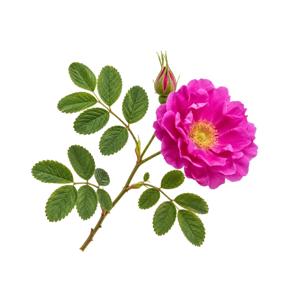 Principal source Rose Rosa spp. Herb monograph →
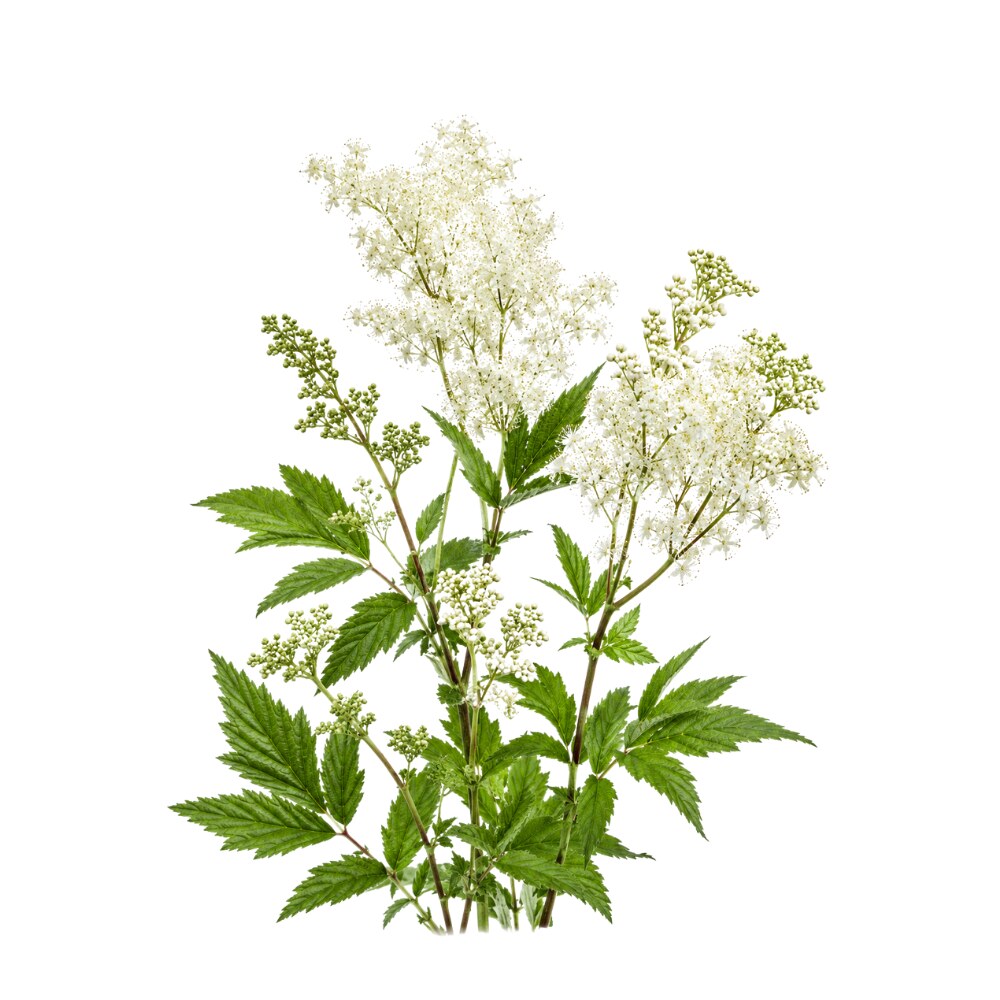 Source Meadowsweet Filipendula ulmaria Herb monograph →
Concentration
Content by Source
Concentrations not reported. No percent-dry-weight or mg/g figures have been published for this compound in its source plants.
Rugosin A at a Glance
Rugosin A is an ellagitannin — a hydrolysable tannin built on a single glucose core carrying galloyl and hexahydroxydiphenoyl (HHDP) esters, plus the distinctive valoneoyl group that names its family. It was described in 1982 from the flower petals of Rosa rugosa, the wrinkled-leaved beach rose, alongside rugosins B and C 6Reference 6Rugosin A, B, C and praecoxin A, tannins having a valoneoyl groupView study →.
Its structural position is the most useful thing about it — and the most easily got wrong. Rugosin A is a monomer: one glucose core. The better-known rugosins — D, E and F (dimers) and G (a trimer) — are built by oxidatively coupling monomer units through a valoneoyl bridge 7Reference 7Rugosin D, E, F and G, dimeric and trimeric hydrolyzable tanninsView study →. But the monomer being coupled is tellimagrandin II, not rugosin A 18Reference 18Biosynthesis of the dimeric ellagitannin, cornusiin E, in Tellima grandifloraView study →. Rugosin A shares the series name, the genus and the 1982 discovery paper with rugosin D — it is not half of it.
That distinction also frames its pharmacology, because in this molecular family size tends to buy potency. Rugosin A is one of four ellagitannins isolated from meadowsweet petal as inhibitors of histidine decarboxylase, the enzyme that makes histamine 1Reference 1Inhibitory activity of Filipendula ulmaria constituents on recombinant human histidine decarboxylaseView study → — but when the same group later measured them individually, rugosin A came out last of the four, weaker even than tellimagrandin II 2Reference 2Inhibition of Morganella morganii histidine decarboxylase activity and histamine accumulation in mackerel muscle derived from Filipendula ulmaria extractsView study →. Its one clear win is elsewhere: among seven rose-bud ellagitannins screened against dipeptidyl peptidase-IV, rugosin A and B were the most potent 3Reference 3Isolation of rugosin A, B and related compounds as dipeptidyl peptidase-IV inhibitors from rose bud extract powderView study →.
Everything below is in-vitro enzymology plus one 1993 mouse experiment. There are no human data on rugosin A, and at 1,107 Da it is far too large to be absorbed intact.
Rugosin A vs. Rugosin D — Same Series, Different Building Block
This page exists mainly to keep rugosin A from being read as a synonym for rugosin D, which is the more abundant, more potent and far more studied molecule. They are not the same thing, and the difference is both structural and measurable.
Structurally, rugosin A (C₄₈H₃₄O₃₁, MW 1,107) is a single-glucose monomer carrying a valoneoyl group. Rugosin D (C₈₂H₅₈O₅₂, MW 1,875) is a dimer of two tellimagrandin II units joined by a single valoneoyl bridge 7,
So the arithmetic that looks tempting here is a trap. Rugosin D’s formula happens to equal rugosin A plus tellimagrandin I minus water, but that is a coincidence of atom counting, not a biosynthetic relationship: rugosin A is not a subunit of rugosin D, and tellimagrandin I is the degalloylated relative of tellimagrandin II rather than an intermediate on the way to it 18Reference 18Biosynthesis of the dimeric ellagitannin, cornusiin E, in Tellima grandifloraView study →.
Functionally, that extra half matters. Nitta and colleagues isolated rugosin D, rugosin A, rugosin A methyl ester and tellimagrandin II from Filipendula ulmaria petal and reported them as non-competitive inhibitors of recombinant human histidine decarboxylase with Kᵢ values of approximately 0.35–1 µM 1Reference 1Inhibitory activity of Filipendula ulmaria constituents on recombinant human histidine decarboxylaseView study →. That widely-quoted figure is a range covering all four compounds — the paper’s abstract does not assign rugosin A its own Kᵢ, so quoting “0.35 µM” for rugosin A specifically is not supported.
The individual ranking only became visible three years later, when the same group tested the four against bacterial (Morganella morganii) histidine decarboxylase and published compound-by-compound IC₅₀ values 2Reference 2Inhibition of Morganella morganii histidine decarboxylase activity and histamine accumulation in mackerel muscle derived from Filipendula ulmaria extractsView study →:
| Compound | Form | M. morganii HDC IC₅₀ |
|---|---|---|
| Rugosin D | dimer | 1.5 µM |
| Rugosin A methyl ester | monomer | 4.4 µM |
| Tellimagrandin II | monomer | 6.1 µM |
| Rugosin A | monomer | 6.8 µM |
Rugosin A is the weakest of the four, roughly 4.5× less potent than rugosin D — and beaten even by its own methyl ester. A later study completed the pattern: the trimer rugosin G inhibits human HDC more strongly than ellagitannin monomers or the macrocyclic dimer oenothein B, which the authors attributed to its linear oligomeric conformation 5Reference 5The ellagitannin trimer rugosin G inhibits recombinant human histidine decarboxylaseView study →.
Read correctly, the ladder is tellimagrandin II (6.1 µM) → its dimer rugosin D (1.5 µM) → the trimer rugosin G: oligomerising the actual monomer unit buys roughly four-fold. Rugosin A is not on that ladder at all. It is a structurally distinct monomer that co-occurs in the same petals, and it is the least potent compound in the set — below even the unit that rugosin D is built from. Its own methyl ester (4.4 µM) outperforms it, which is the sharpest structure–activity signal this molecule generates.
So when meadowsweet’s gastroprotective, histamine-damping reputation is traced to “its ellagitannins”, the molecule doing most of the work is rugosin D. Rugosin A is a real contributor, but a junior one.
A Naming Trap: Rugosin A Is Also a Frog Peptide
Anyone searching this literature should know that “rugosin A” names two completely unrelated substances. Besides this tannin, it is also the name given to a 33-amino-acid antimicrobial peptide isolated in 1995 from the skin of the frog Rana rugosa, along with peptide rugosins B and C 15Reference 15Isolation and characterization of novel antimicrobial peptides, rugosins A, B and C, from the skin of the frog, Rana rugosaView study →. Both sets of names derive independently from the Latin rugosus (“wrinkled”) — Rosa rugosa for the tannin, Rana rugosa for the peptide. Of the fourteen PubMed records matching “rugosin A”, two are about the frog peptide and have nothing to do with plant chemistry.
What It's Used For
Rugosin A’s evidence base is genuinely small: three enzyme-inhibition studies and one mouse tumour study, spread over thirty years. There is no clinical literature, no pharmacokinetics, and no dedicated toxicology.
Support is an experimental score — weighted by study type (human > animal > in vitro) and volume. A beta, not a validated metric. Every score here is low because the entire evidence base is preclinical isolate work with no replication.
The application where rugosin A is the star rather than the understudy. Screening edible-plant extracts for dipeptidyl peptidase-IV inhibitors — the drug target behind the gliptin class of type-2 diabetes medicines, which works by sparing incretin hormones — Kato and colleagues found Rosa gallica bud extract powder highly active, and used activity-guided fractionation to isolate seven ellagitannins. Of those, rugosin A and rugosin B showed the highest inhibitory activities; rugosin B was named the major contributor in the extract by virtue of abundance 3Reference 3Isolation of rugosin A, B and related compounds as dipeptidyl peptidase-IV inhibitors from rose bud extract powderView study →.
Gap: this is a single in-vitro enzyme screen, published as a food-ingredient study. No IC₅₀ for rugosin A is quoted in the abstract, there is no cellular, animal or human follow-up, and a large tannin that does not cross the gut wall could only plausibly reach intestinal DPP-IV, not the circulating enzyme that gliptins target.
The mechanism that put rugosin A here — and the one where the distinction from rugosin D (see the comparison above) matters most. Histidine decarboxylase (HDC) is the sole enzyme making histamine, so inhibiting it is of interest for allergy and for gastric-acid-driven ulceration. Nitta and colleagues screened the ethyl-acetate extract of Filipendula ulmaria petal and found four ellagitannins as the main components — rugosin D, rugosin A, rugosin A methyl ester and tellimagrandin II — all non-competitive inhibitors of recombinant human HDC with Kᵢ ≈ 0.35–1 µM, comparable to the substrate-analogue inhibitor histidine methyl ester (Kᵢ 0.46 µM) 1Reference 1Inhibitory activity of Filipendula ulmaria constituents on recombinant human histidine decarboxylaseView study →.
A follow-up applied the same four to bacterial HDC and to fish spoilage: they inhibited Morganella morganii HDC (rugosin A, IC₅₀ 6.8 µM — the least potent of the four), and 2 wt% meadowsweet extract significantly reduced histamine accumulation in mackerel meat 2Reference 2Inhibition of Morganella morganii histidine decarboxylase activity and histamine accumulation in mackerel muscle derived from Filipendula ulmaria extractsView study →.
Gap: enzyme assays only. The mackerel experiment used whole extract, not isolated rugosin A, so the food-preservation result is an extract result. Nothing tests whether oral rugosin A affects histamine in an animal, let alone a person — and the tannin’s size makes systemic HDC inhibition implausible. There is no animal or human gastroprotection study of any rugosin. The nearest in-vivo bridge belongs to a different molecule in the same petal: tellimagrandin II at 40 mg/kg orally prevented ulceration in the rat ethanol-injury model — an outcome measurement only, with no mechanism attached 19Reference 19Antioxidant, anti-inflammatory and gastroprotective activity of Filipendula ulmaria (L.) Maxim. and Filipendula vulgaris MoenchView study →. Meadowsweet’s gastroprotective reputation otherwise rests on flower decoctions in rats plus this in-vitro enzymology, not on dosing any isolated rugosin.
The only in-vivo data on rugosin A, and it is thirty years old. Miyamoto and colleagues injected fifteen tannins intraperitoneally into mice at 10 mg/kg, four days before inoculation with S-180 sarcoma cells. Rugosin A showed significant antitumour activity, as did tellimagrandin II, hirtellin B and the oenotheins — compounds sharing tellimagrandin I or II structural units. Separately, monomeric ellagitannins including rugosin A increased IL-1β production from human peripheral macrophages by about two-fold over basal, while the oligomeric ellagitannins with stronger antitumour activity stimulated IL-1β more potently 4Reference 4Antitumor activity and interleukin-1 induction by tanninsView study →.
Gap: a single unreplicated 1993 study; a prophylactic intraperitoneal injection into a transplanted-tumour model is a long way from oral use, and the paper’s own conclusion favours the oligomers over the monomers. Two things about this paper get garbled downstream, so both are worth stating plainly: the cytokine is interleukin-1β, not interleukin-2; and rugosin A is one of the fifteen tannins actually tested — the abstract names it explicitly in both the antitumour and the IL-1β result, so this is a finding about this molecule, not a class prediction applied to it 4Reference 4Antitumor activity and interleukin-1 induction by tanninsView study →.
Mechanisms
| Target / pathway | Effect | Relevant to |
|---|---|---|
| Histidine decarboxylase (human, recombinant) | non-competitive inhibition, Kᵢ ≈ 0.35–1 µM reported for the four-ellagitannin group 1Reference 1Inhibitory activity of Filipendula ulmaria constituents on recombinant human histidine decarboxylaseView study → | antiallergicgastroprotective |
| Histidine decarboxylase (M. morganii) | inhibition, IC₅₀ 6.8 µM — weakest of the four meadowsweet ellagitannins 2Reference 2Inhibition of Morganella morganii histidine decarboxylase activity and histamine accumulation in mackerel muscle derived from Filipendula ulmaria extractsView study → | food histamine control |
| Dipeptidyl peptidase-IV | inhibition; highest activity of seven rose-bud ellagitannins tested 3Reference 3Isolation of rugosin A, B and related compounds as dipeptidyl peptidase-IV inhibitors from rose bud extract powderView study → | hypoglycaemic (incretin-sparing) |
| IL-1β release from human macrophages | ~2-fold increase (monomer-level; oligomers stronger) 4Reference 4Antitumor activity and interleukin-1 induction by tanninsView study → | immune / antitumour |
| Protein binding (tannin class) | astringency; complexation of dietary protein and non-heme iron | astringentGI |
The pattern across the rugosin series is conformational, not chemical: HDC potency rises with linear oligomer size — monomer (rugosin A) < linear dimer (rugosin D) < linear trimer (rugosin G) — while the macrocyclic dimer oenothein B underperforms its size 5Reference 5The ellagitannin trimer rugosin G inhibits recombinant human histidine decarboxylaseView study →. Rugosin A’s activity is best read as the baseline unit of that series.
Pharmacokinetics
There is none. A PubMed search for rugosin together with bioavailability, pharmacokinetics or urolithins returns zero records. What can be said is class-level and it is the load-bearing caveat: hydrolysable tannins of this size (rugosin A is 1,107 Da) are not absorbed intact. They are hydrolysed along the gut, releasing ellagic acid and gallic acid, and colonic microbiota convert the ellagic acid to urolithins — the metabolites that actually reach plasma, at levels that vary enormously between people according to their urolithin metabotype (A, B or 0) 17Reference 17Ellagic acid metabolism by human gut microbiota — consistent observation of three urolithin phenotypes in intervention trials, independent of food source, age, and health statusView study →. Free ellagic acid itself peaks in human plasma around 0.06 µmol/L after an ellagitannin-rich drink 16Reference 16Bioavailability of ellagic acid in human plasma after consumption of ellagitannins from pomegranate (Punica granatum L.) juiceView study →.
The practical consequence: the 1.5–7 µM enzyme concentrations in this literature are reachable in the gut lumen of someone drinking a strong rose or meadowsweet infusion, and essentially nowhere else. Whatever a person actually experiences from these plants is more likely astringency and local gut-level effects, plus downstream urolithins, than circulating rugosin A.
Clinical Trials
None. No trial has been registered or published on rugosin A in any form. The nearest human-adjacent evidence is regulatory recognition of the whole herb — the EMA maintains a herbal monograph on Filipendulae ulmariae herba — which is a traditional-use status, not molecule evidence.
| Completed | Planned | Terminated | Preclinical |
|---|---|---|---|
| 0 | 0 | 0 | 4(3 in vitro, 1 mouse) |
Last checked: August 2026.
Isolate vs. Plant Studies
Unusually for this batch, the rugosin A record is mostly isolate work — Kato’s DPP-IV screen, Nitta’s HDC assays and Miyamoto’s tumour study all used purified compound, so the attributions to the molecule are sound as far as they go 1,
Two specific things must not be read off it. First, the mackerel histamine result is an extract result — 2 wt% meadowsweet extract, not rugosin A 2Reference 2Inhibition of Morganella morganii histidine decarboxylase activity and histamine accumulation in mackerel muscle derived from Filipendula ulmaria extractsView study →. Second, meadowsweet’s gastroprotective reputation comes from flower decoctions in rats plus the in-vitro HDC mechanism; no rugosin has ever been given to an animal and had a stomach measured. The only isolated petal tannin with an in-vivo ulcer outcome is tellimagrandin II (40 mg/kg p.o., rat ethanol model) 19Reference 19Antioxidant, anti-inflammatory and gastroprotective activity of Filipendula ulmaria (L.) Maxim. and Filipendula vulgaris MoenchView study → — a neighbour molecule, and an outcome without a mechanism. When a source says “meadowsweet works via HDC-inhibiting ellagitannins”, the enzymology is real, the in-vivo link is inferred, and the most active molecule in the mixture is rugosin D, not rugosin A.
Prevalence in Nature
Rugosin A is a Rosaceae petal tannin first and foremost. Its type source is Rosa rugosa flower petal 6,
Beyond the roses it is sparse but real: it occurs in the spurges Euphorbia prostrata 10Reference 10Chen L, Chen R, Wei K. (1992). [Constituents of tannins from Euphorbia prostrata Ait]. Zhongguo Zhong Yao Za Zhi, 17(4), 225–226. https://pubmed.ncbi.nlm.nih.gov/1418550/View study → and Euphorbia supina (the latter yielding rugosin A methyl ester) 9Reference 9Toxicity of tannins towards the free-living nematode Caenorhabditis elegans and the brine shrimp Artemia salinaView study →, and in the Saharan myrtle Myrtus nivellei, where rugosins A and B are among the compounds distinguishing it from common myrtle 11Reference 11ReviewAntioxidant activity of Myrtus communis L. and Myrtus nivellei Batt. & Trab. extracts — a brief reviewView study →. The pericarp of water caltrop (Trapa bispinosa) yields decarboxylated rugosin A, a derivative with α-glucosidase-inhibitory activity 12Reference 12Characterization and identification of bioactive polyphenols in the Trapa bispinosa Roxb. pericarp extractView study →.
The rugosins are a Rosaceae story, and neighbouring genera are a trap. PubMed returns zero records for rugosin A with Rubus, with Oenothera, or with Eucalyptus. That absence is not an oversight in the literature — those genera carry their own valoneoyl-bridged oligomers (rubusuaviins in Rubus, eucarpanins in Eucalyptus, the macrocyclic oenothein B in Oenothera 5Reference 5The ellagitannin trimer rugosin G inhibits recombinant human histidine decarboxylaseView study →). A shared valoneoyl linkage makes them structural cousins, not the same compound, and none of their data belongs here.
Quantification is a real gap. No source we found reports a mg/g figure for rugosin A in any plant. For scale, its dimer rugosin D reaches 18–41 mg/g in prickly-rose leaf and 4 mg/g in the flower 13Reference 13Metabolites of prickly rose — chemodiversity and digestive-enzyme-inhibiting potential of Rosa acicularis and the main ellagitannin rugosin DView study → — rugosin A is consistently described as a component, never as the dominant one.
Discovery & Synthesis
Rugosin A was isolated in 1982 by Takuo Okuda, Tsutomu Hatano, Kazufumi Yazaki and Noboru Ogawa at Okayama University, from the flower petals of Rosa rugosa, and reported together with rugosins B and C and the related praecoxin A as “tannins having a valoneoyl group” 6Reference 6Rugosin A, B, C and praecoxin A, tannins having a valoneoyl groupView study →. In the same issue of Chemical and Pharmaceutical Bulletin, the group described the dimeric and trimeric members of the series — rugosins D, E, F and G — showing that the valoneoyl group is what links the units together 7Reference 7Rugosin D, E, F and G, dimeric and trimeric hydrolyzable tanninsView study →. A 1990 paper in the same journal’s Tannins of Rosaceous Plants series settled the full structures, including the orientation of the valoneoyl group, by two-dimensional NMR 8Reference 8Tannins of rosaceous plants VIII: hydrolyzable tannin monomers having a valoneoyl group from flower petals of Rosa rugosa ThunbView study →.
Biosynthetically, the series is better understood than the isolation papers alone suggest. Cell-free enzyme preparations oxidise pentagalloylglucose — the branch-point of hydrolysable-tannin metabolism — to the monomeric ellagitannin tellimagrandin II, and a second oxidative coupling joins two tellimagrandin II units through a valoneoyl bridge to give a dimer; the product identified in Tellima grandiflora was cornusiin E, with rugosin D named as its 1,4′,6′ positional isomer 18Reference 18Biosynthesis of the dimeric ellagitannin, cornusiin E, in Tellima grandifloraView study →. Where rugosin A enters this scheme has not been established — no paper traces its biosynthesis, which is part of why its relationship to the dimeric rugosins is so easily misstated.
One point in the Filipendula literature deserves a flag. Nitta and colleagues describe rugosin A methyl ester as “a novel compound” in their 2013 meadowsweet paper 1Reference 1Inhibitory activity of Filipendula ulmaria constituents on recombinant human histidine decarboxylaseView study → — but a compound of that name had already been isolated from Euphorbia supina and reported in 2002 9Reference 9Toxicity of tannins towards the free-living nematode Caenorhabditis elegans and the brine shrimp Artemia salinaView study →. The claim of novelty may be specific to the source or to a stereochemical detail not visible in the abstracts; treat “novel compound” as contested until the full texts are compared.
There is no reported total synthesis of rugosin A, and no commercial production: it is obtained by chromatographic isolation from petal extracts, which is why analytical reference standards of it are scarce and expensive.
Patents: not yet researched.
Toxicity & Safety
Rugosin A carries a low flag — it is a dietary-tannin constituent of rose petals and meadowsweet flowers, both consumed as teas for centuries, and it is not systemically absorbed. That badge reflects the absence of alarm signals, not the presence of a safety dataset: no mammalian toxicology exists for the isolated molecule.
- A food-and-tea tannin. Rose petal and meadowsweet flower are widely consumed; no toxicity has been reported from ordinary infusions.
- Poorly absorbed. At 1,107 Da it does not cross the gut wall intact, so systemic exposure to the molecule itself is minimal.
- Tannin-class iron and protein binding. Like all hydrolysable tannins it complexes dietary protein and non-heme iron; heavy, chronic intake of strong tannin infusions with meals can reduce iron absorption.
- No mammalian safety data on the isolate. No repeat-dose, genotoxicity or reproductive study exists for rugosin A; the one animal exposure on record is a single 10 mg/kg intraperitoneal injection in mice 4Reference 4Antitumor activity and interleukin-1 induction by tanninsView study →.
- Astringency. Concentrated tannin preparations can irritate the stomach and provoke nausea on an empty stomach — a whole-class effect, not specific to this molecule.
Full Safety & Interactions Detail
What exists. The only dedicated toxicity data touching this molecule are invertebrate. Yamasaki and colleagues tested ten ellagitannins against the nematode Caenorhabditis elegans and the brine shrimp Artemia salina, and found rugosin A methyl ester (isolated from Euphorbia supina) among only three of the ten producing significant nematode toxicity, in what they read as a structure-activity relationship for ellagitannins specifically 9Reference 9Toxicity of tannins towards the free-living nematode Caenorhabditis elegans and the brine shrimp Artemia salinaView study →. That is an ecotoxicology finding about a derivative, not a mammalian hazard signal — but it is the closest thing to a toxicology datapoint on record.
What does not exist. No acute or repeat-dose mammalian study, no genotoxicity battery, no organ-toxicity work, no interaction study. The 10 mg/kg intraperitoneal dose in the 1993 mouse tumour experiment was tolerated, with no toxicity reported 4Reference 4Antitumor activity and interleukin-1 induction by tanninsView study → — a single data point, by a route nobody uses.
Reproductive note (prose only). There are no reproductive or developmental data on rugosin A. Decisions about meadowsweet or rose in pregnancy belong on the herb pages, where intake is actually decided; nothing here bears on them.
Class cautions that do carry over. As a hydrolysable tannin, rugosin A shares the tannin profile: protein precipitation (the astringent mouthfeel), reduced non-heme iron absorption when strong infusions are taken with meals, and potential to bind co-administered alkaloid or protein drugs in the gut lumen. These are class effects and apply to any tannin-rich preparation, not to this molecule in particular.
Dosage
There is no dose for rugosin A. It is not sold as an isolate, has never been given to a person deliberately, and would not be absorbed if it were. The figures that appear in this literature are laboratory quantities:
| Context | Figure | Source |
|---|---|---|
| HDC inhibition (human recombinant) | Kᵢ ≈ 0.35–1 µM (four-compound group figure) | 1Reference 1Inhibitory activity of Filipendula ulmaria constituents on recombinant human histidine decarboxylaseView study → |
| HDC inhibition (M. morganii) | IC₅₀ 6.8 µM | 2Reference 2Inhibition of Morganella morganii histidine decarboxylase activity and histamine accumulation in mackerel muscle derived from Filipendula ulmaria extractsView study → |
| Antitumour (mouse S-180) | 10 mg/kg, single intraperitoneal dose | 4Reference 4Antitumor activity and interleukin-1 induction by tanninsView study → |
| Meadowsweet extract on mackerel | 2 wt% whole extract | 2Reference 2Inhibition of Morganella morganii histidine decarboxylase activity and histamine accumulation in mackerel muscle derived from Filipendula ulmaria extractsView study → |
Real human exposure comes only from whole plants — rose-petal tea, meadowsweet flower infusion — where rugosin A is one minor tannin in a mixture dominated by its own larger relatives. None of the above is a recommendation.
References
- Nitta Y, Kikuzaki H, Azuma T, Ye Y, Sakaue M, Higuchi Y, Komori H, Ueno H. (2013). Inhibitory activity of Filipendula ulmaria constituents on recombinant human histidine decarboxylase. Food Chemistry, 138(2–3), 1551–1556. https://pubmed.ncbi.nlm.nih.gov/23411280/
- Nitta Y, Yasukata F, Kitamoto N, Ito M, Sakaue M, Kikuzaki H, Ueno H. (2016). Inhibition of Morganella morganii histidine decarboxylase activity and histamine accumulation in mackerel muscle derived from Filipendula ulmaria extracts. Journal of Food Protection, 79(3), 463–467. https://pubmed.ncbi.nlm.nih.gov/26939657/
- Kato E, Uenishi Y, Inagaki Y, Kurokawa M, Kawabata J. (2016). Isolation of rugosin A, B and related compounds as dipeptidyl peptidase-IV inhibitors from rose bud extract powder. Bioscience, Biotechnology, and Biochemistry, 80(11), 2087–2092. https://pubmed.ncbi.nlm.nih.gov/27477520/
- Miyamoto K, Murayama T, Nomura M, Hatano T, Yoshida T, Furukawa T, Koshiura R, Okuda T. (1993). Antitumor activity and interleukin-1 induction by tannins. Anticancer Research, 13(1), 37–42. https://pubmed.ncbi.nlm.nih.gov/8476228/
- Nitta Y, Ito H, Komori H, Ueno H, Takeshima D, Ito M, Sakaue M, Kikuzaki H. (2019). The ellagitannin trimer rugosin G inhibits recombinant human histidine decarboxylase. Bioscience, Biotechnology, and Biochemistry, 83(7), 1315–1318. https://pubmed.ncbi.nlm.nih.gov/30995171/
- Okuda T, Hatano T, Yazaki K, Ogawa N. (1982). Rugosin A, B, C and praecoxin A, tannins having a valoneoyl group. Chemical and Pharmaceutical Bulletin, 30(11), 4230–4233. https://doi.org/10.1248/cpb.30.4230
- Okuda T, Hatano T, Ogawa N. (1982). Rugosin D, E, F and G, dimeric and trimeric hydrolyzable tannins. Chemical and Pharmaceutical Bulletin, 30(11), 4234–4237. https://doi.org/10.1248/cpb.30.4234
- Hatano T, Ogawa N, Yasuhara T, Okuda T. (1990). Tannins of rosaceous plants VIII: hydrolyzable tannin monomers having a valoneoyl group from flower petals of Rosa rugosa Thunb. Chemical and Pharmaceutical Bulletin, 38(12), 3308–3313. https://doi.org/10.1248/cpb.38.3308
- Yamasaki T, Sato M, Mori T, Mohamed AS, Fujii K, Tsukioka J. (2002). Toxicity of tannins towards the free-living nematode Caenorhabditis elegans and the brine shrimp Artemia salina. Journal of Natural Toxins, 11(3), 165–171. https://pubmed.ncbi.nlm.nih.gov/12182536/
- Chen L, Chen R, Wei K. (1992). [Constituents of tannins from Euphorbia prostrata Ait]. Zhongguo Zhong Yao Za Zhi, 17(4), 225–226. https://pubmed.ncbi.nlm.nih.gov/1418550/
- Hennia A, Miguel MG, Nemmiche S. (2018). Antioxidant activity of Myrtus communis L. and Myrtus nivellei Batt. & Trab. extracts — a brief review. Medicines (Basel), 5(3), 89. https://pubmed.ncbi.nlm.nih.gov/30103510/
- Iwaoka Y, Suzuki S, Kato N, Hayakawa C, Kawabe S, Ganeko N, Uemura T, Ito H. (2021). Characterization and identification of bioactive polyphenols in the Trapa bispinosa Roxb. pericarp extract. Molecules, 26(19), 5802. https://pubmed.ncbi.nlm.nih.gov/34641346/
- Olennikov DN, Chemposov VV, Chirikova NK. (2021). Metabolites of prickly rose — chemodiversity and digestive-enzyme-inhibiting potential of Rosa acicularis and the main ellagitannin rugosin D. Plants (Basel), 10(11), 2525. https://pubmed.ncbi.nlm.nih.gov/34834888/
- Milala J, Kosmala M, Sójka M, Kołodziejczyk K, Klewicki R, Król B, et al. (2023). Rosa rugosa low caloric fiber protein preparations rich in antioxidant flavanols and ellagitannins. Molecules, 28(24), 8021. https://pubmed.ncbi.nlm.nih.gov/38138511/
- Suzuki S, Ohe Y, Okubo T, Kakegawa T, Tatemoto K. (1995). Isolation and characterization of novel antimicrobial peptides, rugosins A, B and C, from the skin of the frog, Rana rugosa. Biochemical and Biophysical Research Communications, 212(1), 249–254. https://pubmed.ncbi.nlm.nih.gov/7612013/
- Seeram NP, Lee R, Heber D. (2004). Bioavailability of ellagic acid in human plasma after consumption of ellagitannins from pomegranate (Punica granatum L.) juice. Clinica Chimica Acta, 348(1–2), 63–68. https://pubmed.ncbi.nlm.nih.gov/15369737/
- Tomás-Barberán FA, García-Villalba R, González-Sarrías A, Selma MV, Espín JC. (2014). Ellagic acid metabolism by human gut microbiota — consistent observation of three urolithin phenotypes in intervention trials, independent of food source, age, and health status. Journal of Agricultural and Food Chemistry, 62(28), 6535–6538. https://pubmed.ncbi.nlm.nih.gov/24976365/
- Niemetz R, Schilling G, Gross GG. (2003). Biosynthesis of the dimeric ellagitannin, cornusiin E, in Tellima grandiflora. Phytochemistry, 64(1), 109–114. https://pubmed.ncbi.nlm.nih.gov/12946409/
- Samardžić S, Arsenijević J, Božić D, Milenković M, Tešević V, Maksimović Z. (2018). Antioxidant, anti-inflammatory and gastroprotective activity of Filipendula ulmaria (L.) Maxim. and Filipendula vulgaris Moench. Journal of Ethnopharmacology, 213, 132–137. https://pubmed.ncbi.nlm.nih.gov/29132911/