Compound Monograph
Tussilagone
Tussilagone is the headline sesquiterpenoid of coltsfoot (Tussilago farfara) — an oplopane-type ester, and neither a pyrrolizidine alkaloid nor a sesquiterpene lactone. Its best evidence is anti-inflammatory (NF-κB suppression and Nrf2/HO-1 induction via covalent modification of KEAP1), alongside a 1980s record as a potent injected cardiovascular-respiratory stimulant.

Sources · 1
Where Does It Come From?
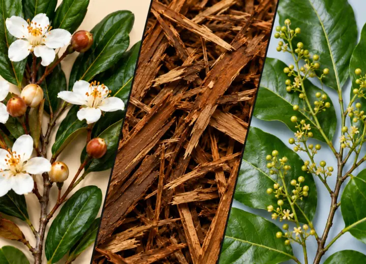
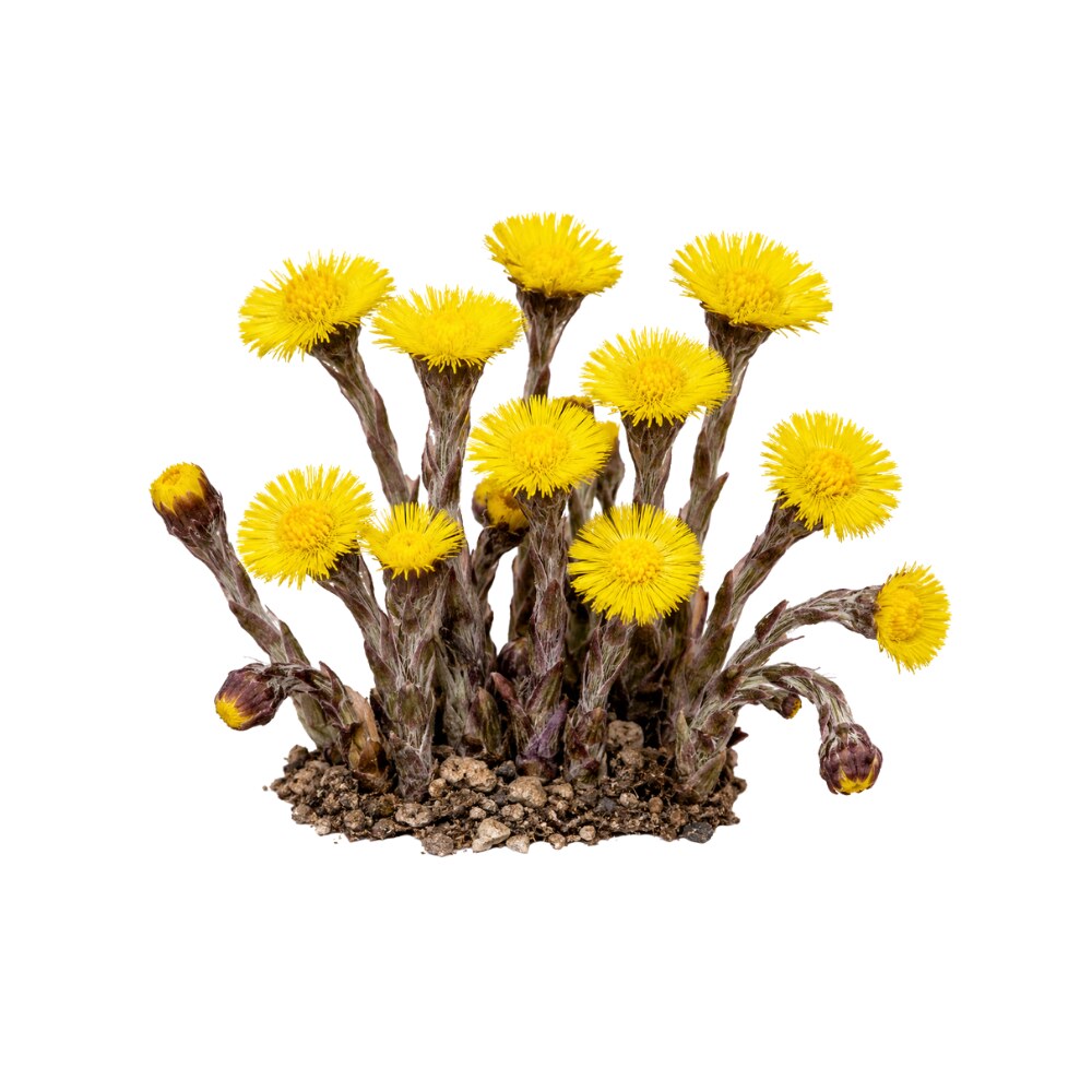
Tussilagone is a naturally occurring sesquiterpenoid ester (oplopane / notonipetranone type), found in Coltsfoot. It is flagged as moderately toxic.
Concentration
Content by Source
Reported concentrations across the plants that contain tussilagone — the bar marks the typical level, the line shows the reported range.
Literature figures for varying plant parts and preparations. Read them as a comparative guide, not exact assays.
Tussilagone at a Glance
Tussilagone is the best-studied single molecule in coltsfoot (Tussilago farfara), the plant Chinese medicine calls kuǎn dōng huā (Farfarae Flos). It is a sesquiterpenoid ester — a bicyclic oplopane-type ketone carrying an acetate and a branched unsaturated ester — and it is concentrated in the flower buds rather than the leaf 24,
Its modern reputation is as an anti-inflammatory. Independent groups, working on macrophages, microglia, dendritic cells, colitis, sepsis, particulate lung injury and cigarette-smoke COPD, keep converging on the same two-armed mechanism: NF-κB suppression and Nrf2/HO-1 induction 5,
Two disciplines govern the account. First, tussilagone is not one of coltsfoot’s pyrrolizidine alkaloids and does not carry their liver toxicity — that belongs to different molecules, and it is spelled out in its own section below. Second, and just as important: nothing here makes the herb safe. Third, there is no human data of any kind — not one clinical trial, not one pharmacokinetic measurement in a person — and the rat oral bioavailability of ~1.3% 28Reference 28AnimalDetermination and pharmacokinetic study of tussilagone in rat plasma by RP-HPLC method — rat pharmacokineticsView study → means most of the literature above is describing an injected compound.
Tussilagone Is Not a Pyrrolizidine Alkaloid
Coltsfoot’s defining safety problem is pyrrolizidine alkaloid (PA) hepatotoxicity. The herb contains senkirkine and senecionine, unsaturated PAs that the liver bioactivates into reactive pyrroles which form DNA and protein adducts. A long-term rat feeding study of pre-blooming coltsfoot flowers produced hepatic haemangioendothelial sarcomas, attributed to senkirkine 38Reference 38AnimalCarcinogenic activity of coltsfoot, Tussilago farfara L. — rat in vivoView study →, and human case reports link coltsfoot ingestion to hepatic veno-occlusive disease, including a fatal infant case — although a systematic review found that species misidentification and substitution repeatedly compromised those reports 39Reference 39Systematic reviewFitoterapia. https://pubmed.ncbi.nlm.nih.gov/32105669/View study →. PA-containing herbs are consequently restricted or capped by daily-intake limits in several countries.
Tussilagone has nothing to do with that. It is a terpenoid, not an alkaloid: it contains no nitrogen at all, no necine base, and no pyrrolizidine ring system. It is not metabolised to a pyrrole. It is not what the rat carcinogenicity study was measuring, and it is not what the intake limits regulate. The two chemistries coexist in the same flower bud and are otherwise unrelated — the PAs are trace-level (typically reported below 0.015% of dried herb, and highly variable by part, chemotype and region) 40Reference 40Journal of Separation Science. https://pubmed.ncbi.nlm.nih.gov/27673325/View study →, while tussilagone runs an order of magnitude higher 32Reference 32Extraction and determination of tussilagone from Farfarae Flos with online solid-phase extraction-high-performance liquid chromatography — analyticalView study →.
The discipline has to run in both directions, and the second one is easier to forget:
- Do not import PA toxicity onto tussilagone. No study attributes hepatotoxicity, DNA adduction or carcinogenicity to this molecule. Its own toxicology is a separate, and much thinner, question (see Toxicity & Safety).
- Do not export tussilagone’s clean pharmacology onto the herb. A favourable isolate profile does not make Tussilago farfara safe to swallow. Coltsfoot’s benefit-risk problem is that the beneficial classes and the harmful one arrive in the same leaf, and purifying tussilagone in a laboratory does nothing about that for someone drinking a tea. That decision belongs on the herb monograph, not here.
A rat study that dosed Farfarae Flos extract by gavage and tracked metabolites of both the active constituents and the hepatotoxic PAs in the same animals is the clearest illustration of the point: in the whole extract the two chemistries travel together, whatever the isolate does on its own 31Reference 31AnimalScreening and identification of metabolites of two kinds of main active ingredients and hepatotoxic pyrrolizidine alkaloids in rat after lavage Farfarae Flos extract by UHPLC-Q-TOF-MS — rat in vivoView study →.
The Oplopane Skeleton & the Missing Lactone Warhead
Tussilagone is routinely filed alongside the Asteraceae sesquiterpene lactones, and it does not belong there. The 1988 characterisation gives it as (7R,14R)-14-acetoxy-7-[(2′E)-3′-methylpent-2′-enoyloxy]-oplopanone 1Reference 1AnimalEvaluation of tussilagone: a cardiovascular-respiratory stimulant isolated from Chinese herbal medicine — dog/cat/rat in vivoView study →; Merck’s independent description, 14-acetoxy-7β-(3′-ethylcrotonoyloxy)-notonipetranone, names the same acyl group on the same skeleton 2Reference 2In vitroL-652,469 — a dual receptor antagonist of platelet activating factor and dihydropyridines from Tussilago farfara L. — in vitroView study →. Formally it is C₂₃H₃₄O₅ — a C₁₅ oplopane (hydrindanone) core plus two ester acyl groups, one acetate and one 3-methylpent-2-enoate.
What that structure contains, and does not:
- A bicyclo[4.3.0] (hydrindane) carbon skeleton — a cyclohexane fused to a cyclopentane. Every ring atom is carbon.
- A cyclopentanone in the five-membered ring.
- An exocyclic methylidene (=CH₂) on the six-membered ring, not conjugated to that ketone — two saturated carbons separate them.
- An allylic ester at the adjacent position, the 3-methylpent-2-enoate.
- A secondary acetate on the side chain.
- No lactone ring of any kind, and therefore no α-methylene-γ-lactone — the thiol-alkylating warhead that defines the reactivity of costunolide, helenalin, parthenolide and the rest of the sesquiterpene-lactone class.
This matters because tussilagone’s headline mechanism is exactly the kind of thing that class is usually invoked to explain. Xu et al. showed by biotin pull-down that tussilagone covalently modifies Cys434 of KEAP1 — a previously unreported high-reactivity cysteine in KEAP1’s Kelch domain — and that this is what activates Nrf2 and, downstream, damps NF-κB and the NLRP3 inflammasome 5Reference 5AnimalTussilagone attenuated cigarette smoke-induced chronic obstructive pulmonary disease through regulating Nrf2 and NF-κB/NLRP3 inflammasome via directly targeting cysteine 434 of KEAP1 — mouse in vivoView study →. A covalent adduct on a cysteine thiol means an electrophilic carbon somewhere on the molecule. It cannot be the class warhead, because the class warhead is absent.
Where it actually comes from is not settled. The published work demonstrates the adduct without assigning the reactive carbon 5Reference 5AnimalTussilagone attenuated cigarette smoke-induced chronic obstructive pulmonary disease through regulating Nrf2 and NF-κB/NLRP3 inflammasome via directly targeting cysteine 434 of KEAP1 — mouse in vivoView study →. On the structure there are two plausible candidates, and this is structural reading rather than published assignment:
- The α,β-unsaturated ester of the 3-methylpent-2-enoyl group is a Michael acceptor in principle — but its β-carbon carries both a methyl and an ethyl substituent, making it a hindered, sluggish one.
- The exocyclic methylidene paired with the allylic ester next to it is the more interesting possibility: that arrangement can in principle accept a thiol at the exocyclic carbon with expulsion of the carboxylate (an addition–elimination at an allylic ester), which would trap a cysteine without ever needing a lactone.
Until someone maps the adduct, treat the mechanism as demonstrated but structurally unexplained. What can be said flatly is the negative: whatever tussilagone is doing to KEAP1, it is not doing it as a sesquiterpene lactone, and reasoning about it from that class’s reactivity will mislead.
L-652,469 — The Molecule’s Second Name
Tussilagone was described twice, under two names, by two groups who were not talking to each other. The Chinese pharmacology (Li and Wang, Shanghai College of TCM) named it tussilagone and studied it as a cardiovascular-respiratory stimulant 1,
- A competitive platelet-activating-factor (PAF) receptor antagonist — inhibiting [³H]PAF binding to rabbit platelet membranes with Kᵢ of 3.2–4.0 µM and a K_B of 5.16 µM.
- A competitive inhibitor of dihydropyridine binding in cardiac sarcolemmal vesicles (Kᵢ 1.2 µM against [³H]nitrendipine) — i.e. an L-type calcium-channel blocker site ligand.
- Functionally, it relaxed Ca²⁺-induced contraction of rat aortic strips by ~60% at 10 µM, inhibited rabbit platelet aggregation, and was orally active against PAF-induced and early-phase carrageenan-induced rat paw oedema.
The dual-antagonist paper is worth knowing about for three reasons. It is the earliest anti-inflammatory data on the molecule and it predates the NF-κB/Nrf2 literature by two decades. It supplies a mechanism — PAF antagonism — that the modern papers rarely reference. And it is the one report of oral activity, which sits awkwardly against the later measurement of ~1.3% oral bioavailability in rats 28Reference 28AnimalDetermination and pharmacokinetic study of tussilagone in rat plasma by RP-HPLC method — rat pharmacokineticsView study →.
It also creates a small literature hazard: searching for “tussilagone” misses the L-652,469 papers entirely, and vice versa. PubChem lists both names on the same compound record (CAS 104012-37-5).
The Antitussive Irony
Tussilago means “cough dispeller.” Coltsfoot has been a cough herb in Europe and in China for well over a thousand years, and the molecule was named tussilagone after the plant. The obvious expectation is that it is the antitussive principle.
There is essentially no evidence that it is. Searching the indexed literature for antitussive testing of isolated tussilagone returns nothing — the antitussive and expectorant work on this plant is on caffeoylquinic acids, where four of them (including chlorogenic acid and the dicaffeoylquinic acids) suppressed ammonia-induced cough and increased phenol-red expectoration in mice at 10–20 mg/kg orally 35,
Meanwhile the molecule’s own documented respiratory effect is the one nobody expected: stimulation of respiration in anaesthetised animals given it intravenously 1Reference 1AnimalEvaluation of tussilagone: a cardiovascular-respiratory stimulant isolated from Chinese herbal medicine — dog/cat/rat in vivoView study →. That is a brainstem/pressor phenomenon, not cough suppression, and it has no bearing on a cup of tea.
So the naming is honorific rather than mechanistic. Tussilagone is coltsfoot’s most pharmacologically interesting molecule; it is not, on current evidence, coltsfoot’s cough molecule.
What It's Used For
The isolate literature is large for a plant sesquiterpenoid — several dozen papers, most since 2008 — and unusually consistent in mechanism. It is also entirely preclinical, heavily weighted to injected dosing, and structured around cell lines and mouse models rather than outcomes.
Support is an experimental score I’m building — a composite weighted by study type (human > animal > in vitro > review) and study volume. It’s a beta: a fast way to rank strength of evidence at a glance, not a validated metric. Every row here is preclinical, so no score is high; the ranking is about depth and replication within that ceiling. Each application links down to its write-up.
The deepest and most reproducible part of the record, and all of it isolate work. Tussilagone induces heme oxygenase-1 in murine macrophages and suppresses NO, TNF-α and PGE₂ 6Reference 6In vitroThe anti-inflammatory effect of tussilagone, from Tussilago farfara, is mediated by the induction of heme oxygenase-1 in murine macrophages — in vitroView study →; it inhibits NO and PGE₂ production in LPS-activated BV-2 microglia with IC₅₀ values of 8.67 µM and 14.1 µM, suppressing iNOS and COX-2 protein and mRNA through blocked IκBα degradation and p65 nuclear translocation 7Reference 7In vitroSuppression of inducible nitric oxide synthase and cyclooxygenase-2 expression by tussilagone from Farfarae flos in BV-2 microglial cells — in vitroView study →; it suppresses dendritic-cell function via HO-1 induction 9Reference 9In vitroTussilagone inhibits dendritic cell functions via induction of heme oxygenase-1 — in vitroView study →; and it ameliorates dextran-sulphate-sodium colitis in mice 8Reference 8AnimalTussilagone, a major active component in Tussilago farfara, ameliorates inflammatory responses in dextran sulphate sodium-induced murine colitis — mouse in vivoView study →.
The 2026 COPD study supplies the missing target: biotin-labelled tussilagone pulls down KEAP1, modifies Cys434 covalently, and thereby activates Nrf2, which in turn suppresses NF-κB and the NLRP3 inflammasome 5Reference 5AnimalTussilagone attenuated cigarette smoke-induced chronic obstructive pulmonary disease through regulating Nrf2 and NF-κB/NLRP3 inflammasome via directly targeting cysteine 434 of KEAP1 — mouse in vivoView study →.
The convergence of unrelated groups on one axis, from one purified molecule, is what puts this row on top.
Gap: in vitro and mouse only, with no human anti-inflammatory data; the reactive carbon responsible for the KEAP1 adduct is unassigned; and the effective in-vitro range (roughly 5–20 µM) has never been reconciled with the ~1.3% oral bioavailability measured in rats 28Reference 28AnimalDetermination and pharmacokinetic study of tussilagone in rat plasma by RP-HPLC method — rat pharmacokineticsView study →.
Coltsfoot’s traditional territory, approached through injury models rather than cough. Isolated tussilagone attenuated cigarette-smoke-induced COPD in mice through the KEAP1/Nrf2 and NF-κB/NLRP3 axis 5Reference 5AnimalTussilagone attenuated cigarette smoke-induced chronic obstructive pulmonary disease through regulating Nrf2 and NF-κB/NLRP3 inflammasome via directly targeting cysteine 434 of KEAP1 — mouse in vivoView study →, protected against PM2.5-induced acute lung injury via Hif-1α/NF-κB 11Reference 11AnimalTussilagone protects acute lung injury from PM2.5 via alleviating Hif-1α/NF-κB-mediated inflammatory response — mouse in vivoView study →, improved survival and reduced inflammation in CLP-induced sepsis 12Reference 12AnimalTussilagone inhibits the inflammatory response and improves survival in CLP-induced septic mice — mouse in vivo (published correction 2020)View study →, and mitigated sepsis-induced acute lung injury by suppressing RIPK1 13Reference 13AnimalTussilagone mitigates sepsis-induced acute lung injury in mice by suppressing RIPK1 expressionView study →.
In airway epithelial cells it suppressed production and gene expression of the mucin MUC5AC through NF-κB 10Reference 10In vitroTussilagone suppressed the production and gene expression of MUC5AC mucin via regulating nuclear factor-kappa B signaling pathway in airway epithelial cells — in vitroView study →.
A separate ethanol extract of the flower buds — plant, not isolate — reached the same NLRP3/Nrf2/NF-κB endpoints in cigarette-smoke lung inflammation 14Reference 14AnimalThe ethanol extract of flower buds of Tussilago farfara L. attenuates cigarette smoke-induced lung inflammation through regulating NLRP3 inflammasome, Nrf2, and NF-κB — mouse in vivoView study →.
Gap: the strong models use isolated tussilagone by injection, so they speak to the molecule rather than to an infusion; MUC5AC suppression is mucoregulatory and does not demonstrate the traditional expectorant action; no human respiratory data exist.
The oldest pharmacology, and the only place the compound has been given to a large animal. Intravenous tussilagone produced an instant, dose-dependent pressor effect in anaesthetised dogs (0.02–0.3 mg/kg), cats (0.02–0.5 mg/kg) and rats (0.4–4 mg/kg), described as similar in character to dopamine but without tachyphylaxis, together with significant respiratory stimulation; the same paper reports an acute intravenous LD₅₀ in mice of 28.9 mg/kg 1Reference 1AnimalEvaluation of tussilagone: a cardiovascular-respiratory stimulant isolated from Chinese herbal medicine — dog/cat/rat in vivoView study →.
Companion Chinese-language work examined the pressor mechanism 3Reference 3AnimalLi YP, Wang YM (1986). [Pressor mechanism of tussilagone] — animal model. Zhongguo Yao Li Xue Bao (Acta Pharmacologica Sinica). https://pubmed.ncbi.nlm.nih.gov/2954393/View study → and the haemodynamic effects in conscious dogs and in dogs during haemorrhagic shock 4Reference 4AnimalLi YP, Wang YM (1987). [The effects of tussilagone on the hemodynamics of conscious dogs and dogs during hemorrhagic shock] — animal model. Yao Xue Xue Bao (Acta Pharmaceutica Sinica). https://pubmed.ncbi.nlm.nih.gov/3450146/View study →.
Cutting the other way, the Merck characterisation found the same molecule relaxed Ca²⁺-induced aortic contraction by ~60% at 10 µM through dihydropyridine-site antagonism 2Reference 2In vitroL-652,469 — a dual receptor antagonist of platelet activating factor and dihydropyridines from Tussilago farfara L. — in vitroView study → — so the intact-animal pressor response is unlikely to be direct vasoconstriction.
Gap: all isolate, all intravenous, all 1980s, and never replicated with modern methods; the mechanism papers are Chinese-language and were not read in full here; and a pressor agent with an IV LD₅₀ of 28.9 mg/kg is a hazard signal as much as a therapeutic one.
Isolate work across four unrelated pathways. Tussilagone suppressed colon-cancer cell proliferation by promoting β-catenin degradation 15Reference 15In vitroTussilagone suppresses colon cancer cell proliferation by promoting the degradation of β-catenin — in vitroView study → and reduced tumour burden in a colitis-associated colon-cancer mouse model by damping inflammation 16Reference 16Tussilagone Reduces Tumorigenesis by Diminishing Inflammation in Experimental Colitis-Associated Colon CancerView study →; it suppressed angiogenesis through VEGFR2 signalling 17Reference 17In vitroFrontiers in Pharmacology. https://pubmed.ncbi.nlm.nih.gov/31333473/View study →; and it inhibited triple-negative breast-cancer progression via the TLR4/NF-κB pathway while enhancing antitumour immunity 18Reference 18In vitroImmunopharmacology and Immunotoxicology. https://pubmed.ncbi.nlm.nih.gov/41029481/View study →.
The pathway diversity is a mild strength — these are not one finding reported four times.
Gap: cell lines plus a single mouse model, with no dose-matched comparison across the pathways and no human data. This row also carries the herb-level contradiction stated above: the whole plant is hepatocarcinogenic in rodents through its pyrrolizidine alkaloids 38Reference 38AnimalCarcinogenic activity of coltsfoot, Tussilago farfara L. — rat in vivoView study →, so nothing here transfers to coltsfoot as a plant.
An unexpectedly well-replicated application, all isolate. Tussilagone inhibited osteoclastogenesis and wear-particle-induced periprosthetic osteolysis by suppressing NF-κB and p38 MAPK, confirmed in a murine calvarial osteolysis model 25Reference 25In vitroFrontiers in Pharmacology. https://pubmed.ncbi.nlm.nih.gov/32317967/View study →.
It promoted osteoclast apoptosis and prevented ovariectomy-induced (oestrogen-deficiency) osteoporosis in mice 26Reference 26AnimalTussilagone promotes osteoclast apoptosis and prevents estrogen deficiency-induced osteoporosis in miceView study →.
A third group found it inhibits osteoclastogenesis by modulating mitochondrial function and ROS production through Nrf2 activation 27Reference 27In vitroTussilagone inhibits osteoclastogenesis by modulating mitochondrial function and ROS production involved Nrf2 activation — mouse in vivo / in vitroView study →.
Three groups, two disease models, and a mechanism continuous with the anti-inflammatory story.
Gap: no human or large-animal bone data, no dose-ranging beyond the model doses, no fracture or bone-mineral-density endpoint, and no test of whether the ~1.3% oral bioavailability permits bone exposure by mouth.
Three separate strands, four decades apart. As L-652,469 the molecule is a competitive PAF-receptor antagonist (Kᵢ 3.2–4.0 µM) that inhibited PAF-induced rat paw oedema after oral dosing 2Reference 2In vitroL-652,469 — a dual receptor antagonist of platelet activating factor and dihydropyridines from Tussilago farfara L. — in vitroView study →.
Isolated tussilagone reduced symptoms and serum histamine, IgE and cytokines in ovalbumin-sensitised allergic-rhinitis guinea pigs and stabilised IgE-stimulated RBL-2H3 mast cells 21Reference 21In vitroFitoterapia. https://pubmed.ncbi.nlm.nih.gov/32058052/View study →.
Most recently it was shown to inhibit MRGPRX2-mediated mast-cell degranulation, suppressing pseudo-allergic (non-IgE) reactions 22Reference 22In vitroToxicology and Applied Pharmacology. https://pubmed.ncbi.nlm.nih.gov/41707990/View study → — a distinct receptor from the IgE route.
Gap: one species per strand, no cross-validation between the PAF, IgE and MRGPRX2 mechanisms, and no human allergy data. The PAF work has not been revisited since 1987.
Isolate data at two levels. In bioassay-guided fractionation of coltsfoot flower buds, tussilagone was the most potent of four sesquiterpenoids against microsomal DGAT1 — IC₅₀ 18.8 µM against rat liver microsomes and 49.1 µM against HepG2 microsomes, comfortably ahead of tussilagonone at 99.2 µM and >1 mM respectively 24Reference 24In vitroSesquiterpenoids isolated from the flower buds of Tussilago farfara L. inhibit diacylglycerol acyltransferase — in vitroView study →.
In vivo, tussilagone ameliorated high-fat-diet-induced hepatic steatosis in mice by enhancing energy metabolism and antioxidant activity 23Reference 23Tussilagone ameliorates high-fat diet-induced hepatic steatosis by enhancing energy metabolism and antioxidant activityView study →.
Gap: a single NAFLD mouse model plus an enzyme assay in the tens-of-micromolar range — well above any plausible oral plasma level 28Reference 28AnimalDetermination and pharmacokinetic study of tussilagone in rat plasma by RP-HPLC method — rat pharmacokineticsView study →; no glycaemic endpoint, no lipid-panel data, no human work.
Isolate again, and mechanistically split. Tussilagone inhibits platelet activation by blocking GPVI downstream signalling in vitro 19Reference 19In vitroFrontiers in Medicine. https://pubmed.ncbi.nlm.nih.gov/32850895/View study →, and it attenuated atherosclerotic lesion formation in ApoE-deficient mice by suppressing MAPK-mediated macrophage inflammation 20Reference 20Tussilagone attenuates atherosclerosis through inhibiting MAPKs-mediated inflammation in macrophagesView study →.
The 1987 Merck paper independently reported inhibition of gel-filtered rabbit platelet aggregation (pA₂ 5.79), attributed there to the dual PAF/calcium-channel antagonism rather than to GPVI 2Reference 2In vitroL-652,469 — a dual receptor antagonist of platelet activating factor and dihydropyridines from Tussilago farfara L. — in vitroView study →.
Gap: one in-vitro platelet study and one mouse model, with two different proposed antiplatelet mechanisms that have never been tested against each other. An antiplatelet signal is also a theoretical bleeding-interaction concern as much as a benefit, and it has not been evaluated clinically.
The row that exists to record an absence. Despite the molecule being named for coltsfoot’s cough reputation, no antitussive assay of isolated tussilagone appears in the indexed literature. The mouse cough and expectoration data for this plant belong to the caffeoylquinic acids — chlorogenic acid and three dicaffeoylquinic acids, active orally at 10–20 mg/kg 35,
Tussilagone’s only airway-secretion finding is MUC5AC suppression 10Reference 10In vitroTussilagone suppressed the production and gene expression of MUC5AC mucin via regulating nuclear factor-kappa B signaling pathway in airway epithelial cells — in vitroView study →, which is mucoregulatory rather than expectorant, and its own documented respiratory effect is intravenous respiratory stimulation 1Reference 1AnimalEvaluation of tussilagone: a cardiovascular-respiratory stimulant isolated from Chinese herbal medicine — dog/cat/rat in vivoView study →.
Gap: the isolate has not been put through a cough model. Until someone does, the antitussive attribution is etymology, not pharmacology.
Mechanisms
| Target / pathway | Effect | Relevant to |
|---|---|---|
| KEAP1 Cys434 (covalent modification) | frees Nrf2 from KEAP1-mediated degradation → Nrf2 nuclear translocation; the one named molecular target for this molecule 5Reference 5AnimalTussilagone attenuated cigarette smoke-induced chronic obstructive pulmonary disease through regulating Nrf2 and NF-κB/NLRP3 inflammasome via directly targeting cysteine 434 of KEAP1 — mouse in vivoView study → | anti-inflammatoryrespiratorycytoprotectivebone |
| Nrf2 → HO-1, NQO1 | antioxidant/cytoprotective gene induction; HO-1 also mediates the dendritic-cell and macrophage effects 6, | anti-inflammatorylung protectionosteoclast inhibition |
| NF-κB (IκBα degradation ↓, p65 translocation ↓) | suppresses iNOS, COX-2, TNF-α, IL-6, MUC5AC transcription 7, | anti-inflammatoryrespiratoryanticancer |
| NLRP3 inflammasome | downstream of Nrf2 activation; damped in cigarette-smoke COPD 5Reference 5AnimalTussilagone attenuated cigarette smoke-induced chronic obstructive pulmonary disease through regulating Nrf2 and NF-κB/NLRP3 inflammasome via directly targeting cysteine 434 of KEAP1 — mouse in vivoView study → | respiratory |
| p38 / MAPK | suppressed in macrophages and osteoclast precursors 20, | atherosclerosisbone |
| PAF receptor | competitive antagonism, Kᵢ 3.2–4.0 µM (as L-652,469) 2Reference 2In vitroL-652,469 — a dual receptor antagonist of platelet activating factor and dihydropyridines from Tussilago farfara L. — in vitroView study → | anti-allergicantiplateletanti-oedema |
| Dihydropyridine site / L-type Ca²⁺ channel | competitive inhibition of [³H]nitrendipine binding, Kᵢ 1.2 µM; relaxes Ca²⁺-induced aortic contraction 2Reference 2In vitroL-652,469 — a dual receptor antagonist of platelet activating factor and dihydropyridines from Tussilago farfara L. — in vitroView study → | cardiovascular |
| GPVI downstream signalling | platelet activation ↓ in vitro 19Reference 19In vitroFrontiers in Medicine. https://pubmed.ncbi.nlm.nih.gov/32850895/View study → | antiplatelet (and a theoretical bleeding risk) |
| β-catenin degradation; VEGFR2; TLR4 | antiproliferative and antiangiogenic effects in tumour models 15, | anticancer |
| DGAT1 | inhibition, IC₅₀ 18.8 µM (rat liver microsomes) 24Reference 24In vitroSesquiterpenoids isolated from the flower buds of Tussilago farfara L. inhibit diacylglycerol acyltransferase — in vitroView study → | metabolic |
| Unidentified pressor mechanism | dose-dependent IV pressor + respiratory stimulation in dogs, cats, rats, no tachyphylaxis 1, | cardiovascular — and a safety signal |
The load-bearing point is the first row. A covalent cysteine adduct is a specific, checkable claim — and because tussilagone has no α-methylene-γ-lactone, the usual class explanation for sesquiterpenoid thiol trapping does not apply here (see “The Oplopane Skeleton” above). The reactive carbon remains unassigned.
Pharmacokinetics
This is where most of the page’s optimism is spent. In rats, tussilagone given intravenously at 5 mg/kg shows biphasic kinetics — rapid distribution then slower elimination — while intragastric dosing at 200 mg/kg fits a one-compartment model; absolute oral bioavailability is about 1.31% 28Reference 28AnimalDetermination and pharmacokinetic study of tussilagone in rat plasma by RP-HPLC method — rat pharmacokineticsView study →. That single number reframes the rest of the literature: the in-vitro effective concentrations (typically 5–20 µM) and the intraperitoneal in-vivo doses are not achievable by swallowing the molecule, let alone by drinking a tea in which it is a fraction of a percent of dry weight.
Metabolism is well described in vitro. Nine metabolites were identified across rat and human liver microsomes; only one was common to both, with eight seen in human microsomes only — a genuine species difference that undercuts direct rat-to-human extrapolation. The routes are hydrolysis of the C-14 ester and hydroxylation of the side chains at C-12, C-5′ and C-6′, and CYP3A4 is the dominant P450 for the hydroxylation steps (two of the three hydroxy metabolites formed only with CYP3A4) 29Reference 29In vitroComparative metabolism of tussilagone in rat and human liver microsomes using UHPLC coupled with high-resolution LTQ-Orbitrap mass spectrometry — in vitroView study →. The molecule is lipophilic (XLogP ≈ 4.1), consistent with ester hydrolysis being the main clearance step.
In the whole-extract setting, an LC-MS/MS study dosing rats with Farfarae Flos extract found tussilagone reached T_max within roughly 0.2–0.7 h alongside the phenolic acids and flavonoids, but with the lowest C_max and AUC and the shortest terminal half-life of the ten compounds measured 30Reference 30AnimalSimultaneous determination of seven phenolic acids, two flavonoids, and tussilagone in rat plasma after administration of Farfarae Flos extract by LC-MS/MS — rat pharmacokineticsView study →. A separate gavage study tracked metabolites of the active constituents and the hepatotoxic pyrrolizidine alkaloids in the same animals 31Reference 31AnimalScreening and identification of metabolites of two kinds of main active ingredients and hepatotoxic pyrrolizidine alkaloids in rat after lavage Farfarae Flos extract by UHPLC-Q-TOF-MS — rat in vivoView study →.
What is missing: no human pharmacokinetic data of any kind, no plasma protein binding, no tissue distribution, no absolute half-life figure in hours, and no measurement of whether the C-14-hydrolysed metabolite is active.
Clinical Trials
There are none. No registered trial evaluates tussilagone in humans for any indication, no human pharmacokinetic study exists, and the molecule has no regulatory status as a drug or supplement anywhere. The 1987 report of oral activity against rat paw oedema 2Reference 2In vitroL-652,469 — a dual receptor antagonist of platelet activating factor and dihydropyridines from Tussilago farfara L. — in vitroView study → is the closest thing to a translational signal, and it is thirty-nine years old.
| Completed | Planned | Terminated | Preclinical |
|---|---|---|---|
| 0 | 0 | 0 | ~40–50 |
Last checked: July 2026.
Isolate vs. Plant Studies
Unusually for a plant molecule, the evidence here is overwhelmingly isolate, and that is a genuine methodological strength as far as it goes. Almost every entry above — the KEAP1/Nrf2 COPD work 5Reference 5AnimalTussilagone attenuated cigarette smoke-induced chronic obstructive pulmonary disease through regulating Nrf2 and NF-κB/NLRP3 inflammasome via directly targeting cysteine 434 of KEAP1 — mouse in vivoView study →, the macrophage and microglial NF-κB work 6,
The strength ends there, for three reasons.
Route. The isolate studies overwhelmingly use intraperitoneal or intravenous dosing. Coltsfoot is drunk, not injected. With ~1.3% oral bioavailability in rats 28Reference 28AnimalDetermination and pharmacokinetic study of tussilagone in rat plasma by RP-HPLC method — rat pharmacokineticsView study →, the isolate literature describes an exposure that a tea cannot produce.
Dose. Effective concentrations sit in the 5–20 µM range in vitro and in the mg/kg range in vivo. Flower buds assay at roughly 0.74 mg/g 32Reference 32Extraction and determination of tussilagone from Farfarae Flos with online solid-phase extraction-high-performance liquid chromatography — analyticalView study →, so a conventional herbal dose delivers single-digit milligrams of tussilagone at best, before absorption.
The plant is not the molecule. The handful of whole-plant studies — the cigarette-smoke flower-bud extract 14Reference 14AnimalThe ethanol extract of flower buds of Tussilago farfara L. attenuates cigarette smoke-induced lung inflammation through regulating NLRP3 inflammasome, Nrf2, and NF-κB — mouse in vivoView study →, the focal-cerebral-ischaemia rat study 45Reference 45AnimalEffects of Flower Buds Extract of Tussilago farfara on Focal Cerebral Ischemia in Rats and Inflammatory Response in BV2 MicrogliaView study →, the antitussive caffeoylquinic-acid work 35,
The honest summary: strong molecular attribution, no route or dose bridge to how anyone actually uses the plant, and no human step at either level.
Prevalence in Nature
Tussilagone appears to be effectively confined to Tussilago farfara. A search of the indexed literature for the molecule outside coltsfoot returns nothing — no other genus, no other family, no non-plant source. That is unusual and worth stating plainly: most named plant sesquiterpenoids turn up across a genus at least, and this one has not.
Within the plant it is a flower-bud compound. Farfarae Flos (kuǎn dōng huā, the dried flower buds) is the tissue every isolation study starts from, and it is the part Chinese medicine uses; leaf material is dominated by mucilage and the phenolic fraction instead 35,
Chemically it sits in a cluster of closely related coltsfoot sesquiterpenoids, not alone. Bioassay-guided work has repeatedly pulled tussilagone out alongside tussilagonone, 7β-(3-ethyl-cis-crotonoyloxy)-1α-(2-methylbutyryloxy)-3,14-dehydro-Z-notonipetranone, and bisabolane-type ketones — an oplopane/bisabolane suite that shares the anti-inflammatory and Nrf2 activity to varying degrees 24,
Biosynthesis and ecological role are unknown. No pathway has been mapped, no enzyme identified, and no defence or signalling function demonstrated for this compound in the plant.
Discovery & Synthesis
The compound emerged from two independent lines of work in the mid-1980s.
Naming and first pharmacology (China). Li Yuping and Wang Yumin at the Shanghai College of Traditional Chinese Medicine isolated the active principle from Kuandong Hua and named it tussilagone, after the genus. Their published sequence runs from the pressor mechanism (1986) 3Reference 3AnimalLi YP, Wang YM (1986). [Pressor mechanism of tussilagone] — animal model. Zhongguo Yao Li Xue Bao (Acta Pharmacologica Sinica). https://pubmed.ncbi.nlm.nih.gov/2954393/View study → to the haemodynamic work in conscious and shocked dogs (1987) 4Reference 4AnimalLi YP, Wang YM (1987). [The effects of tussilagone on the hemodynamics of conscious dogs and dogs during hemorrhagic shock] — animal model. Yao Xue Xue Bao (Acta Pharmaceutica Sinica). https://pubmed.ncbi.nlm.nih.gov/3450146/View study → to the summary evaluation in General Pharmacology (1988), which gives the full structure — (7R,14R)-14-acetoxy-7-[(2′E)-3′-methylpent-2′-enoyloxy]-oplopanone — the species-by-species pressor doses, and the intravenous mouse LD₅₀ of 28.9 mg/kg 1Reference 1AnimalEvaluation of tussilagone: a cardiovascular-respiratory stimulant isolated from Chinese herbal medicine — dog/cat/rat in vivoView study →. Earlier Chinese pharmacology on coltsfoot cardiovascular extracts (1979) predates the isolation and is the likely origin of the search.
Independent rediscovery (Merck). In 1987 Hwang and colleagues at Merck Sharp & Dohme Research Laboratories isolated the same molecule from methylene-chloride extracts of coltsfoot buds while screening for platelet-activating-factor antagonists, coded it L-652,469, and described it under the alternative skeletal name 14-acetoxy-7β-(3′-ethylcrotonoyloxy)-notonipetranone 2Reference 2In vitroL-652,469 — a dual receptor antagonist of platelet activating factor and dihydropyridines from Tussilago farfara L. — in vitroView study →. The two names have since been consolidated onto one compound record (CAS 104012-37-5).
Etymology. Tussilago from Latin tussis (cough) + agere (to drive away) — the plant’s name, transferred to the molecule. As the antitussive section notes, the isolate has never been shown to earn it.
Total synthesis. No total synthesis of tussilagone is documented in the indexed literature — searches of PubMed and Crossref return nothing. This is plausible for an oplopanone-type ester with four stereocentres and low commercial pull; the semisynthetic route via esterification of an oplopanone core has likewise not been reported. Related oplopanone chemistry exists, but not for this target.
Supply today is therefore extraction only. Research-grade tussilagone is isolated from Farfarae Flos, historically by bioassay-guided solvent fractionation 7,
Patents: not yet researched.
Toxicity & Safety
The moderate flag here is a judgement about the isolate, and it is deliberately not the herb’s flag. It is not driven by hepatotoxicity — that is the pyrrolizidine alkaloids’ problem, and no PA-type finding attaches to this molecule (see “Tussilagone Is Not a Pyrrolizidine Alkaloid”). It is driven by two other things: a potent, dose-dependent pressor and respiratory-stimulant effect at intravenous doses as low as 0.02 mg/kg in dogs and cats 1Reference 1AnimalEvaluation of tussilagone: a cardiovascular-respiratory stimulant isolated from Chinese herbal medicine — dog/cat/rat in vivoView study →, and an acute intravenous LD₅₀ in mice of 28.9 mg/kg 1Reference 1AnimalEvaluation of tussilagone: a cardiovascular-respiratory stimulant isolated from Chinese herbal medicine — dog/cat/rat in vivoView study → — meaning the therapeutic and lethal ranges by injection are separated by roughly two orders of magnitude, which is narrow for a compound with no clinical monitoring behind it. Against that sits ~1.3% oral bioavailability 28Reference 28AnimalDetermination and pharmacokinetic study of tussilagone in rat plasma by RP-HPLC method — rat pharmacokineticsView study →, which limits systemic exposure from swallowing it.
- Not a pyrrolizidine alkaloid. Coltsfoot’s hepatotoxicity and carcinogenicity belong to senkirkine and senecionine, not to this molecule; no hepatotoxic or genotoxic finding is attributed to tussilagone.
- Very low oral absorption. ~1.3% bioavailability in rats caps systemic exposure from ingestion — the same fact that undercuts most of its claimed benefits.
- Cardiovascular pressor effect. Dose-dependent blood-pressure rise and respiratory stimulation in dogs, cats and rats from 0.02 mg/kg intravenously, without tachyphylaxis; an acute IV LD₅₀ of 28.9 mg/kg in mice. Injected routes are not benign.
- This says nothing about coltsfoot’s safety. A clean isolate profile does not clear the herb — coltsfoot carries hepatotoxic pyrrolizidine alkaloids in the same tissue, is restricted in several countries, and that assessment lives on the herb monograph.
- Theoretical bleeding interaction. Two independent antiplatelet mechanisms in vitro (GPVI blockade; PAF antagonism) raise a possible interaction with anticoagulant or antiplatelet drugs — never studied clinically.
- CYP3A4 substrate. Hydroxylation is CYP3A4-dominant in human liver microsomes; whether tussilagone also inhibits or induces CYP3A4 has not been tested, so a drug-interaction potential is unassessed rather than excluded.
- No human data at all. No trial, no human PK, no adverse-event series, no repeat-dose or genotoxicity package. Absence of reported harm here reflects absence of human exposure, not established safety.
Full Safety & Interactions Detail
The cardiovascular signal is the real one. The 1988 evaluation is unambiguous: intravenous tussilagone produced an instant, dose-dependent pressor effect in anaesthetised dogs (0.02–0.3 mg/kg), cats (0.02–0.5 mg/kg) and rats (0.4–4 mg/kg), likened to dopamine but without tachyphylaxis, together with significant respiratory stimulation, and with an acute IV LD₅₀ in mice of 28.9 mg/kg 1Reference 1AnimalEvaluation of tussilagone: a cardiovascular-respiratory stimulant isolated from Chinese herbal medicine — dog/cat/rat in vivoView study →. A compound that raises blood pressure reliably and does not desensitise is pharmacologically interesting and clinically hazardous in equal measure, and the mechanism has never been characterised in a language-accessible modern paper 3,
Interactions — named where possible. CYP3A4 is the dominant P450 in the hydroxylation of tussilagone in human liver microsomes, with two of three hydroxy metabolites formed exclusively by that isoform 29Reference 29In vitroComparative metabolism of tussilagone in rat and human liver microsomes using UHPLC coupled with high-resolution LTQ-Orbitrap mass spectrometry — in vitroView study →; the compound has not been tested as a CYP3A4 inhibitor or inducer, and no transporter (P-gp, BCRP) work exists. Antiplatelet drugs and anticoagulants are the plausible pharmacodynamic interaction: tussilagone blocks GPVI downstream signalling in vitro 19Reference 19In vitroFrontiers in Medicine. https://pubmed.ncbi.nlm.nih.gov/32850895/View study → and, as L-652,469, antagonises the PAF receptor and inhibits rabbit platelet aggregation 2Reference 2In vitroL-652,469 — a dual receptor antagonist of platelet activating factor and dihydropyridines from Tussilago farfara L. — in vitroView study →. Both are in-vitro findings and neither has been clinically evaluated. Antihypertensives deserve a theoretical flag in the opposite direction given the pressor data 1Reference 1AnimalEvaluation of tussilagone: a cardiovascular-respiratory stimulant isolated from Chinese herbal medicine — dog/cat/rat in vivoView study →, though only by an injected route.
Species differences complicate extrapolation. Nine microsomal metabolites were identified across rat and human preparations, with only one shared and eight human-specific 29Reference 29In vitroComparative metabolism of tussilagone in rat and human liver microsomes using UHPLC coupled with high-resolution LTQ-Orbitrap mass spectrometry — in vitroView study →. Rat toxicology therefore transfers to humans poorly, and the existing toxicology is rat and mouse.
Reproductive and developmental findings. No reproductive- or developmental-toxicity study of isolated tussilagone was located — not a negative result, an absent one. No teratogenicity, fertility, or lactation-transfer data exist for the molecule. Decisions about intake in pregnancy or breastfeeding are made at the herb or product level, where the pyrrolizidine-alkaloid contraindication dominates the assessment; that determination belongs on the coltsfoot monograph, not on this molecule page.
This covers the isolated compound only. No repeat-dose toxicity study, no genotoxicity battery, no carcinogenicity study, no oral LD₅₀ and no human safety data were located for tussilagone. Absence of adverse-event reports reflects the absence of human use, not demonstrated safety. Nothing here modifies the safety assessment of coltsfoot the herb.
Dosage
There is no human dose for tussilagone, and no basis for setting one. It is not a drug, not a supplement, and has never been administered to a person in a published study. What follows is a record of what has been given to animals and cells, presented so the numbers can be checked against each other — not as guidance.
| Context | Route | Dose / concentration |
|---|---|---|
| Pressor effect, anaesthetised dogs 1Reference 1AnimalEvaluation of tussilagone: a cardiovascular-respiratory stimulant isolated from Chinese herbal medicine — dog/cat/rat in vivoView study → | intravenous | 0.02–0.3 mg/kg |
| Pressor effect, anaesthetised cats 1Reference 1AnimalEvaluation of tussilagone: a cardiovascular-respiratory stimulant isolated from Chinese herbal medicine — dog/cat/rat in vivoView study → | intravenous | 0.02–0.5 mg/kg |
| Pressor effect, rats 1Reference 1AnimalEvaluation of tussilagone: a cardiovascular-respiratory stimulant isolated from Chinese herbal medicine — dog/cat/rat in vivoView study → | intravenous | 0.4–4 mg/kg |
| Acute lethality, mice 1Reference 1AnimalEvaluation of tussilagone: a cardiovascular-respiratory stimulant isolated from Chinese herbal medicine — dog/cat/rat in vivoView study → | intravenous | LD₅₀ 28.9 mg/kg |
| Pharmacokinetic study, rats 28Reference 28AnimalDetermination and pharmacokinetic study of tussilagone in rat plasma by RP-HPLC method — rat pharmacokineticsView study → | intravenous / intragastric | 5 mg/kg IV; 200 mg/kg PO |
| NO and PGE₂ inhibition, BV-2 microglia 7Reference 7In vitroSuppression of inducible nitric oxide synthase and cyclooxygenase-2 expression by tussilagone from Farfarae flos in BV-2 microglial cells — in vitroView study → | in vitro | IC₅₀ 8.67 µM and 14.1 µM |
| PAF-receptor antagonism (as L-652,469) 2Reference 2In vitroL-652,469 — a dual receptor antagonist of platelet activating factor and dihydropyridines from Tussilago farfara L. — in vitroView study → | in vitro | Kᵢ 3.2–4.0 µM; K_B 5.16 µM |
| DGAT1 inhibition, rat liver microsomes 24Reference 24In vitroSesquiterpenoids isolated from the flower buds of Tussilago farfara L. inhibit diacylglycerol acyltransferase — in vitroView study → | in vitro | IC₅₀ 18.8 µM |
Two figures should be read together before any of the others. Oral bioavailability in rats is ~1.31% 28Reference 28AnimalDetermination and pharmacokinetic study of tussilagone in rat plasma by RP-HPLC method — rat pharmacokineticsView study → — so the 200 mg/kg oral dose and the 5 mg/kg intravenous dose in that same study are roughly comparable in exposure terms, and the in-vitro micromolar potencies are not reachable by mouth at anything like a plant-derived intake. And flower buds assay at about 0.74 mg/g 32Reference 32Extraction and determination of tussilagone from Farfarae Flos with online solid-phase extraction-high-performance liquid chromatography — analyticalView study →, so a conventional 3–9 g dose of Farfarae Flos contains on the order of 2–7 mg of tussilagone before absorption losses — which, at ~1% absorption, is tens of micrograms systemically. That arithmetic, more than any single study, is why the isolate literature should not be read as an account of what coltsfoot does in a person.
These are doses studied in research and are not a personal recommendation.
References
- Li YP, Wang YM (1988). Evaluation of tussilagone: a cardiovascular-respiratory stimulant isolated from Chinese herbal medicine — dog/cat/rat in vivo. General Pharmacology. https://pubmed.ncbi.nlm.nih.gov/3350333/
- Hwang SB, Chang MN, Garcia ML, et al. (1987). L-652,469 — a dual receptor antagonist of platelet activating factor and dihydropyridines from Tussilago farfara L. — in vitro. European Journal of Pharmacology. https://pubmed.ncbi.nlm.nih.gov/2824219/
- Li YP, Wang YM (1986). [Pressor mechanism of tussilagone] — animal model. Zhongguo Yao Li Xue Bao (Acta Pharmacologica Sinica). https://pubmed.ncbi.nlm.nih.gov/2954393/
- Li YP, Wang YM (1987). [The effects of tussilagone on the hemodynamics of conscious dogs and dogs during hemorrhagic shock] — animal model. Yao Xue Xue Bao (Acta Pharmaceutica Sinica). https://pubmed.ncbi.nlm.nih.gov/3450146/
- Xu LT, Han QT, Xu ZP, et al. (2026). Tussilagone attenuated cigarette smoke-induced chronic obstructive pulmonary disease through regulating Nrf2 and NF-κB/NLRP3 inflammasome via directly targeting cysteine 434 of KEAP1 — mouse in vivo. Journal of Advanced Research. https://pubmed.ncbi.nlm.nih.gov/40669599/
- Hwangbo C, Lee HS, Park J, et al. (2009). The anti-inflammatory effect of tussilagone, from Tussilago farfara, is mediated by the induction of heme oxygenase-1 in murine macrophages — in vitro. International Immunopharmacology. https://pubmed.ncbi.nlm.nih.gov/19800419/
- Lim HJ, Lee HS, Ryu JH (2008). Suppression of inducible nitric oxide synthase and cyclooxygenase-2 expression by tussilagone from Farfarae flos in BV-2 microglial cells — in vitro. Archives of Pharmacal Research. https://pubmed.ncbi.nlm.nih.gov/18481023/
- Cheon HJ, Nam SH, Kim JK (2018). Tussilagone, a major active component in Tussilago farfara, ameliorates inflammatory responses in dextran sulphate sodium-induced murine colitis — mouse in vivo. Chemico-Biological Interactions. https://pubmed.ncbi.nlm.nih.gov/30142311/
- Park Y, Ryu HS, Lee HK, et al. (2014). Tussilagone inhibits dendritic cell functions via induction of heme oxygenase-1 — in vitro. International Immunopharmacology. https://pubmed.ncbi.nlm.nih.gov/25091622/
- Choi BS, Kim YJ, Yoon YP, et al. (2018). Tussilagone suppressed the production and gene expression of MUC5AC mucin via regulating nuclear factor-kappa B signaling pathway in airway epithelial cells — in vitro. Korean Journal of Physiology & Pharmacology. https://pubmed.ncbi.nlm.nih.gov/30402027/
- Lin H, Chen M, Gao Y, et al. (2022). Tussilagone protects acute lung injury from PM2.5 via alleviating Hif-1α/NF-κB-mediated inflammatory response — mouse in vivo. Environmental Toxicology. https://pubmed.ncbi.nlm.nih.gov/35112795/
- Kim YK, Yeo MG, Oh BK, et al. (2017). Tussilagone inhibits the inflammatory response and improves survival in CLP-induced septic mice — mouse in vivo (published correction 2020). International Journal of Molecular Sciences. https://pubmed.ncbi.nlm.nih.gov/29258263/
- Gu J, Cai Y, Yin J, et al. (2026). Tussilagone mitigates sepsis-induced acute lung injury in mice by suppressing RIPK1 expression. Hereditas. https://pubmed.ncbi.nlm.nih.gov/42015326/
- Xu LT, Wang T, Fang KL, et al. (2022). The ethanol extract of flower buds of Tussilago farfara L. attenuates cigarette smoke-induced lung inflammation through regulating NLRP3 inflammasome, Nrf2, and NF-κB — mouse in vivo. Journal of Ethnopharmacology. https://pubmed.ncbi.nlm.nih.gov/34601084/
- Li H, Lee HJ, Ahn YH, et al. (2014). Tussilagone suppresses colon cancer cell proliferation by promoting the degradation of β-catenin — in vitro. Biochemical and Biophysical Research Communications. https://pubmed.ncbi.nlm.nih.gov/24269588/
- Nam SH, Kim JK (2020). Tussilagone Reduces Tumorigenesis by Diminishing Inflammation in Experimental Colitis-Associated Colon Cancer. Biomedicines. https://pubmed.ncbi.nlm.nih.gov/32290483/
- Tussilagone suppresses angiogenesis by inhibiting the VEGFR2 signaling pathway — in vitro. (2019). Frontiers in Pharmacology. https://pubmed.ncbi.nlm.nih.gov/31333473/
- Tussilagone suppresses triple-negative breast cancer progression by inhibiting the TLR4/NF-κB pathway and enhancing anti-tumor immunity — in vitro. (2025). Immunopharmacology and Immunotoxicology. https://pubmed.ncbi.nlm.nih.gov/41029481/
- Antiplatelet activity of tussilagone via inhibition of the GPVI downstream signaling pathway in platelets — in vitro. (2020). Frontiers in Medicine. https://pubmed.ncbi.nlm.nih.gov/32850895/
- Shen S, Huang Z, Lin L, et al. (2023). Tussilagone attenuates atherosclerosis through inhibiting MAPKs-mediated inflammation in macrophages. Int Immunopharmacol. https://pubmed.ncbi.nlm.nih.gov/37058752/
- Tussilagone inhibits allergic responses in OVA-induced allergic rhinitis guinea pigs and IgE-stimulated RBL-2H3 cells — animal / in vitro. (2020). Fitoterapia. https://pubmed.ncbi.nlm.nih.gov/32058052/
- Tussilagone inhibits MRGPRX2-mediated mast cell degranulation and suppresses pseudo-allergic reactions — in vitro. (2026). Toxicology and Applied Pharmacology. https://pubmed.ncbi.nlm.nih.gov/41707990/
- Sun M, Li Y, Su S, et al. (2024). Tussilagone ameliorates high-fat diet-induced hepatic steatosis by enhancing energy metabolism and antioxidant activity. Phytother Res. https://pubmed.ncbi.nlm.nih.gov/37010930/
- Park HR, Yoo MY, Seo JH, et al. (2008). Sesquiterpenoids isolated from the flower buds of Tussilago farfara L. inhibit diacylglycerol acyltransferase — in vitro. Journal of Agricultural and Food Chemistry. https://pubmed.ncbi.nlm.nih.gov/18937486/
- Tussilagone inhibits osteoclastogenesis and periprosthetic osteolysis by suppressing the NF-κB and P38 MAPK signaling pathways — mouse in vivo / in vitro. (2020). Frontiers in Pharmacology. https://pubmed.ncbi.nlm.nih.gov/32317967/
- Ryoo GH, Moon YJ, Choi S, et al. (2020). Tussilagone promotes osteoclast apoptosis and prevents estrogen deficiency-induced osteoporosis in mice. Biochem Biophys Res Commun. https://pubmed.ncbi.nlm.nih.gov/32807498/
- Feng X, Liu Z, Su Y, et al. (2023). Tussilagone inhibits osteoclastogenesis by modulating mitochondrial function and ROS production involved Nrf2 activation — mouse in vivo / in vitro. Biochemical Pharmacology. https://pubmed.ncbi.nlm.nih.gov/38084677/
- Liu YF, Yang XW, Lu W, Xin XL (2008). Determination and pharmacokinetic study of tussilagone in rat plasma by RP-HPLC method — rat pharmacokinetics. Biomedical Chromatography. https://pubmed.ncbi.nlm.nih.gov/18651585/
- Zhang XS, Ren W, Bian BL, et al. (2015). Comparative metabolism of tussilagone in rat and human liver microsomes using UHPLC coupled with high-resolution LTQ-Orbitrap mass spectrometry — in vitro. Rapid Communications in Mass Spectrometry. https://pubmed.ncbi.nlm.nih.gov/26467116/
- Yu G, Fang D (2022). Simultaneous determination of seven phenolic acids, two flavonoids, and tussilagone in rat plasma after administration of Farfarae Flos extract by LC-MS/MS — rat pharmacokinetics. Evidence-Based Complementary and Alternative Medicine. https://pubmed.ncbi.nlm.nih.gov/35368762/
- Cheng X, Liao M, Diao X, et al. (2018). Screening and identification of metabolites of two kinds of main active ingredients and hepatotoxic pyrrolizidine alkaloids in rat after lavage Farfarae Flos extract by UHPLC-Q-TOF-MS — rat in vivo. Biomedical Chromatography. https://pubmed.ncbi.nlm.nih.gov/28702943/
- Wang L, Liu M, Sun F, et al. (2021). Extraction and determination of tussilagone from Farfarae Flos with online solid-phase extraction-high-performance liquid chromatography — analytical. Journal of Separation Science. https://pubmed.ncbi.nlm.nih.gov/34687475/
- Quality evaluation and pattern recognition analyses of bioactive marker compounds from Farfarae Flos using HPLC/PDA — analytical. (2015). Chemical and Pharmaceutical Bulletin. https://pubmed.ncbi.nlm.nih.gov/25971744/
- Application of microscopy technique and high-performance liquid chromatography for quality assessment of the flower bud of Tussilago farfara L. (Kuandonghua) — analytical. (2015). Pharmacognosy Magazine. https://pubmed.ncbi.nlm.nih.gov/26246737/
- Liu C, Wu H, Wang L, et al. (2020). Farfarae Flos: a review of botany, traditional uses, phytochemistry, pharmacology, and toxicology — review. Journal of Ethnopharmacology. https://pubmed.ncbi.nlm.nih.gov/32526340/
- Chen S, Dong L, Quan H, et al. (2021). A review of the ethnobotanical value, phytochemistry, pharmacology, toxicity and quality control of Tussilago farfara L. (coltsfoot) — review. Journal of Ethnopharmacology. https://pubmed.ncbi.nlm.nih.gov/33069788/
- Coltsfoot (Tussilago farfara L.; Asteraceae): modern methods of extraction, phytochemistry, nanoparticles synthesis, ethnopharmacology, and biological activities — review. (2025). Natural Product Research. https://pubmed.ncbi.nlm.nih.gov/40892112/
- Hirono I, Mori H, Culvenor CC (1976). Carcinogenic activity of coltsfoot, Tussilago farfara L. — rat in vivo. Gann (Japanese Journal of Cancer Research). https://pubmed.ncbi.nlm.nih.gov/1269853/
- A systematic review and quality assessment of case reports of adverse events for borage (Borago officinalis), coltsfoot (Tussilago farfara) and comfrey (Symphytum officinale) — systematic review. (2020). Fitoterapia. https://pubmed.ncbi.nlm.nih.gov/32105669/
- Sensitive determination of pyrrolizidine alkaloids in Tussilago farfara L. by field-amplified sample-stacking sweeping micellar electrokinetic chromatography — analytical. (2016). Journal of Separation Science. https://pubmed.ncbi.nlm.nih.gov/27673325/
- Lee KM, Kang JH, Yun M, Lee SB (2017). Tussilagonone-induced Nrf2 pathway activation protects HepG2 cells from oxidative injury — in vitro. Food and Chemical Toxicology. https://pubmed.ncbi.nlm.nih.gov/28733231/
- Lim HJ, Dong GZ, Lee HJ, Ryu JH (2015). In vitro neuroprotective activity of sesquiterpenoids from the flower buds of Tussilago farfara — in vitro. Journal of Enzyme Inhibition and Medicinal Chemistry. https://pubmed.ncbi.nlm.nih.gov/25291008/
- Sesquiterpenoids from Tussilago farfara: full structure elucidation, anti-diabetic and anti-inflammatory signaling pathways — in vitro. (2026). Chinese Journal of Natural Medicines. https://pubmed.ncbi.nlm.nih.gov/41781124/
- Lee J, Song K, Huh E, et al. (2018). Neuroprotection against 6-OHDA toxicity in PC12 cells and mice through the Nrf2 pathway by a sesquiterpenoid from Tussilago farfara — mouse in vivo / in vitro. Redox Biology. https://pubmed.ncbi.nlm.nih.gov/29890337/
- Hwang JH, Kumar VR, Kang SY, et al. (2018). Effects of Flower Buds Extract of Tussilago farfara on Focal Cerebral Ischemia in Rats and Inflammatory Response in BV2 Microglia. Chin J Integr Med. https://pubmed.ncbi.nlm.nih.gov/30090976/