
Condition
Macular Degeneration
Age-related macular degeneration — dry versus wet, the Amsler-grid warning signs, and what the AREDS trials really showed about zinc, beta-carotene and smokers.

Age-related macular degeneration (AMD) is the leading cause of irreversible central vision loss in people over 50 in the developed world. It destroys the macula — the small central region of the retina responsible for reading, faces and fine detail — while sparing peripheral vision, so it rarely causes total blindness but takes away exactly the vision people rely on most.
AMD has both a supplement formulation with a replicated benefit and supplements with clear null results.
Presentation
Types & Variants
- Dry (non-exudative) AMD — around 85–90% of cases. Characterized by drusen, yellowish extracellular deposits beneath the retinal pigment epithelium, with progressive thinning and pigmentary change. It advances slowly, over years.
- Geographic atrophy — the advanced form of dry AMD: sharply demarcated patches where the retinal pigment epithelium and overlying photoreceptors have died. Until very recently there was no treatment at all.
- Wet (neovascular, exudative) AMD — 10–15% of cases but the majority of severe vision loss. Abnormal new vessels grow from the choroid through Bruch’s membrane, leaking fluid and blood. It can destroy central vision within weeks, which is why sudden distortion is an emergency.
Staging matters for treatment decisions: early AMD (medium drusen, no vision loss), intermediate (large drusen or pigment changes), and advanced (geographic atrophy or neovascular disease). The supplement evidence applies specifically to intermediate AMD and to advanced disease in one eye — not to early AMD, and not to prevention in people who do not have it.
Overview
General Pathology
The retinal pigment epithelium (RPE) sits between the photoreceptors and their blood supply, performing the daily work of recycling shed photoreceptor outer segments. That job places it under exceptional oxidative load: high oxygen tension, intense light exposure and a membrane rich in polyunsaturated fats. Over decades, incompletely digested material accumulates as lipofuscin inside RPE cells and as drusen beneath them.
Drusen impair transport between the choroidal circulation and the photoreceptors, and they trigger local complement-mediated inflammation — the strongest genetic risk factors for AMD are variants in complement pathway genes, chiefly CFH, which is what makes the complement system a rational drug target and underlies the recently approved treatments for geographic atrophy.
In wet AMD, hypoxia and inflammation drive vascular endothelial growth factor (VEGF) expression, prompting choroidal neovascularization. Those vessels are structurally abnormal and leak, producing the sudden distortion and central blur that characterize the wet form. VEGF is the target of the injections that transformed the prognosis of wet AMD.
Smoking is by far the largest modifiable risk factor, roughly doubling to quadrupling risk and accelerating progression; age, family history, pale iris colour and cardiovascular risk factors contribute. Diet matters at the margins.
Diagnosis & Warning Signs
Diagnosis is by dilated fundus examination plus optical coherence tomography (OCT), which images the retinal layers and detects fluid, with fluorescein angiography where neovascularization is suspected.
The standard self-monitoring tool is the Amsler grid — a simple square grid with a central dot. Looking at it one eye at a time, new distortion of straight lines (metamorphopsia), a blurred or missing central patch, or lines that appear wavy or bent is the classic early sign of wet AMD.
Sudden distortion, a new central blind spot, or rapid deterioration of central vision needs same-week — ideally same-day — ophthalmology assessment. Wet AMD treated early preserves far more vision than wet AMD treated late, and the window is measured in weeks.
Treatment
Conventional Treatment
For wet AMD, intravitreal anti-VEGF injections — ranibizumab, aflibercept, bevacizumab, faricimab — are the standard of care and one of the great successes of modern ophthalmology. Before them, most people with wet AMD progressed to legal blindness in the affected eye; with them, the majority maintain or improve vision. Treatment is ongoing, typically every 4–12 weeks depending on the agent and regimen.
For geographic atrophy, complement inhibitors (pegcetacoplan, avacincaptad pegol) are the first treatments to slow lesion growth, though they slow progression rather than restoring vision, and their benefit-to-burden balance is actively debated.
Stopping smoking is the single most valuable thing anyone with AMD can do, and it remains valuable at any stage.
What Helps — the AREDS Trials
The original AREDS trial tested a specific high-dose formulation — vitamin C 500 mg, vitamin E 400 IU, beta-carotene 15 mg, zinc 80 mg and copper 2 mg — and found that antioxidants plus zinc reduced progression to advanced AMD by about 28% (OR 0.72) in people at higher risk, meaning those with intermediate AMD or advanced disease in one eye 10Reference 10RCTA randomized, placebo-controlled clinical trial of high-dose supplementation with vitamins C and E, beta carotene, and zinc for age-related macular degeneration and vision loss — AREDS report noView study →. That is a real, replicated, clinically meaningful result, and it remains the strongest supplement finding in AMD.
AREDS2 then refined it, with a finding that matters for safety. Adding lutein/zeaxanthin or omega-3 fatty acids did not significantly reduce progression on their own (lutein/zeaxanthin HR 0.90, P=.12; omega-3 HR 0.97, P=.70). But the trial found more lung cancers in the beta-carotene group than without it — 23 (2.0%) versus 11 (0.9%) — mostly in former smokers 11Reference 11RCTLutein + zeaxanthin and omega-3 fatty acids for age-related macular degeneration — the AREDS2 randomized clinical trialView study →. The conclusion was that lutein and zeaxanthin are an appropriate carotenoid substitute for beta-carotene, which is why the modern AREDS2 formulation contains no beta-carotene at all.
In practice:
- The benefit applies to intermediate or advanced-in-one-eye AMD. It has not been shown to prevent AMD in people who do not have it, or to help early AMD.
- Anyone who smokes or has smoked should avoid beta-carotene formulations. Older AREDS-1-style products are still sold. Check the label.
- The doses are high and specific — 80 mg of zinc is far above the 40 mg upper limit for general use, which is why copper is included (high-dose zinc induces copper deficiency) and why this is a targeted medical formulation rather than a general supplement.
Lutein and zeaxanthin are the carotenoids that replaced beta-carotene in the AREDS2 formulation.
Bilberry is often cited for AMD, but its own evidence does not support the claim. The AMD and presbyopia trials behind it tested multi-ingredient berry-and-lutein formulas and cannot attribute benefit to bilberry alone. Its only positive randomised ocular data are for eye fatigue, a different endpoint.
What Does Not Help
Cacao has a large randomised trial behind it, and the result is null: a pre-specified ancillary analysis of the large COSMOS randomized trial found cocoa-flavanol supplementation did not reduce AMD incidence — consistent with COSMOS being null on its primary cardiovascular endpoint too. Cacao has real flavanol vascular pharmacology; it does not translate to AMD.
Ginkgo, long marketed for eye health on circulatory reasoning, likewise reads as negative on the current evidence.
Saffron has the strongest positive signal among the botanicals. Small crossover and extension trials report gains in retinal function on top of standard AREDS supplementation, but the endpoints are electrophysiological and functional rather than long-term vision preservation, and the samples are small.
Materia Medica
Materia Medica
The herbs used for macular degeneration range from small randomised trials to single cell studies. None of them is a substitute for professional care.
 SaffronCrocus sativusStrong
A randomised placebo-controlled crossover trial of oral saffron 20 mg/day in mild/moderate AMD found small but significant gains in best-corrected visual acuity and multifocal ERG response on top of standard AREDS supplements 7Reference 7RCTSaffron therapy for the treatment of mild/moderate age-related macular degeneration: a randomised clinical trialView study →, a 12-month open-label extension reported sustained mfERG gains 8Reference 8Saffron therapy for the ongoing treatment of age-related macular degenerationView study →, and an earlier crossover RCT improved retinal flicker sensitivity in early AMD 9Reference 9Influence of saffron supplementation on retinal flicker sensitivity in early age-related macular degenerationView study →.
The endpoints are largely functional/electrophysiological rather than long-term vision preservation, samples are small, and effects on progression to advanced AMD are untested.
Full monograph Gotu KolaCentella asiaticaModerate
A 2026 randomised, double-blind, placebo-controlled trial found an oral Centella asiatica extract (CA-HE50) raised macular pigment optical density, a nutrition-responsive ocular antioxidant marker associated with lower AMD risk 6Reference 6RCTLong-term supplementation with Centella asiatica extract (CA-HE50) on macular pigment optical density — randomised, double-blind, placebo-controlled trialView study →.
MPOD is a surrogate biomarker, not a clinical AMD or visual-acuity endpoint, the result is for one standardized extract (not tea or raw herb), and no AMD-progression data exist. The signal for retinal protection is suggestive only.
Full monograph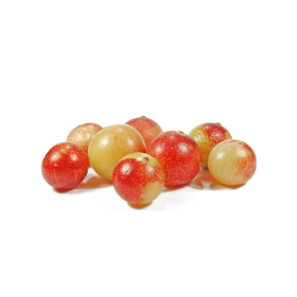 Camu-camuMyrciaria dubiaPreclinical
A single in-vitro study found camu-camu downregulated HIF-1α and VEGF in retinal-pigment-epithelium cells (ARPE-19 and fetal human RPE) 2Reference 2Modulation of hypoxia-inducible factors and VEGF expression by camu-camu in ARPE-19 and fetal human RPE cellsView study →, an anti-angiogenic mechanism relevant to neovascular (wet) AMD. This is one cell-based study, with no animal or human ocular data.
Full monograph ChrysanthemumChrysanthemum morifoliumPreclinical
Tied to the traditional Chinese “brighten the eyes” (ju hua) use, a C. morifolium extract suppressed pathological angiogenesis in an AMD model via VEGF and NF-κB — the herb’s strongest ocular signal to date 3Reference 3AnimalC. morifolium extract attenuates AMD pathological angiogenesis via VEGF / NF-κB (in vivo)View study → — supported by a flavonoid review noting luteolin and apigenin protect retinal cells from oxidative injury 4Reference 4ReviewPharmacological effects of Chrysanthemum flavonoids on liver and eye diseases (review)View study →. Entirely preclinical: no human trial for any eye condition, and effects vary with cultivar and preparation.
Full monograph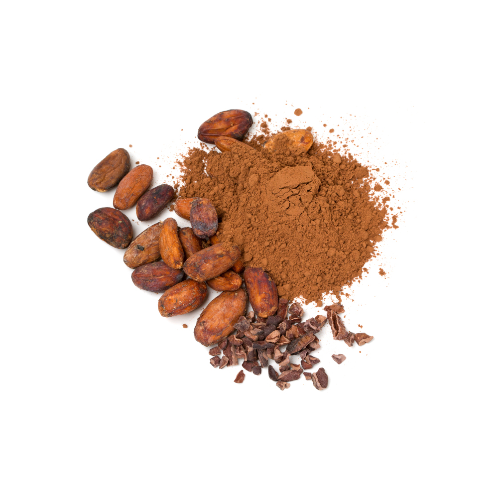 CacaoTheobroma cacaoNo benefit shown
A pre-specified ancillary analysis of the large COSMOS randomised trial found cocoa-flavanol supplementation (500 mg/day) did not reduce the incidence of age-related macular degeneration 1Reference 1RCTCocoa flavanol supplementation and risk of age-related macular degeneration: an ancillary study of the COSMOS randomised controlled trialView study → — a high-quality null result, consistent with COSMOS being null on its primary cardiovascular and diabetes endpoints. Despite cacao’s genuine flavanol/nitric-oxide vascular pharmacology, the best AMD evidence says it does not help.
Full monograph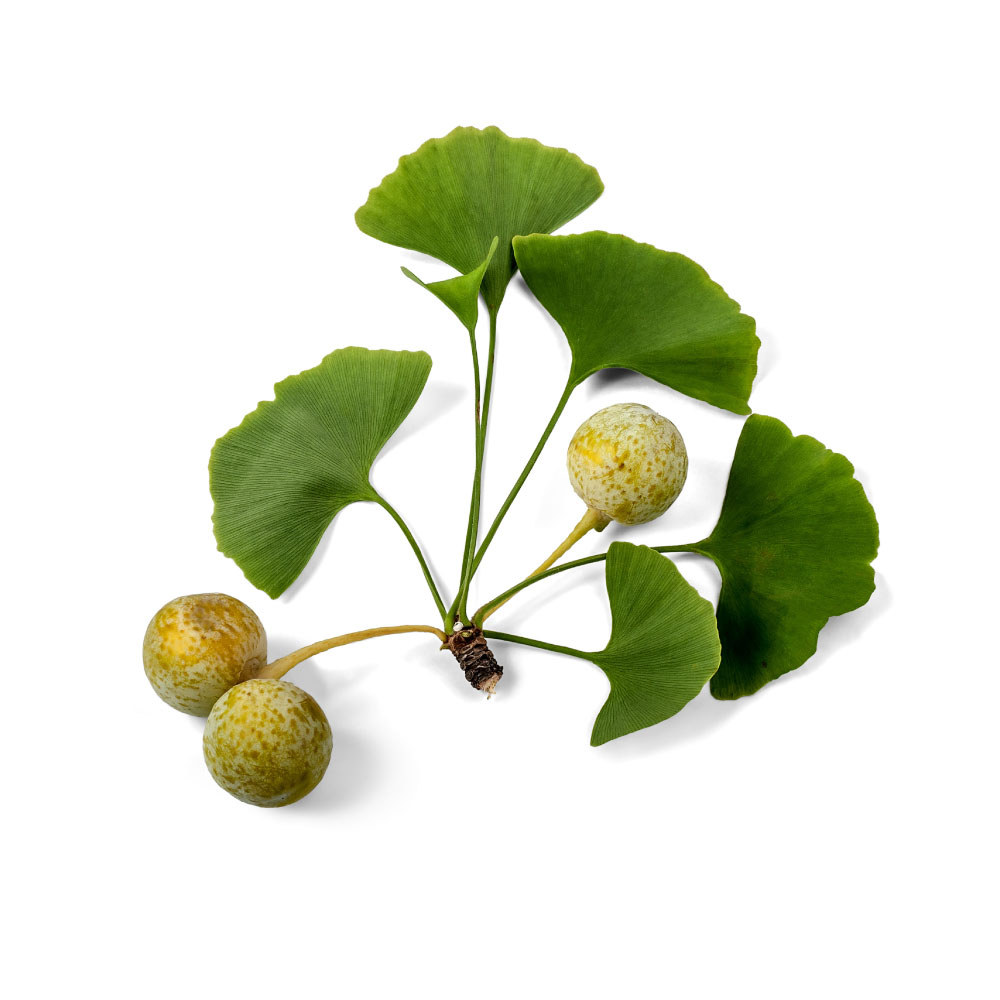 GinkgoGinkgo bilobaNo benefit shown
The ocular rationale (improved retinal blood flow, antioxidant retinal protection) is mechanistically reasonable, but the Cochrane review identified only two small trials and concluded there is insufficient evidence to determine whether standardized EGb 761 benefits AMD 5Reference 5Systematic reviewGinkgo biloba extract for age-related macular degeneration — systematic reviewView study →. Ginkgo for AMD is a hypothesis awaiting a proper trial, not an established use. It is frequently marketed for eye health on evidence that does not yet exist.
Full monographSupplementation
Supplementation
References
- Christen WG, Rist PM, et al. (2025). Cocoa flavanol supplementation and risk of age-related macular degeneration: an ancillary study of the COSMOS randomised controlled trial. JAMA Ophthalmol. https://pubmed.ncbi.nlm.nih.gov/40146119/
- Nakai A, et al. Modulation of hypoxia-inducible factors and VEGF expression by camu-camu in ARPE-19 and fetal human RPE cells. Journal of Ophthalmology. 2023. https://pubmed.ncbi.nlm.nih.gov/38187496/
- Jiang et al. (2025). C. morifolium extract attenuates AMD pathological angiogenesis via VEGF / NF-κB (in vivo). Journal of Ethnopharmacology. https://pubmed.ncbi.nlm.nih.gov/41016543/
- Xiong et al. (2024). Pharmacological effects of Chrysanthemum flavonoids on liver and eye diseases (review). Journal of Ethnopharmacology. https://pubmed.ncbi.nlm.nih.gov/39532220/
- Evans JR (2013). Ginkgo biloba extract for age-related macular degeneration — systematic review. The Cochrane database of systematic reviews. https://pubmed.ncbi.nlm.nih.gov/23440785/
- Baek, H. I., et al. (2026). Long-term supplementation with Centella asiatica extract (CA-HE50) on macular pigment optical density — randomised, double-blind, placebo-controlled trial. Nutrients. https://pubmed.ncbi.nlm.nih.gov/41901080/
- Broadhead, G. K., et al. (2019). Saffron therapy for the treatment of mild/moderate age-related macular degeneration: a randomised clinical trial. Graefe’s Archive for Clinical and Experimental Ophthalmology. https://pubmed.ncbi.nlm.nih.gov/30343354/
- Broadhead GK, Grigg J, McCluskey PJ, et al. (2024). Saffron therapy for the ongoing treatment of age-related macular degeneration. BMJ Open Ophthalmology. https://pubmed.ncbi.nlm.nih.gov/38485112/
- Falsini, B., et al. (2010). Influence of saffron supplementation on retinal flicker sensitivity in early age-related macular degeneration. Investigative Ophthalmology & Visual Science. https://pubmed.ncbi.nlm.nih.gov/20688744/
- Age-Related Eye Disease Study Research Group. (2001). A randomized, placebo-controlled clinical trial of high-dose supplementation with vitamins C and E, beta carotene, and zinc for age-related macular degeneration and vision loss — AREDS report no. 8. Archives of Ophthalmology, 119(10), 1417–1436. https://pubmed.ncbi.nlm.nih.gov/11594942/
- Age-Related Eye Disease Study 2 (AREDS2) Research Group. (2013). Lutein + zeaxanthin and omega-3 fatty acids for age-related macular degeneration — the AREDS2 randomized clinical trial. JAMA, 309(19), 2005–2015. https://pubmed.ncbi.nlm.nih.gov/23644932/