Compound Monograph
Miquelianin
Miquelianin is quercetin-3-O-glucuronide — a flavonol glucuronide with a dual identity. It is a genuine plant constituent of St John's wort, meadowsweet, gotu kola and lotus leaf, and simultaneously one of the principal forms in which quercetin actually circulates in human blood. That makes it the rare conjugate that pharmacokinetics treats as the "inactivated" metabolite while pharmacology studies it as an active in its own right.

Sources · 9
Where Does It Come From?
Miquelianin is a naturally occurring (and made in the body) flavonol glycoside (quercetin o-glucuronide), found in St John's wort, Meadowsweet, Gotu kola and 6 other sources. It is well tolerated orally (low toxicity).
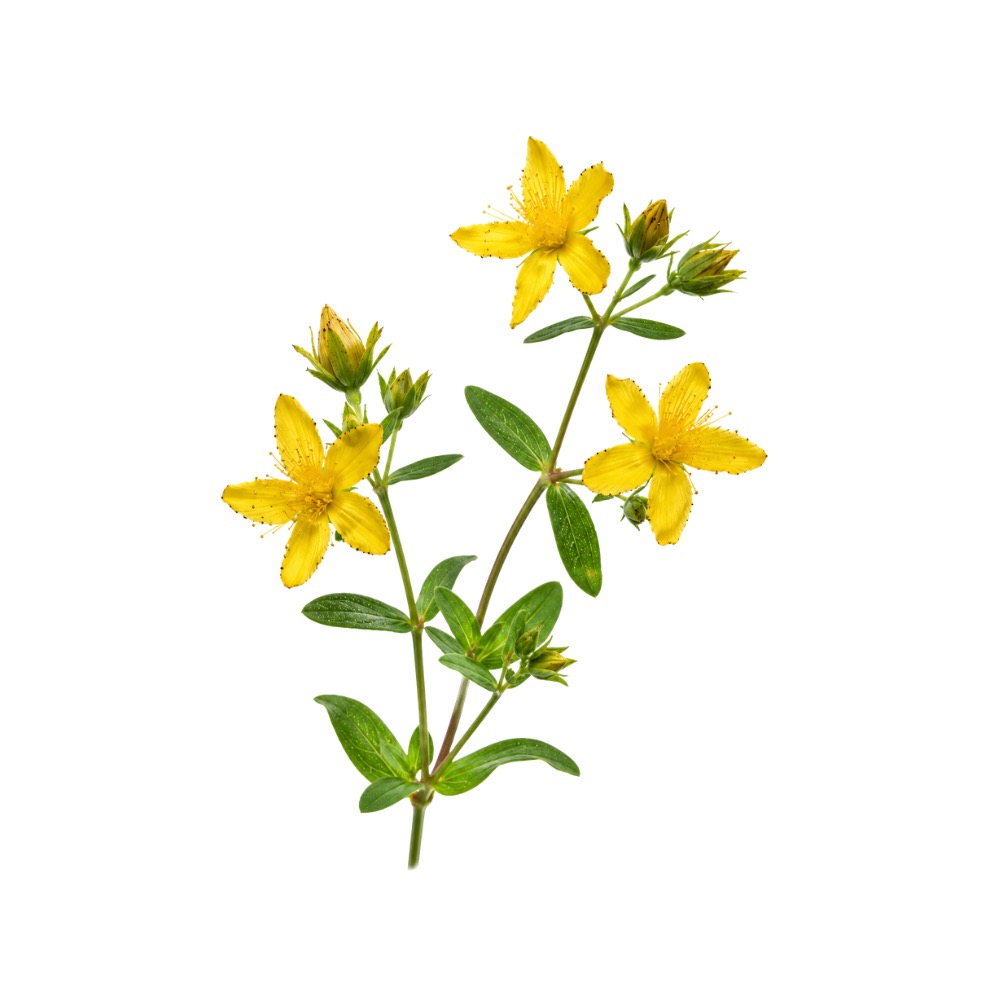 Principal source St. John's Wort Hypericum perforatum Herb monograph →
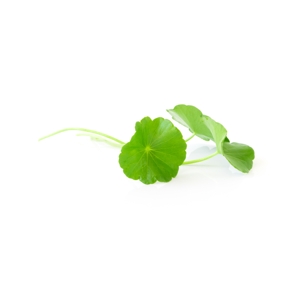 Source Gotu Kola Centella asiatica Herb monograph →
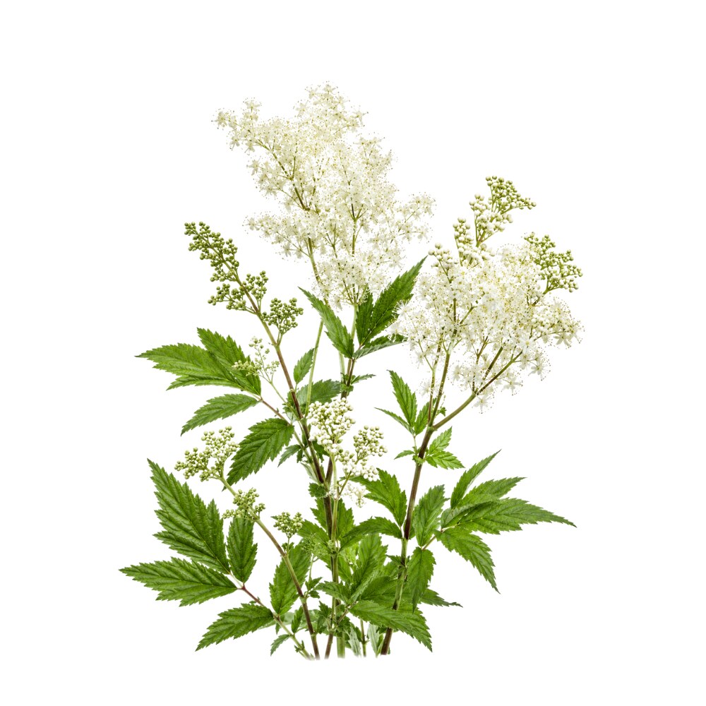 Source Meadowsweet Filipendula ulmaria Herb monograph →
Concentration
Content by Source
Reported concentrations across the plants that contain miquelianin — the bar marks the typical level, the line shows the reported range.
Literature figures for varying plant parts and preparations. Read them as a comparative guide, not exact assays.
Miquelianin at a Glance
Miquelianin is quercetin-3-O-glucuronide — the flavonol quercetin carrying a single glucuronic acid at its 3-position. Structurally it is an unremarkable member of a crowded family: the 3-O-glucoside is isoquercitrin, the 3-O-galactoside is hyperoside, the 3-O-rhamnoside is quercitrin, and the 3-O-rutinoside is rutin. Miquelianin is the one carrying a sugar acid.
What makes it worth its own page is that it has two separate lives. It is a real plant constituent, named among the major flavonoids of St John’s wort 17Reference 17Identification of the major constituents of Hypericum perforatum by LC/SPE/NMR and/or LC/MSView study → and identified in the flavonoid fraction of meadowsweet 1Reference 1Miquelianin and spiraeoside from Filipendula ulmaria mitigate α-synuclein accumulation in C. elegans and reduce the expression of neuroinflammatory cytokines in human microgliaView study →. It is also one of the principal forms in which quercetin actually circulates in human blood — when you eat onions, apples, capers or tea, the quercetin released in your gut is glucuronidated during absorption, and quercetin-3-glucuronide is among the handful of conjugates that show up in plasma 7,
That double identity has an awkward implication. Pharmacology conventionally treats a glucuronide as the inactivated, ready-for-excretion form. But if the aglycone is essentially absent from plasma and the glucuronide is what circulates, then a great deal of what has been attributed to “quercetin” in living animals may in fact be the work of its conjugates — and miquelianin is the main one 7,
The honest frame: this is a preclinical molecule. Nothing below is a human trial of miquelianin.
The Metabolite Identity
This is the load-bearing point of the page, so it is worth stating precisely.
Quercetin glucosides do not survive absorption intact. The β-glucosidase capacity of the small intestine and liver is large enough that quercetin glycosides are deglycosylated before or during uptake, and the compounds that appear in blood are phase-II conjugates — glucuronides, sulfates and methylated forms — not the original plant glycosides 21Reference 21ReviewBiomarkers for exposure to dietary flavonoids: a review of the current evidence for identification of quercetin glycosides in plasmaView study →. When lightly fried onions were fed to human volunteers, the quantifiable plasma metabolites were quercetin-3’-sulfate, quercetin-3-glucuronide, isorhamnetin-3-glucuronide, a quercetin diglucuronide and a quercetin glucuronide sulfate. Peak concentrations were sub-micromolar and arrived 0.6–0.8 h after ingestion; the half-life of quercetin-3-glucuronide was 2.33 h 7Reference 7Absorption, excretion and metabolite profiling of methyl-, glucuronyl-, glucosyl- and sulpho-conjugates of quercetin in human plasma and urine after ingestion of onionsView study →. The free aglycone was not a meaningful circulating species.
The authors of that study drew the conclusion themselves: if the samples had been hydrolysed first, as most earlier work did, “only quercetin and isorhamnetin would have been detected”, and the bioactivity of the metabolites should be considered 7Reference 7Absorption, excretion and metabolite profiling of methyl-, glucuronyl-, glucosyl- and sulpho-conjugates of quercetin in human plasma and urine after ingestion of onionsView study →. Later reviews of quercetin bioavailability reach the same place — the bioavailable species are the conjugates, and interindividual variation in which conjugates you form is substantial 19Reference 19Bioavailability of quercetin in humans with a focus on interindividual variationView study →.
Two consequences follow, and they pull in opposite directions:
- Attribution. In-vitro experiments that dose cells with 10–50 µM quercetin aglycone are not modelling what happens after a meal. If an in-vivo effect of dietary quercetin is real, the molecule present at the site is far more likely to be miquelianin than quercetin.
- Deconjugation. The glucuronide is not necessarily the endpoint. β-Glucuronidase activity — which rises at sites of inflammation — can strip the sugar acid back off and release the far more reactive aglycone locally 8,
10Reference 8Macrophage as a target of quercetin glucuronides in human atherosclerotic arteries: implication in the anti-atherosclerotic mechanism of dietary flavonoidsView study →Reference 10Comparison of metabolism and biological properties among positional isomers of quercetin glucuronide in LPS- and RANKL-challenged RAW264.7 cellsView study → . On that reading miquelianin is a circulating reservoir that becomes quercetin where quercetin is wanted.
Both stories are supported, and they are not mutually exclusive. Which one dominates for any given effect is, at present, unresolved.
Crossing the Blood-Brain Barrier
Glucuronides are polar, charged at physiological pH, and are the textbook example of a metabolite designed not to cross membranes. Miquelianin is one of the few for which that assumption has been tested directly, and it did better than expected.
Using three in-vitro barrier systems, a Münster group found that in Caco-2 monolayers miquelianin showed higher uptake than either hyperoside or quercitrin (1.93 vs 0.55 vs 0.22 pmol·min⁻¹·cm⁻²), with a permeability coefficient (Pc ≈ 0.4 × 10⁻⁶ cm/s) “in the range of orally available drugs”. Uptake rose when the MRP-2 efflux inhibitor MK-571 was added, indicating the barrier is actively pumping some of it back. In porcine brain-capillary endothelial cells (a blood-brain-barrier model) and choroid-plexus epithelium (a blood-CSF model), permeability was moderate but real — Pc ≈ 1.34 and 2.0 × 10⁻⁶ cm/s respectively — with no evidence of directional active transport 2Reference 2In vitroIn vitro studies indicate that miquelianin (quercetin 3-O-beta-D-glucuronopyranoside) is able to reach the CNS from the small intestineView study →.
The paper’s conclusion is deliberately careful, and so is ours: these coefficients “indicate the ability of miquelianin to cross both barriers to finally reach the CNS” 2Reference 2In vitroIn vitro studies indicate that miquelianin (quercetin 3-O-beta-D-glucuronopyranoside) is able to reach the CNS from the small intestineView study →. That is a permeability argument, not a measurement of brain miquelianin.
The in-vivo picture is less tidy. When rats were dosed with St John’s wort extract and their brains analysed, what was found there was quercetin and its methylated forms (isorhamnetin/tamarixetin) — 7 ng/g and 35 ng/g of brain after a single dose, rising with repeated dosing — rather than the intact glucuronide 20,
So: miquelianin can cross the barriers in vitro, and quercetin-derived material does reach rodent brain in vivo. Whether the molecule arrives as miquelianin, or is deconjugated en route, is not settled.
What It's Used For
Miquelianin’s research base is small, entirely preclinical, and unusually scattered across organ systems — a consequence of it being picked up separately by St John’s wort pharmacognosists, quercetin-bioavailability chemists, and food scientists standardising herbal juices.
Support is an experimental score — weighted by study type (human > animal > in vitro) and volume. A beta, not a validated metric. Every row here is preclinical; the scores rank thin evidence against thinner, and none of them imply clinical usefulness.
The newest and most interesting thread. In a 2025 study of a hydroethanolic Filipendula ulmaria extract, biochemometric fractionation assigned the activity to two flavonoid glycosides — miquelianin and spiraeoside. In transgenic NL5901 C. elegans, which express human α-synuclein fused to YFP, miquelianin reduced α-synuclein fluorescence by 35% at 200 µg/mL (spiraeoside, 32%) while increasing thermotolerance; in LPS-stimulated human HMC3 microglia both compounds suppressed pro-inflammatory gene expression, mirroring the parent extract’s downregulation of IL-1β, IL-6 and MCP-1 1Reference 1Miquelianin and spiraeoside from Filipendula ulmaria mitigate α-synuclein accumulation in C. elegans and reduce the expression of neuroinflammatory cytokines in human microgliaView study →.
The authors position them as “hit candidates” for early neurodegenerative mechanisms — deliberately modest language.
A separate line offers a possible mechanism for why the conjugate might outperform the aglycone in neurons. Quercetin and its glucuronide and sulfate conjugates all bind the 67-kDa laminin receptor (67LR) with high affinity, and in Neuroscreen-1 cells a pretreatment with 1–10 nM of the conjugates protected against serum-starvation death better than quercetin or EGCG, with protection abolished by a 67LR-blocking antibody 11Reference 11Flavonoid quercetin and its glucuronide and sulfate conjugates bind to 67-kDa laminin receptor and prevent neuronal cell death induced by serum starvationView study →.
At the low nanomolar concentrations conjugates actually reach in brain, receptor binding — not antioxidant chemistry — is the plausible route.
Isolate vs. plant: 1Reference 1Miquelianin and spiraeoside from Filipendula ulmaria mitigate α-synuclein accumulation in C. elegans and reduce the expression of neuroinflammatory cytokines in human microgliaView study → tested both the whole meadowsweet extract and the isolated compounds, which is the strong design; 11Reference 11Flavonoid quercetin and its glucuronide and sulfate conjugates bind to 67-kDa laminin receptor and prevent neuronal cell death induced by serum starvationView study → is isolate-only cell work. Neither involves a mammal with Parkinson’s disease.
Gap: a worm expressing human α-synuclein and an immortalised microglial line are screening tools, not disease models. There is no rodent Parkinson’s study of miquelianin, no pharmacokinetic bridge showing brain exposure at an effective concentration, and no clinical work of any kind.
Miquelianin’s oldest research identity is as a St John’s wort flavonoid. Fractionating a Hypericum perforatum extract for activity in the rat forced-swimming test, the Münster group narrowed the active flavonoid fraction to one containing hyperoside, isoquercitrin, miquelianin and quercitrin — and on testing the isolates found miquelianin active, alongside hyperoside and isoquercitrin, while quercetin itself, quercitrin and astilbin were not 3Reference 3Flavonoids from Hypericum perforatum show antidepressant activity in the forced swimming testView study →.
That the aglycone was inactive where three of its glycosides were active is a striking result for a family usually treated as interchangeable.
Two follow-ups extended it. Daily gavage of miquelianin at 0.6 mg/kg for two weeks downregulated plasma ACTH and corticosterone by 40–70% in rats — comparable to imipramine and hypericin — although the effect disappeared after eight weeks of dosing 4Reference 4AnimalFlavonoids of StView study →.
And in a mouse stress-induced-hyperthermia model, miquelianin (1.2 mg/kg orally) was the most potent compound tested among the St John’s wort flavonoids, outperforming hyperoside, isoquercitrin, quercitrin and rutin, with only the biflavonoid amentoflavone also active 5Reference 5AnimalEffects of StView study →.
More recently, in human SH-SY5Y neuronal cells stressed with dexamethasone, miquelianin at 10 µM reversed the glucocorticoid-induced rise in FKBP5 — an HPA-axis biomarker of antidepressant response — as did hyperforin and citalopram, at the 48-hour timepoint 6Reference 6Hyperforin and miquelianin from StView study →.
Isolate vs. plant: unusually for this database, these are isolate studies. The caveat runs the other way — St John’s wort’s clinical antidepressant record belongs to standardised whole extracts, in which the phloroglucinol hyperforin and the naphthodianthrone hypericin are the conventional actives, and no trial has isolated a flavonoid contribution.
Gap: all rodent behavioural pharmacology, in models (FST, stress hyperthermia) with well-known translational limits; the HPA effect faded with chronic dosing 4Reference 4AnimalFlavonoids of StView study →; and no human has ever taken isolated miquelianin.
Two independent threads converge here. First, a direct comparison of the four quercetin monoglucuronide positional isomers in activated RAW264.7 macrophages found Q3G (miquelianin) the most effective at inhibiting LPS-dependent IL-6 induction and RANKL-dependent osteoclast-marker activation. It also showed the highest deconjugation efficacy of the four — the most readily converted back to aglycone by β-glucuronidase — which the authors link directly to its activity 10Reference 10Comparison of metabolism and biological properties among positional isomers of quercetin glucuronide in LPS- and RANKL-challenged RAW264.7 cellsView study →.
Second, food-science work on gotu kola (Centella asiatica) juice identified miquelianin by UHPLC-MS² as the dominant constituent and functional mediator behind attenuation of IL-1β-driven inflammation in intestinal epithelial cells via MAPK and NF-κB signalling 15Reference 15Attenuation of IL-1β-induced inflammation in intestinal epithelial cells by Centella asiatica juice via MAPK and NF-κB signaling cascades and characterization of its key bioactive compoundView study →; a companion study in DSS-induced colitis in mice used miquelianin as the validated HPLC marker for standardising the same juice, which improved clinical colitis scores, epithelial integrity and mucin production 14Reference 14Anti-colitic effects of Centella asiatica (L.) Urb. juice via ERK/p38 and NF-κB signaling modulation and the characterization of a key marker compoundView study →.
Isolate vs. plant: 10Reference 10Comparison of metabolism and biological properties among positional isomers of quercetin glucuronide in LPS- and RANKL-challenged RAW264.7 cellsView study → is isolate work and is the stronger evidence for the molecule. 14Reference 14Anti-colitic effects of Centella asiatica (L.) Urb. juice via ERK/p38 and NF-κB signaling modulation and the characterization of a key marker compoundView study → and 15Reference 15Attenuation of IL-1β-induced inflammation in intestinal epithelial cells by Centella asiatica juice via MAPK and NF-κB signaling cascades and characterization of its key bioactive compoundView study → are whole-juice studies in which miquelianin is the analytical marker — the compound was measured, not administered alone. Marker status is a quality-control claim, not a proof of activity.
Gap: the gut is where an unabsorbed or biliary-recycled glucuronide plausibly reaches useful concentrations, which makes this the most mechanistically credible application — and there is no animal study of the isolate in colitis, and no human data at all.
The most elegant experiment on this molecule. A Japanese group raised a monoclonal antibody (14A2) against quercetin-3-glucuronide and used it to ask where dietary quercetin metabolites actually go. Staining appeared specifically in human atherosclerotic lesions and not in normal aorta, concentrated in macrophage-derived foam cells. In cultured macrophages, Q3GA was taken up and deconjugated to the more active aglycone (some of it then methylated), and treatment suppressed mRNA for the class-A scavenger receptor and CD36, the receptors that drive foam-cell formation 8Reference 8Macrophage as a target of quercetin glucuronides in human atherosclerotic arteries: implication in the anti-atherosclerotic mechanism of dietary flavonoidsView study →.
That is the “circulating reservoir” model made visible: the conjugate travels in blood, accumulates preferentially at inflamed sites, and is unmasked there.
A separate finding sharpens the aglycone/conjugate distinction. Quercetin, isorhamnetin and quercetin-3’-sulfate all auto-oxidise in aqueous buffer and generate superoxide, and quercetin scavenges nitric oxide and blunts its biological activity in rat aortic rings. Quercetin-3-glucuronide does none of this 9Reference 9Glucuronidated metabolites of the flavonoid quercetin do not auto-oxidise, do not generate free radicals and do not decrease nitric oxide bioavailabilityView study →. Whatever the glucuronide is doing in the vasculature, it is not the pro-oxidant chemistry the free flavonol is capable of.
Isolate vs. plant: both 8Reference 8Macrophage as a target of quercetin glucuronides in human atherosclerotic arteries: implication in the anti-atherosclerotic mechanism of dietary flavonoidsView study → and 9Reference 9Glucuronidated metabolites of the flavonoid quercetin do not auto-oxidise, do not generate free radicals and do not decrease nitric oxide bioavailabilityView study → test the isolated conjugate — good design, no plant-extract confound.
Gap: immunohistochemistry establishes localisation, not causation; the macrophage work is in murine cell lines; and nothing here has been tested as an intervention in an animal or human with atherosclerosis.
The one result that puts miquelianin ahead of a licensed drug, and it is worth stating exactly what was measured. Screening 49 plants with ethnobotanical records for wounds, burns, skin disease, rheumatism and bone conditions, a Leiden–Bologna group combined ¹H-NMR profiling with in-vitro collagenase inhibition and an orthogonal partial-least-squares model. Bioactivity tracked phenolic content — but phenolics include tannins, which inhibit enzymes non-specifically. After a tannin-removal step, Alchemilla vulgaris was the most persistently active extract, proving its activity did not rest on tannins. Bioassay-guided fractionation of that extract yielded a single flavonoid, confirmed by NMR and HRMS as quercetin-3-O-β-glucuronide, which inhibited collagenase more potently than other flavonoids carrying the same quercetin aglycone, and more potently than doxycycline — the only collagenase inhibitor with FDA approval, used as the positive control 24Reference 24In vitroIdentification of a collagenase-inhibiting flavonoid from Alchemilla vulgaris using NMR-based metabolomics — in vitroView study →.
Collagenases are matrix metalloproteinases that break down dermal collagen, so the finding is the molecular hook behind lady’s mantle’s traditional wound-repair and skin reputation. It also fits the enzyme-inhibition profile reported for the flavonol glycosides of fireweed, where anti-collagenase and anti-hyaluronidase activity carry the wound-healing signal.
Isolate vs. plant: the strong design here — whole extracts screened, tannins removed to exclude the obvious confounder, then the active molecule isolated and tested against a drug comparator.
Gap: a cell-free enzyme assay. Nothing shows the molecule reaching dermal collagenase at an inhibitory concentration through skin or from the gut, and there is no cell, animal or human wound-healing study of isolated miquelianin. A compound that beats doxycycline in a cuvette has cleared the first hurdle only 24Reference 24In vitroIdentification of a collagenase-inhibiting flavonoid from Alchemilla vulgaris using NMR-based metabolomics — in vitroView study →.
The thinnest row, included because it is the only non-neurodegenerative CNS claim. An ethanolic sacred lotus leaf (Nelumbo nucifera) extract increased pentobarbital-induced sleep duration and EEG theta power in rodents, and recovered caffeine-disrupted sleep. A [³H]-flumazenil ligand-binding assay attributed the effect to the quercetin-3-O-glucuronide in the extract — present at 77.27 µg per mg of extract — acting at GABAA receptors 13Reference 13AnimalQuercetin-3-O-glucuronide in the ethanol extract of lotus leaf (Nelumbo nucifera) enhances sleep quantity and quality in a rodent model via a GABAergic mechanismView study →.
Isolate vs. plant: the behavioural work is whole extract; only the binding assay addresses the molecule. Attribution by displacement assay is suggestive, not demonstrative.
Gap: single study, single group, no isolate dosing in vivo, and no independent replication. Read it as a lead.
Mechanisms
| Target / pathway | Effect | Relevant to |
|---|---|---|
| α-Synuclein proteostasis (NL5901 C. elegans) | α-syn::YFP aggregation ↓ ~35% at 200 µg/mL; thermotolerance ↑ | neuroprotection |
| Microglial pro-inflammatory transcription (HMC3) | IL-1β, IL-6, MCP-1 expression ↓ under LPS | neuroinflammation |
| 67-kDa laminin receptor (67LR) | high-affinity binding; neuroprotection at 1–10 nM, abolished by blocking antibody | neuroprotection (the low-concentration route) |
| HPA axis (ACTH / corticosterone) | 40–70% ↓ after 2 weeks at 0.6 mg/kg in rats; lost by 8 weeks | antidepressant-like |
| FKBP5 expression (SH-SY5Y under dexamethasone) | glucocorticoid-induced rise reversed at 10 µM | stress-axis / antidepressant biomarker |
| NF-κB and ERK/p38 MAPK signalling (intestinal epithelium) | inflammatory mediator output ↓ (as juice marker compound) | intestinal inflammation |
| Scavenger receptor class A / CD36 (macrophages) | mRNA ↓; foam-cell formation pathway suppressed | atherosclerosis |
| β-Glucuronidase (inflamed tissue, activated macrophages) | deconjugation → local release of quercetin aglycone | the “reservoir/prodrug” model |
| GABAA receptor (benzodiazepine site) | flumazenil displacement in lotus-leaf extract | sedation / sleep |
| MRP-2 efflux transporter (intestinal epithelium) | pumps miquelianin back into the lumen, limiting net absorption | pharmacokinetics |
| Collagenase (matrix metalloproteinase) | inhibited in vitro more potently than doxycycline, the only FDA-approved collagenase inhibitor 24Reference 24In vitroIdentification of a collagenase-inhibiting flavonoid from Alchemilla vulgaris using NMR-based metabolomics — in vitroView study → | skin / wound repair |
| Auto-oxidation / superoxide generation | absent, in contrast to quercetin and quercetin-3’-sulfate | why the conjugate is not simply “quercetinweaker” |
The last two rows are the ones to hold onto. The 3-O-glucuronide is not a diluted quercetin: it lacks the aglycone’s redox reactivity entirely 9Reference 9Glucuronidated metabolites of the flavonoid quercetin do not auto-oxidise, do not generate free radicals and do not decrease nitric oxide bioavailabilityView study →, yet it binds a receptor the aglycone also binds, at concentrations the aglycone never reaches in vivo 11Reference 11Flavonoid quercetin and its glucuronide and sulfate conjugates bind to 67-kDa laminin receptor and prevent neuronal cell death induced by serum starvationView study →.
Pharmacokinetics
Miquelianin sits on both sides of the pharmacokinetic ledger, which is what makes its PK confusing to read.
As a metabolite, it is formed rather than absorbed. Dietary quercetin glycosides are deglycosylated at the intestinal brush border and the liberated aglycone is glucuronidated by UDP-glucuronosyltransferases in the enterocyte and hepatocyte; quercetin-3-glucuronide is one of the resulting conjugates released into portal and systemic blood 19,
As an ingested compound, it is poorly bioavailable in the usual glycoside way. A 2026 review of dietary quercetin-3-O-glucuronide summarises it as rapidly absorbed and widely distributed but with limited bioavailability 12Reference 12Dietary quercetin 3-O-glucuronide: current research on its extraction, biosynthesis, pharmacokinetics, health benefits, and potential food applicationsView study →, and the in-vitro barrier work shows why part of that is active: MRP-2 pumps it back across the intestinal epithelium, with uptake and permeability both rising when MRP-2 is inhibited 2Reference 2In vitroIn vitro studies indicate that miquelianin (quercetin 3-O-beta-D-glucuronopyranoside) is able to reach the CNS from the small intestineView study →.
Deconjugation is the wildcard. β-Glucuronidase — abundant in activated macrophages and upregulated at inflamed sites — hydrolyses the conjugate back to quercetin, and among the four monoglucuronide isomers the 3-O form is the most readily deconjugated 10Reference 10Comparison of metabolism and biological properties among positional isomers of quercetin glucuronide in LPS- and RANKL-challenged RAW264.7 cellsView study →, with direct evidence of this happening inside macrophages in atherosclerotic tissue 8Reference 8Macrophage as a target of quercetin glucuronides in human atherosclerotic arteries: implication in the anti-atherosclerotic mechanism of dietary flavonoidsView study →. Any effect measured for miquelianin in a system with β-glucuronidase activity may partly be quercetin’s.
Clinical Trials
None. There are no clinical trials of isolated miquelianin — no completed, ongoing or terminated human studies of the molecule. The closest human data are (a) pharmacokinetic, where miquelianin appears as an endogenous metabolite measured in plasma and urine after ordinary flavonoid-containing meals 7,
| Completed | Planned | Terminated | Preclinical |
|---|---|---|---|
| —(human data are metabolite PK only) | — | — | Neuroprotection, antidepressant-like, anti-inflammatory, vascular, sleep |
Last checked: July 2026.
Isolate vs. Plant Studies
This molecule scores better than most on the isolate question, and worse than most on everything downstream of it.
In its favour: the St John’s wort behavioural work 3,
Against it: the intestinal 14,
The deeper confound is unique to this compound. Because β-glucuronidase can regenerate quercetin from miquelianin inside cells and tissues 8,
Prevalence in Nature
Miquelianin is widely but thinly distributed — a recurring minor-to-moderate member of flavonol fractions rather than a signature compound of any one plant.
In this database it matters chiefly in two herbs. In St John’s wort it is one of the flavonoids catalogued alongside quercetin, quercitrin, isoquercitrin, hyperoside, astilbin and biapigenin in the definitive LC/SPE/NMR characterisation of the herb’s major constituents 17Reference 17Identification of the major constituents of Hypericum perforatum by LC/SPE/NMR and/or LC/MSView study → — a flavonoid-fraction member, not a headline active like hyperforin or hypericin. In meadowsweet it sits in the flavonoid fraction beside the herb’s signature spiraeoside, and the two together were identified as the constituents carrying the extract’s neuroprotective activity 1Reference 1Miquelianin and spiraeoside from Filipendula ulmaria mitigate α-synuclein accumulation in C. elegans and reduce the expression of neuroinflammatory cytokines in human microgliaView study →.
A third herb is now attached to it by activity rather than abundance. In lady’s mantle (Alchemilla vulgaris), bioassay-guided fractionation of the tannin-depleted extract isolated miquelianin as the compound responsible for the plant’s collagenase inhibition 24Reference 24In vitroIdentification of a collagenase-inhibiting flavonoid from Alchemilla vulgaris using NMR-based metabolomics — in vitroView study → — the reverse of the usual pattern, in which a compound is found first and a use is inferred afterwards. It is also named among the flavonol glycosides of fireweed (Chamerion angustifolium), whose anti-collagenase and anti-hyaluronidase flavonoid fraction carries that herb’s wound-healing signal.
Beyond those, it turns up in gotu kola juice, where it is reported as the dominant flavonoid and is used as the standardisation marker 14,
What it is not is a signature compound. On no plant here is miquelianin the marker constituent people reach for — it is always in the second rank, behind hyperforin in St John’s wort, behind spiraeoside and the salicylates in meadowsweet, behind the triterpene asiaticoside in gotu kola. Its importance comes from the metabolic route, not the botanical one.
Major Natural Sources
| Source | Part | Reported level | Note |
|---|---|---|---|
| Cuphea glutinosa | leaf | ~1.03% dry wt | Validated UPLC assay; used as the species’ chemical marker 16Reference 16Ultrasound-assisted extraction optimization and validation of ultra-performance liquid chromatographic method for the quantification of miquelianin in Cuphea glutinosa leavesView study → |
| Sacred lotus (Nelumbo nucifera) | leaf (ethanol extract) | 77.27 µg/mg extract | Extract basis, not plant dry weight 13Reference 13AnimalQuercetin-3-O-glucuronide in the ethanol extract of lotus leaf (Nelumbo nucifera) enhances sleep quantity and quality in a rodent model via a GABAergic mechanismView study → |
| Gotu kola (Centella asiatica) | juice | dominant flavonoid (not quantified here) | HPLC-UVD marker for juice standardisation 14, |
| St John’s wort (Hypericum perforatum) | flowering tops | qualitative — a major catalogued flavonoid | LC/SPE/NMR constituent survey 17Reference 17Identification of the major constituents of Hypericum perforatum by LC/SPE/NMR and/or LC/MSView study → |
| Meadowsweet (Filipendula ulmaria) | aerial parts | qualitative — flavonoid-fraction constituent | Bioactivity-guided fractionation 1Reference 1Miquelianin and spiraeoside from Filipendula ulmaria mitigate α-synuclein accumulation in C. elegans and reduce the expression of neuroinflammatory cytokines in human microgliaView study → |
| Lady’s mantle (Alchemilla vulgaris) | aerial parts | qualitative — isolated from the tannin-depleted extract | Bioassay-guided fractionation; the collagenase inhibitor 24Reference 24In vitroIdentification of a collagenase-inhibiting flavonoid from Alchemilla vulgaris using NMR-based metabolomics — in vitroView study → |
| Human plasma (endogenous) | — | sub-micromolar Cmax, t½ ≈ 2.3 h | Formed from dietary quercetin, not ingested 7Reference 7Absorption, excretion and metabolite profiling of methyl-, glucuronyl-, glucosyl- and sulpho-conjugates of quercetin in human plasma and urine after ingestion of onionsView study → |
That last row is not a joke. For most people, the largest single exposure to miquelianin comes not from any of these plants but from their own glucuronidation of dietary quercetin.
Discovery & Synthesis
Miquelianin’s trivial name is long-established in the flavonoid literature, well ahead of any interest in its pharmacology; it has been identified in maidenhair fern fronds, Salvia, common bean and many other plants over decades of phytochemical survey work. No single first-isolation report could be verified, and the name’s specific commemorative origin is not documented in the sources checked — both are honest gaps rather than settled facts.
Its research career, by contrast, has a clear starting point: the St John’s wort flavonoid programme at Münster in the late 1990s and early 2000s, which fractionated Hypericum extract for antidepressant activity, identified miquelianin as one of the active isolates 3Reference 3Flavonoids from Hypericum perforatum show antidepressant activity in the forced swimming testView study →, and then asked the question that defines the molecule — whether a glucuronide could reach the brain at all 2Reference 2In vitroIn vitro studies indicate that miquelianin (quercetin 3-O-beta-D-glucuronopyranoside) is able to reach the CNS from the small intestineView study →. Everything since has either extended that CNS thread or come at the compound from the opposite direction, as a quercetin metabolite 7,
Preparation. There is no dominant total synthesis in the literature reviewed here. In practice the compound is obtained by isolation from plant material — though not easily: high-speed counter-current chromatography of H. perforatum extract yields miquelianin only as part of a co-eluting mixture with hyperoside and isoquercitrin, all three being quercetin 3-O-glycosides of near-identical polarity, requiring a further preparative-HPLC step to resolve 22Reference 22Separation of epigallocatechin and flavonoids from Hypericum perforatum L. by high-speed counter-current chromatography and preparative high-performance liquid chromatographyView study →. A validated ultrasound-assisted extraction and UPLC quantification method exists for Cuphea glutinosa leaf, the richest reported plant source 16Reference 16Ultrasound-assisted extraction optimization and validation of ultra-performance liquid chromatographic method for the quantification of miquelianin in Cuphea glutinosa leavesView study →. A 2026 review surveys extraction and biosynthetic routes — the plant pathway is enzymatically and genetically regulated and can be modulated by external factors — and frames enzymatic/biotechnological production as the direction of travel for food and nutraceutical use 12Reference 12Dietary quercetin 3-O-glucuronide: current research on its extraction, biosynthesis, pharmacokinetics, health benefits, and potential food applicationsView study →.
Patents: not yet researched.
Toxicity & Safety
Miquelianin carries a low flag, and for an unusual reason: it is not a foreign substance. Anyone eating a normal diet containing onions, apples, tea, capers or berries manufactures miquelianin continuously, in the gut wall and liver, and clears it within hours 7,
- A normal human metabolite. Everyone forms quercetin-3-glucuronide from dietary flavonols; plasma levels are sub-micromolar and cleared with a ~2-hour half-life.
- Not pro-oxidant. Unlike quercetin and quercetin-3’-sulfate, the 3-glucuronide does not auto-oxidise, does not generate superoxide, and does not scavenge nitric oxide in vitro.
- No isolate safety dataset. No repeat-dose toxicology, genotoxicity package or human tolerability study exists for purified miquelianin at supra-dietary doses. Absence of findings is not evidence of safety.
- Not a supplement, and not sold as one in any meaningful way. There is no established human dose, no dosing history and no product tradition to fall back on.
Full Safety & Interactions Detail
Baseline exposure. The strongest safety argument for miquelianin is that it is endogenously generated at low micromolar-to-nanomolar concentrations by ordinary flavonol intake, with rapid renal clearance — plasma Cmax below 1 µM and a half-life near 2 h after a substantial onion meal, with roughly 4.7% of ingested flavonol recovered in urine as metabolites 7Reference 7Absorption, excretion and metabolite profiling of methyl-, glucuronyl-, glucosyl- and sulpho-conjugates of quercetin in human plasma and urine after ingestion of onionsView study →. Nothing in that pattern suggests accumulation.
The conjugate is chemically gentler than the aglycone. Quercetin can behave as a pro-oxidant: it auto-oxidises in aqueous buffer, generates superoxide, scavenges nitric oxide, and blunts NO’s biological activity in rat aortic rings. Quercetin-3-glucuronide did none of these in a head-to-head comparison 9Reference 9Glucuronidated metabolites of the flavonoid quercetin do not auto-oxidise, do not generate free radicals and do not decrease nitric oxide bioavailabilityView study →. To the extent that quercetin’s high-dose safety concerns are redox-driven, they do not transfer straightforwardly to its 3-glucuronide.
But deconjugation complicates that. β-Glucuronidase activity at inflamed sites and inside activated macrophages regenerates the aglycone from the glucuronide 8Reference 8Macrophage as a target of quercetin glucuronides in human atherosclerotic arteries: implication in the anti-atherosclerotic mechanism of dietary flavonoidsView study →, and the 3-O isomer is the most readily deconjugated of the four monoglucuronides 10Reference 10Comparison of metabolism and biological properties among positional isomers of quercetin glucuronide in LPS- and RANKL-challenged RAW264.7 cellsView study →. Locally, therefore, miquelianin can become quercetin — which means quercetin’s own cautions (high-dose supplement contexts, its documented pro-oxidant chemistry, its interactions with drug-metabolising enzymes and transporters) cannot be dismissed entirely.
Cytotoxicity across the isomers. In the direct comparison of quercetin monoglucuronides in macrophages, the aglycone and the 7-O-glucuronide were the cytotoxic species, tracking their lower stability at neutral pH; the 3-O form (miquelianin) was not flagged as the cytotoxic one and was simultaneously the most active anti-inflammatory isomer 10Reference 10Comparison of metabolism and biological properties among positional isomers of quercetin glucuronide in LPS- and RANKL-challenged RAW264.7 cellsView study →. Position matters within this family.
Herbal context. In practice, miquelianin is encountered inside herbs whose own safety profiles dominate — most importantly St John’s wort, where the clinically consequential issues (CYP3A4/P-glycoprotein induction, serotonergic interactions, photosensitivity) belong to hyperforin and hypericin, not to a flavonol glucuronide. Safety decisions belong on the herb page, where the actual exposure is decided.
Reproductive note (prose only). No dedicated reproductive-toxicity dataset exists for isolated miquelianin. Because it is a normal metabolite of dietary flavonols, ordinary dietary exposure is not a distinct concern; concentrated-isolate exposure is simply uncharacterised. Pregnancy and lactation judgements belong to the herb or product page, not to this molecule page.
Dosage
There is no established human dose of miquelianin, and no product for which one would apply. The figures below are the research doses at which the effects described above were produced — all in rodents or cell culture.
| Context | Form | Dose / concentration |
|---|---|---|
| HPA-axis suppression (rats, 2 weeks) | isolated miquelianin, oral gavage | 0.6 mg/kg/day 4Reference 4AnimalFlavonoids of StView study → |
| Stress-induced hyperthermia (mice) | isolated miquelianin, oral | 1.2 mg/kg 5Reference 5AnimalEffects of StView study → |
| FKBP5 reversal (human SH-SY5Y cells) | isolated miquelianin | 10 µM 6Reference 6Hyperforin and miquelianin from StView study → |
| α-Synuclein reduction (C. elegans) | isolated miquelianin | 200 µg/mL 1Reference 1Miquelianin and spiraeoside from Filipendula ulmaria mitigate α-synuclein accumulation in C. elegans and reduce the expression of neuroinflammatory cytokines in human microgliaView study → |
| Neuroprotection via 67LR (Neuroscreen-1 cells) | quercetin-3-O-glucuronide | 1–10 nM 11Reference 11Flavonoid quercetin and its glucuronide and sulfate conjugates bind to 67-kDa laminin receptor and prevent neuronal cell death induced by serum starvationView study → |
| Dietary (human, endogenous) | formed from dietary quercetin | sub-micromolar plasma Cmax, t½ ≈ 2.3 h 7Reference 7Absorption, excretion and metabolite profiling of methyl-, glucuronyl-, glucosyl- and sulpho-conjugates of quercetin in human plasma and urine after ingestion of onionsView study → |
The contrast between the last two rows is the most useful number here. The concentration at which the 67LR-mediated neuroprotection appears — low nanomolar — is within reach of ordinary dietary metabolite levels, while the concentrations used in most cell work (10 µM and up) are one to four orders of magnitude above what circulates. These are research figures, not a personal recommendation.
References
- Redl M, Weisenburger S, Wasilewicz A, Grienke U, Lehner MD, Bredenbröker D, Rollinger JM (2025). Miquelianin and spiraeoside from Filipendula ulmaria mitigate α-synuclein accumulation in C. elegans and reduce the expression of neuroinflammatory cytokines in human microglia. Frontiers in Pharmacology. https://pubmed.ncbi.nlm.nih.gov/41756116/
- Juergenliemk G, Boje K, Huewel S, Lohmann C, Galla HJ, Nahrstedt A (2003). In vitro studies indicate that miquelianin (quercetin 3-O-beta-D-glucuronopyranoside) is able to reach the CNS from the small intestine. Planta Medica. https://pubmed.ncbi.nlm.nih.gov/14735439/
- Butterweck V, Jürgenliemk G, Nahrstedt A, Winterhoff H (2000). Flavonoids from Hypericum perforatum show antidepressant activity in the forced swimming test. Planta Medica. https://pubmed.ncbi.nlm.nih.gov/10705724/
- Butterweck V, Hegger M, Winterhoff H (2004). Flavonoids of St. John’s Wort reduce HPA axis function in the rat. Planta Medica. https://pubmed.ncbi.nlm.nih.gov/15490333/
- Grundmann O, Kelber O, Butterweck V (2006). Effects of St. John’s wort extract and single constituents on stress-induced hyperthermia in mice. Planta Medica. https://pubmed.ncbi.nlm.nih.gov/17051457/
- Verjee S, Weston A, Kolb C, Kalbhenn-Aziz H, Butterweck V (2018). Hyperforin and miquelianin from St. John’s wort attenuate gene expression in neuronal cells after dexamethasone-induced stress. Planta Medica. https://pubmed.ncbi.nlm.nih.gov/29499586/
- Mullen W, Edwards CA, Crozier A (2006). Absorption, excretion and metabolite profiling of methyl-, glucuronyl-, glucosyl- and sulpho-conjugates of quercetin in human plasma and urine after ingestion of onions. British Journal of Nutrition. https://pubmed.ncbi.nlm.nih.gov/16869998/
- Kawai Y, Nishikawa T, Shiba Y, Saito S, Murota K, Shibata N, Kobayashi M, Kanayama M, Uchida K, Terao J (2008). Macrophage as a target of quercetin glucuronides in human atherosclerotic arteries: implication in the anti-atherosclerotic mechanism of dietary flavonoids. The Journal of Biological Chemistry. https://pubmed.ncbi.nlm.nih.gov/18199750/
- Lodi F, Jiménez R, Menendez C, Needs PW, Duarte J, Perez-Vizcaino F (2008). Glucuronidated metabolites of the flavonoid quercetin do not auto-oxidise, do not generate free radicals and do not decrease nitric oxide bioavailability. Planta Medica. https://pubmed.ncbi.nlm.nih.gov/18484521/
- Nishikawa M, Kada Y, Kimata M, Sakaki T, Ikushiro S (2022). Comparison of metabolism and biological properties among positional isomers of quercetin glucuronide in LPS- and RANKL-challenged RAW264.7 cells. Bioscience, Biotechnology, and Biochemistry. https://pubmed.ncbi.nlm.nih.gov/36085182/
- Gopalakrishna R, Oh A, Hou L, Lee E, Aguilar J, Li A, Mack WJ (2023). Flavonoid quercetin and its glucuronide and sulfate conjugates bind to 67-kDa laminin receptor and prevent neuronal cell death induced by serum starvation. Biochemical and Biophysical Research Communications. https://pubmed.ncbi.nlm.nih.gov/37300941/
- Wang Z, Yang J, Liu X, Yu H, Dai X (2026). Dietary quercetin 3-O-glucuronide: current research on its extraction, biosynthesis, pharmacokinetics, health benefits, and potential food applications. Journal of Agricultural and Food Chemistry. https://pubmed.ncbi.nlm.nih.gov/41808541/
- Kim S, Hong KB, Jo K, Suh HJ (2021). Quercetin-3-O-glucuronide in the ethanol extract of lotus leaf (Nelumbo nucifera) enhances sleep quantity and quality in a rodent model via a GABAergic mechanism. Molecules. https://pubmed.ncbi.nlm.nih.gov/34069439/
- Shin HY, Jeong YY, Kim JE, Shin KS, Yu KW (2026). Anti-colitic effects of Centella asiatica (L.) Urb. juice via ERK/p38 and NF-κB signaling modulation and the characterization of a key marker compound. Journal of Ethnopharmacology. https://pubmed.ncbi.nlm.nih.gov/41016540/
- Shin HY, Jeong WB, Kim WJ, Joung MY, Shin KS, Yu KW (2026). Attenuation of IL-1β-induced inflammation in intestinal epithelial cells by Centella asiatica juice via MAPK and NF-κB signaling cascades and characterization of its key bioactive compound. Food Science and Biotechnology. https://pubmed.ncbi.nlm.nih.gov/41523689/
- Santos MC, Koetz M, Mendez ASL, Henriques AT (2020). Ultrasound-assisted extraction optimization and validation of ultra-performance liquid chromatographic method for the quantification of miquelianin in Cuphea glutinosa leaves. Talanta. https://pubmed.ncbi.nlm.nih.gov/32456919/
- Tatsis EC, Boeren S, Exarchou V, Troganis AN, Vervoort J, Gerothanassis IP (2007). Identification of the major constituents of Hypericum perforatum by LC/SPE/NMR and/or LC/MS. Phytochemistry. https://pubmed.ncbi.nlm.nih.gov/17196625/
- Ishisaka A, Ichikawa S, Sakakibara H, Piskula MK, Nakamura T, Kato Y, Ito M, Miyamoto K, Tsuji A, Kawai Y, Terao J (2011). Accumulation of orally administered quercetin in brain tissue and its antioxidative effects in rats. Free Radical Biology & Medicine. https://pubmed.ncbi.nlm.nih.gov/21741473/
- Almeida AF, Borge GIA, Piskula M, Tudose A, Tudoreanu L, Valentová K, Williamson G, Santos CN (2018). Bioavailability of quercetin in humans with a focus on interindividual variation. Comprehensive Reviews in Food Science and Food Safety. https://pubmed.ncbi.nlm.nih.gov/33350133/
- Paulke A, Nöldner M, Schubert-Zsilavecz M, Wurglics M (2008). St. John’s wort flavonoids and their metabolites show antidepressant activity and accumulate in brain after multiple oral doses. Pharmazie. https://pubmed.ncbi.nlm.nih.gov/18468390/
- Day AJ, Williamson G (2001). Biomarkers for exposure to dietary flavonoids: a review of the current evidence for identification of quercetin glycosides in plasma. British Journal of Nutrition. https://pubmed.ncbi.nlm.nih.gov/11520427/
- Wei Y, Xie Q, Dong W, Ito Y (2009). Separation of epigallocatechin and flavonoids from Hypericum perforatum L. by high-speed counter-current chromatography and preparative high-performance liquid chromatography. Journal of Chromatography A. https://pubmed.ncbi.nlm.nih.gov/19150073/
- Sakib MS, Eruygur N, Ahmed S, Yilmaz MA, Mannan Siam SM, Moniruzzaman M, Naim Uddin SM, Tarhan A, Rahman MN, Shahid Ud Daula AFM (2026). Polyphenols of Zingiber roseum leaf as a potential source of α-glucosidase inhibitors for type-2 diabetes management: LC-MS/MS, pharmacological and computational analysis. ACS Omega. https://pubmed.ncbi.nlm.nih.gov/42222821/
- Mandrone M, Coqueiro A, Poli F, Antognoni F, Choi YH (2018). Identification of a collagenase-inhibiting flavonoid from Alchemilla vulgaris using NMR-based metabolomics — in vitro. Planta Medica. https://pubmed.ncbi.nlm.nih.gov/29797306/