Compound Monograph
Protogracillin
Protogracillin is the furostanol precursor of gracillin — a large steroidal saponin best known as an analytical marker in Dioscorea and Paris. Its whole literature is 30 papers, almost all of them chromatography; where it has been tested against its own spirostanol product it usually loses.

Sources · 10
Where Does It Come From?
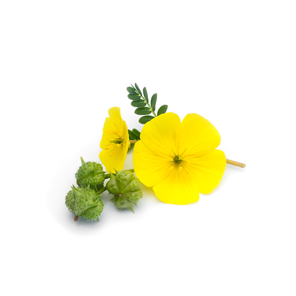
Protogracillin is a naturally occurring steroidal saponin (furostanol glycoside; gracillin series), found in Puncture vine and 9 other sources. It is well tolerated orally (low toxicity).
Concentration
Content by Source
Reported concentrations across the plants that contain protogracillin — the bar marks the typical level, the line shows the reported range.
Literature figures for varying plant parts and preparations. Read them as a comparative guide, not exact assays.
Protogracillin at a Glance
Protogracillin is a furostanol steroidal saponin — C₅₁H₈₄O₂₃, MW 1,065 Da, PubChem CID 441892, CAS 54848-30-5. Structurally it is diosgenin carrying a branched trisaccharide at C-3 (rhamnose→glucose plus a second glucose) and a further glucose at C-26, with the F-ring held open as a 22-hydroxy furostane. Remove the C-26 glucose and the F-ring snaps shut: you get gracillin, the spirostanol. That single sugar is the whole story of this molecule.
It reaches this site through tribulus, where it is listed as a co-occurring saponin alongside protodioscin. Two facts should frame everything below.
First, this molecule is barely studied on its own. A PubMed search for protogracillin returns 30 records in total. Twenty of them involve Dioscorea; zero of them involve Tribulus. There is no review article about protogracillin, no dedicated pharmacology paper, no toxicology study, and no human data. The great majority of the 30 are analytical chemistry — fingerprinting, quality control, species discrimination — in which protogracillin appears as a peak, not as a subject.
Second, and more important: almost every claim you will read about protogracillin belongs to a different molecule. It is the permanent companion of protodioscin in Tribulus reviews and of gracillin and dioscin in Dioscorea papers, and the habit of naming the pair together has quietly transferred protodioscin’s aphrodisiac reputation and gracillin’s cytotoxicity onto a molecule that has rarely been tested alone. Where it has been tested alone, the result is usually modest — and in the two studies that compared it head-to-head with its own spirostanol product, protogracillin lost.
The Attribution Problem: What Actually Belongs to Protogracillin
This is the single most useful thing to do, so it goes first. Findings in the literature that are attached to the word “protogracillin” fall into three very different buckets.
| Bucket | What it means | Examples |
|---|---|---|
| Its own measurements | The purified compound (≥95% pure standard) was put in an assay and a number came out | Xanthine oxidase IC₅₀ 0.19 mM 8Reference 8Chemical comparison and identification of xanthine oxidase inhibitors of Dioscoreae Hypoglaucae Rhizoma and Dioscoreae Spongiosae Rhizoma by chemometric analysis and spectrum-effect relationshipView study →; osteosarcoma IC₅₀ 10.61 µM 7Reference 7Development of an oriental medicine discrimination method through analysis of steroidal saponins in Dioscorea nipponica Makino and their anti-osteosarcoma effectsView study →; F26G substrate Kᵢₙₑₜᵢcs Kₘ 50 µM 13Reference 13Purification and characterization of furostanol glycoside 26-O-β-glucosidase from Costus speciosus rhizomesView study →; rat plasma PK 16Reference 16AnimalSimultaneous determination of four furostanol glycosides in rat plasma by UPLC-MS/MS and its application to PK study after oral administration of Dioscorea nipponica extractsView study → |
| A fraction containing it | A saponin fraction, total-saponin extract or whole herb was tested; protogracillin was one of several components identified in it | Anti-inflammatory activity of purified D. hypoglauca saponins 29Reference 29Purification of steroidal saponins from Dioscoreae hypoglaucae rhizoma by macroporous resin and evaluation of their anti-inflammatory effectView study →; total-saponin anti-gout and anti-thrombotic work; every “Tribulus saponin fraction” study |
| A sibling’s result, transferred by association | The measurement was made on protodioscin, gracillin or methyl protogracillin, and protogracillin was named alongside it | ”Protodioscin and protogracillin are responsible for the aphrodisiac activity” 3Reference 3Phytopharmacological overview of Tribulus terrestrisView study →; the NCI 60-cell-line anticancer data 19Reference 19Methyl protogracillin (NSC-698792): the spectrum of cytotoxicity against 60 human cancer cell lines in the National Cancer Institute’s anticancer drug screen panelView study → |
The third bucket deserves naming. The widely repeated claim that Tribulus’s two main saponins, “protodioscin and protogracillin, are responsible for the observed biological aphrodisiac activity,” appears in a 2014 review 3Reference 3Phytopharmacological overview of Tribulus terrestrisView study → and traces back to a 2001 narrative review 4Reference 4History of herbal medicines with an insight on the pharmacological properties of Tribulus terrestrisView study → — not to any experiment in which protogracillin was administered to anything. No study has ever tested protogracillin for effects on erectile function, testosterone, luteinising hormone or libido, in any species.
Similarly, the impressive-looking National Cancer Institute 60-cell-line screen 19Reference 19Methyl protogracillin (NSC-698792): the spectrum of cytotoxicity against 60 human cancer cell lines in the National Cancer Institute’s anticancer drug screen panelView study → was run on methyl protogracillin (NSC-698792), which is a different compound — the 22-O-methyl ether that forms when furostanol saponins are extracted with methanol, and which the group that isolated it explicitly called an artefact 18Reference 18Antineoplastic agentsView study →. Its GI₅₀ ≤ 2.0 µM against seven cell lines is a real result about methyl protogracillin. It is not a result about protogracillin.
Protogracillin → Gracillin: The Furostanol Bond
Furostanol saponins are the plant’s bidesmosidic storage form. Two sugar chains, one at C-3 and one at C-26; the C-26 glucose keeps the F-ring open and the molecule biologically quiet. When the tissue is damaged — or dried, stored or processed — a specific enzyme cleaves that C-26 glucose, the F-ring cyclises, and the monodesmosidic spirostanol is released. The conversion was characterised directly on protogracillin: furostanol glycoside 26-O-β-glucosidase (F26G), purified to homogeneity from Costus speciosus rhizomes in 1996, is highly specific for the C-26 glucose and has a Kₘ of 50 µM for protogracillin 13Reference 13Purification and characterization of furostanol glycoside 26-O-β-glucosidase from Costus speciosus rhizomesView study →. It is a two-component chemical defence — an inert precursor plus a segregated activating enzyme, the same architecture as the mustard-oil and cyanogenic-glycoside bombs.
Three practical consequences follow, and they shape everything that comes after:
- Analytical. The protogracillin/gracillin ratio in a sample is partly a record of how the material was harvested and dried, not only of what the plant made. The same is true for protodioscin/dioscin.
- Pharmacological. In direct comparisons, the spirostanol is usually the active species. In a Paris polyphylla gastroprotection study, four spirostanol saponins strongly inhibited ethanol- and indomethacin-induced gastric lesions in rats at 1.25–10 mg/kg, and the authors concluded that the spirostanol structure was essential for the activity — protogracillin, isolated from the same active fraction, was among the furostanols that did not carry it 12Reference 12AnimalProtective effects of steroid saponins from Paris polyphylla var. yunnanensis on ethanol- or indomethacin-induced gastric mucosal lesions in rats: structural requirement for activity and mode of actionView study →. In a Dioscorea nipponica lipase study, protodioscin and protogracillin appeared to inhibit pancreatic lipase, but stability testing showed the effect was attributable to their conversion to dioscin and gracillin, which were confirmed as the true inhibitors 10Reference 10Screening and identification of lipase inhibitors extracted from Dioscorea nipponica Makino by UV-vis and HPLC coupled to UPLC-Q-TOF-MS/MSView study →. A 2003 structure–activity comparison put it plainly: the F-ring is the feature that separates furostanol from spirostanol activity in this series 20Reference 20In vitroThe cytotoxicity of methyl protoneogracillin (NSC-698793) and gracillin (NSC-698787), two steroidal saponins from the rhizomes of Dioscorea collettii var. hypoglauca, against human cancer cells in vitroView study →.
- Metabolic. Both routes converge on the same aglycone. In rats given Dioscorea saponins, furostanes including protogracillin were absorbed fast (<1 h) and cleared fast (mostly <8 h), while the spirostanol and the free sapogenin appeared later and persisted beyond 36 h; desugarisation to diosgenin was the main metabolic pathway for the whole class 17Reference 17AnimalUPLC-QTOF-MS identification of metabolites in rat biosamples after oral administration of Dioscorea saponins: a comparative studyView study →.
⚠️ And diosgenin is where the most persistent myth in this category lives. Diosgenin is the industrial feedstock from which progesterone and cortisone were semi-synthesised by the Marker degradation — a multi-step factory process involving acetic anhydride, chromium oxidation and high heat. The human body has no enzyme that performs it. There is no metabolic route from protogracillin, gracillin or diosgenin to progesterone, DHEA or testosterone in a person. Topical “natural progesterone” wild-yam creams that do anything do so because progesterone was added to them. Nothing here supports a hormonal claim of any kind.
Protogracillin as a Chemotype & Quality Marker
This is the role protogracillin genuinely plays, and it is the reason it has 30 papers rather than three. Because it co-elutes near protodioscin but is cleanly resolvable, and because its ratio to gracillin encodes both genetics and processing, it has become a standard marker in Chinese and Korean pharmacopoeial analysis of Dioscorea:
- Species discrimination. Quantified by validated HPLC-UV across three commonly confused rhizomes: Dioscorea nipponica 4.25 ± 0.02 mg/g, D. quinquelobata 5.95 ± 0.02 mg/g, D. septemloba 9.90 ± 0.06 mg/g — while protodioscin swung from 160 mg/g in D. nipponica to 3.5 mg/g in D. quinquelobata, which is what makes the pair discriminating rather than either alone 7Reference 7Development of an oriental medicine discrimination method through analysis of steroidal saponins in Dioscorea nipponica Makino and their anti-osteosarcoma effectsView study →.
- Drug-material discrimination. Protodioscin, protogracillin, dioscin and gracillin were the four chemical markers separating Dioscoreae Hypoglaucae Rhizoma from Dioscoreae Spongiosae Rhizoma; the two furostanols run significantly higher in the former, the two spirostanols in the latter 8Reference 8Chemical comparison and identification of xanthine oxidase inhibitors of Dioscoreae Hypoglaucae Rhizoma and Dioscoreae Spongiosae Rhizoma by chemometric analysis and spectrum-effect relationshipView study →.
- Wild vs. cultivated. In Dioscoreae Nipponicae Rhizoma, protodioscin and protogracillin were the two compounds that reliably separated wild from cultivated material 15Reference 15Quality control of Dioscoreae Nipponicae Rhizoma based on fingerprint and quantitative analysisView study →.
- Habitat. Protogracillin is one of six compounds quantified as habitat markers for Dioscorea septemloba 23Reference 23Strategy of combining offline 2D LC-MS with LC-DIA-MS/MS to accurately identify chemical compounds and for quality control of Dioscorea septemloba ThunbView study →, and in Asparagus cochinchinensis its content (0.044%, against protodioscin’s 0.373%) tracked with climate and cultivation site 14Reference 14Assessment of suitable region of Asparagus cochinchinensis (Lour.) Merr. under different climatic conditions in China by the MaxEnt model and HPLC analysisView study →.
- Genus-level chemotyping. A metabolomic survey of 26 Paris species found P. tengchongensis distinguished by relatively high gracillin and protogracillin 22Reference 22UHPLC-Q-TOF/MS unveils metabolic diversity in Paris L. and highlights medicinal potential of five newly identified speciesView study →.
The one place this does not work is Tribulus. The definitive geographic-chemotype survey — nine countries, LC-ESI/MS/MS — quantified protodioscin, prototribestin, pseudoprotodioscin, dioscin, tribestin, tribulosin and rutin. Protogracillin was not among the compounds measured 5Reference 5Distribution of steroidal saponins in Tribulus terrestris from different geographical regionsView study →. No published analysis has ever reported a protogracillin figure for Tribulus terrestris from any origin. Whatever “co-marker in Tribulus chemotyping” means, it is not supported by the analytical record.
What It's Used For
Everything below is in vitro except the rat plasma work. There is no animal efficacy study and no human study of this molecule.
Support is an experimental score — weighted by study type (human > animal > in vitro) and volume. A beta, not a validated metric. With a 30-paper literature that is mostly chromatography, and zero animal-efficacy or human studies, every score here is capped low by design. Each application links to its write-up.
The one positive isolate result. A 2024 Dioscorea spongiosa study purified twelve compounds to >95% by counter-current chromatography and assayed each for anti-inflammatory, urate-lowering and anti-diabetic activity in vitro; protogracillin (compound 6) was one of three saponins reported active in all three assays 11Reference 11Efficient discovery of active isolates from Dioscorea spongiosa by the combination of bioassay-guided macroporous resin column chromatography and high-speed counter-current chromatographyView study →.
No IC₅₀ for protogracillin was given in the abstract, and the paper’s purpose was method development rather than pharmacology.
Against this sits a directly contradictory result. A 2022 study of Dioscorea nipponica set out to identify which of its saponins drive its anti-inflammatory activity, quantified seven of them — protogracillin included — and used correlation and partial-least-squares regression to select the actives. Protodioscin, dioscin and gracillin were selected; protogracillin was not. The three selected compounds were then confirmed against LPS-stimulated RAW264.7 macrophages with NO-inhibition IC₅₀ values of 0.712, 0.469 and 0.815 µM and significant reductions in TNF-α, IL-1β and IL-6 9Reference 9Identification of anti-inflammatory components in Dioscorea nipponica Makino based on HPLC-MS/MS, quantitative analysis of multiple components by single marker and chemometric methodsView study →.
A 2025 paper likewise showed that a purified Dioscorea hypoglauca saponin fraction containing protogracillin out-performed the crude extract in the same macrophage model — a fraction result, not a molecule result 29Reference 29Purification of steroidal saponins from Dioscoreae hypoglaucae rhizoma by macroporous resin and evaluation of their anti-inflammatory effectView study →.
Gap: no molecular target, no dose–response curve, no cellular mechanism, and no in-vivo model for protogracillin specifically. The class mechanism (NF-κB and iNOS/COX-2 suppression) is documented for dioscin and gracillin, not for this molecule.
Two genuine isolate measurements exist. In 2019, protogracillin from Dioscorea nipponica gave IC₅₀ 10.61 µM against osteosarcoma cells — alongside protodioscin at 6.43 µM, pseudoprotodioscin at 10.48 µM and dioscin at 6.90 µM. The study’s conclusion was that the herb’s anti-osteosarcoma effect is attributable to protodioscin and dioscin, “which is also related to the quantitative results” — that is, to the compounds present at 160 and 23 mg/g, not to the one present at 4.25 mg/g 7Reference 7Development of an oriental medicine discrimination method through analysis of steroidal saponins in Dioscorea nipponica Makino and their anti-osteosarcoma effectsView study →.
In 1997, protogracillin and seven related furostanols from Dioscorea collettii var. hypoglauca all caused morphological abnormality in Pyricularia oryzae mycelia and showed cytotoxicity against the K562 leukaemia line, reported as a group rather than compound-by-compound 18Reference 18Antineoplastic agentsView study →.
Two cautions apply and both are load-bearing. Neither study reported a selectivity index against normal cells. Steroidal saponins are surfactants that permeabilise cholesterol-containing membranes, and a large share of “cytotoxic at X µM” results in this class do not discriminate tumour from normal cells 21Reference 21ReviewSaponins as cytotoxic agents: a reviewView study →.
And the frequently cited NCI 60-cell-line data belong to methyl protogracillin, a methanol-extraction artefact and a different molecule 19Reference 19Methyl protogracillin (NSC-698792): the spectrum of cytotoxicity against 60 human cancer cell lines in the National Cancer Institute’s anticancer drug screen panelView study → — as does the related work on methyl protoneogracillin, the 25S epimer, where the authors found that C-25 configuration and the presence or absence of the F-ring were both critical to selectivity 20Reference 20In vitroThe cytotoxicity of methyl protoneogracillin (NSC-698793) and gracillin (NSC-698787), two steroidal saponins from the rhizomes of Dioscorea collettii var. hypoglauca, against human cancer cells in vitroView study →.
Gap: no selectivity index, no mechanism, no animal tumour model, no follow-up. Given the µM potencies and the ~1,065 Da mass, systemic exposure at these concentrations is implausible in any case.
Dioscorea rhizomes are used in Chinese medicine for gout, and a 2023 spectrum–effect study asked which constituents inhibit xanthine oxidase. Protodioscin, protogracillin, methyl protodioscin and pseudoprogracillin were selected by correlation analysis, then validated with a purified reference standard: protogracillin’s IC₅₀ was 0.19 mM, against 0.20, 0.22 and 0.25 mM for the others and 0.17 mM for the allopurinol positive control. Docking placed it in the enzyme’s hydrophobic FAD-binding pocket with a binding affinity of −8.9 kcal/mol — the weakest of the four 8Reference 8Chemical comparison and identification of xanthine oxidase inhibitors of Dioscoreae Hypoglaucae Rhizoma and Dioscoreae Spongiosae Rhizoma by chemometric analysis and spectrum-effect relationshipView study →.
The paper reads this as “similar to allopurinol.” It is worth being clearer than that. Allopurinol’s IC₅₀ against xanthine oxidase is normally reported in the low micromolar range; an assay in which it needs 170 µM is an insensitive assay, and a compound that matches it there has not shown meaningful potency. A 190 µM plasma concentration of a 1,065 Da saponin is not reachable by any oral dose.
Gap: in-vitro enzyme plus docking only. No urate measurement in any animal, no cell model, and no comparison against a compound of known clinical potency in a validated assay.
Instructive as a piece of methodology. A 2023 study screened Dioscorea nipponica for pancreatic-lipase inhibitors and fished out four saponins — protodioscin, protogracillin, dioscin and gracillin. But stability testing showed that the furostanols convert to the spirostanols under the assay conditions, and the authors concluded that dioscin and gracillin were the major inhibitors, with kinetics and docking performed on those two rather than on the furostanols 10Reference 10Screening and identification of lipase inhibitors extracted from Dioscorea nipponica Makino by UV-vis and HPLC coupled to UPLC-Q-TOF-MS/MSView study →.
This is the clearest published demonstration of the trap that runs through the whole class: a furostanol saponin can score in an assay because of what it turns into, and reporting that score as the furostanol’s activity is a category error.
Gap: protogracillin’s own contribution, if any, was never separated out. The honest reading is that this is a gracillin result.
A negative result worth stating, because it is rare to have one this clean. In a bioassay-guided study of Paris polyphylla var. yunnanensis, seven saponins were isolated from the fraction that inhibited ethanol-induced gastric lesions in rats. The four spirostanol saponins strongly inhibited both ethanol- and indomethacin-induced lesions at 1.25–10 mg/kg orally. The three furostanols — including protogracillin — did not, and the authors concluded that the 3-O-glycoside moiety and the spirostanol structure were both required, with the 17-hydroxyl enhancing the effect 12Reference 12AnimalProtective effects of steroid saponins from Paris polyphylla var. yunnanensis on ethanol- or indomethacin-induced gastric mucosal lesions in rats: structural requirement for activity and mode of actionView study →.
Gap: none needed. This is a properly designed in-vivo comparison in which protogracillin was tested and found inactive, and it is the single most informative pharmacology datum here.
Mechanisms
| Target / pathway | Effect | Relevant to | Evidence |
|---|---|---|---|
| Furostanol 26-O-β-glucosidase (F26G) | substrate, Kₘ 50 µM — cleavage converts it to gracillin | plant defenceprocessing chemistry | purified enzyme 13Reference 13Purification and characterization of furostanol glycoside 26-O-β-glucosidase from Costus speciosus rhizomesView study → |
| Xanthine oxidase | inhibition, IC₅₀ 0.19 mM; docking −8.9 kcal/mol at the FAD pocket | urate production | isolate assay + docking 8Reference 8Chemical comparison and identification of xanthine oxidase inhibitors of Dioscoreae Hypoglaucae Rhizoma and Dioscoreae Spongiosae Rhizoma by chemometric analysis and spectrum-effect relationshipView study → |
| Osteosarcoma cell viability | IC₅₀ 10.61 µM; weaker than protodioscin and dioscin in the same run | cytotoxicity | isolate assay, no selectivity index 7Reference 7Development of an oriental medicine discrimination method through analysis of steroidal saponins in Dioscorea nipponica Makino and their anti-osteosarcoma effectsView study → |
| NO / iNOS in LPS-stimulated macrophages | active in one isolate panel; not selected as an active in a second, better-controlled study | inflammation | conflicting 9, |
| Pancreatic lipase | apparent inhibition attributable to conversion into gracillin | lipid digestion | attributed away 10Reference 10Screening and identification of lipase inhibitors extracted from Dioscorea nipponica Makino by UV-vis and HPLC coupled to UPLC-Q-TOF-MS/MSView study → |
| Gastric mucosal protection | inactive; spirostanol ring closure required | gastroprotection | in-vivo rat comparison 12Reference 12AnimalProtective effects of steroid saponins from Paris polyphylla var. yunnanensis on ethanol- or indomethacin-induced gastric mucosal lesions in rats: structural requirement for activity and mode of actionView study → |
| Cell membranes (class effect) | surfactant permeabilisation — a general confounder in saponin cytotoxicity assays | interpretation of all of the above | class review 21Reference 21ReviewSaponins as cytotoxic agents: a reviewView study → |
Pharmacokinetics
Unusually for a molecule this obscure, protogracillin has been measured in plasma. A validated UPLC-MS/MS method quantified four furostanol glycosides in rat plasma after intragastric Dioscorea nipponica extract, with a calibrated range of 2–4,000 ng/mL for protogracillin; the saponins reached “relatively high” plasma concentrations, showed rapid excretion, and both t½ and AUC₀₋ₜ scaled with dose across low, medium and high dosing 16Reference 16AnimalSimultaneous determination of four furostanol glycosides in rat plasma by UPLC-MS/MS and its application to PK study after oral administration of Dioscorea nipponica extractsView study →. A companion metabolic study using UPLC-QTOF-MS put numbers on the shape: furostanes including protogracillin were absorbed into blood within 1 hour but largely gone by 8 hours, whereas the spirostanol and the free sapogenin appeared more slowly and persisted past 36 hours. The dominant transformation for the whole class was desugarisation to diosgenin, found in plasma and faeces after every treatment 17Reference 17AnimalUPLC-QTOF-MS identification of metabolites in rat biosamples after oral administration of Dioscorea saponins: a comparative studyView study →.
Two things follow. First, “poorly absorbed” is too crude a description here — some intact furostanol does get in, at least in rats given a concentrated extract. Second, it does not stay, and the species with the long residence time is the aglycone, not the parent. So the micromolar in-vitro numbers above are being compared against a short, low, rapidly cleared plasma exposure of the intact molecule, and against a much longer exposure to a different molecule, diosgenin. No human pharmacokinetic study of protogracillin exists, and no absolute oral bioavailability figure has been published for it in any species.
Clinical Trials
None. There are no clinical trials of protogracillin and no registered study of it under any name. Every human exposure to date has been incidental, through Dioscorea, Paris or Tribulus preparations.
| Completed | Planned | Terminated | Preclinical |
|---|---|---|---|
| —(none) | — | — | ~4isolate assays; 1 rat PK study |
Last checked: August 2026.
Isolate vs. Plant
The split here is lopsided in an unusual direction. Nearly every study that touches protogracillin used a purified reference standard — bought commercially at ≥95–98% purity, or isolated by counter-current chromatography — because the studies were analytical, and analysis requires standards 7,
The blurring happens afterwards, in prose. A finding about a Dioscorea total-saponin fraction, or a Tribulus furostanol fraction, gets restated in a review as a property of the named constituents — and protogracillin is almost always one of the names. The corrective is simple: none of the whole-extract literature on Tribulus sexual function, Dioscorea gout, or Paris haemostasis contains a single experiment in which protogracillin was the variable.
There is also a species caution specific to this site. Protogracillin’s Dioscorea record is entirely Asian species — D. nipponica, D. septemloba, D. spongiosa, D. hypoglauca/collettii, D. quinquelobata, D. zingiberensis, D. tokoro, D. opposita, D. pseudojaponica 7,
Prevalence in Nature
Protogracillin is a saponin of monocot rhizomes and tubers, concentrated in the underground storage organs where furostanol glycosides accumulate. Its recorded distribution spans three families: Dioscoreaceae (Dioscorea), Melanthiaceae (Paris), Asparagaceae (Asparagus, and Costus speciosus in Costaceae), with the outlying Zygophyllaceae report from Tribulus terrestris.
Where it has been quantified, it sits in the 4–10 mg/g dry weight band in Dioscorea rhizomes 7Reference 7Development of an oriental medicine discrimination method through analysis of steroidal saponins in Dioscorea nipponica Makino and their anti-osteosarcoma effectsView study → and around 0.44 mg/g in Asparagus cochinchinensis root tuber 14Reference 14Assessment of suitable region of Asparagus cochinchinensis (Lour.) Merr. under different climatic conditions in China by the MaxEnt model and HPLC analysisView study → — a real but secondary component, typically an order of magnitude below protodioscin in the same tissue. In Dioscorea opposita (Chinese yam, a food crop) it concentrates in the peel, along with protodioscin and dioscin, which is a reason peeling changes a yam’s saponin load 27Reference 27Separation and structural elucidation of C25 epimers of furostanol saponin from aerial parts of Paris polyphylla var. chinensisView study →.
Major Natural Sources
| Source | Part | Reported level | Basis |
|---|---|---|---|
| Dioscorea septemloba | Rhizome | 9.90 ± 0.06 mg/g | Validated HPLC-UV quantification 7Reference 7Development of an oriental medicine discrimination method through analysis of steroidal saponins in Dioscorea nipponica Makino and their anti-osteosarcoma effectsView study →; also isolated directly 24Reference 24Study on steroidal saponins of Dioscorea septemloba ThunbView study → |
| Dioscorea quinquelobata | Rhizome | 5.95 ± 0.02 mg/g | Validated HPLC-UV quantification 7Reference 7Development of an oriental medicine discrimination method through analysis of steroidal saponins in Dioscorea nipponica Makino and their anti-osteosarcoma effectsView study → |
| Dioscorea nipponica | Rhizome | 4.25 ± 0.02 mg/g | Validated HPLC-UV 7Reference 7Development of an oriental medicine discrimination method through analysis of steroidal saponins in Dioscorea nipponica Makino and their anti-osteosarcoma effectsView study →; also a wild-vs-cultivated marker 15Reference 15Quality control of Dioscoreae Nipponicae Rhizoma based on fingerprint and quantitative analysisView study → |
| Dioscorea hypoglauca / D. collettii var. hypoglauca | Rhizome | Higher than in D. spongiosa (relative) | Chemometric marker 8Reference 8Chemical comparison and identification of xanthine oxidase inhibitors of Dioscoreae Hypoglaucae Rhizoma and Dioscoreae Spongiosae Rhizoma by chemometric analysis and spectrum-effect relationshipView study →; isolated 1997 18Reference 18Antineoplastic agentsView study → |
| Dioscorea spongiosa | Rhizome | Not quantified | Purified to >95% by counter-current chromatography 11Reference 11Efficient discovery of active isolates from Dioscorea spongiosa by the combination of bioassay-guided macroporous resin column chromatography and high-speed counter-current chromatographyView study → |
| Asparagus cochinchinensis | Root tuber | 0.044% (≈0.44 mg/g) | HPLC, varying by cultivation site 14Reference 14Assessment of suitable region of Asparagus cochinchinensis (Lour.) Merr. under different climatic conditions in China by the MaxEnt model and HPLC analysisView study → |
| Paris polyphylla var. chinensis | Aerial parts | Not quantified | Isolated with its 25S epimer neoprotogracillin 27Reference 27Separation and structural elucidation of C25 epimers of furostanol saponin from aerial parts of Paris polyphylla var. chinensisView study → |
| Paris spp. (incl. P. tengchongensis) | Rhizome | Relatively high (metabolomic) | 26-species survey 22Reference 22UHPLC-Q-TOF/MS unveils metabolic diversity in Paris L. and highlights medicinal potential of five newly identified speciesView study → |
| Costus speciosus | Rhizome | Not quantified as such | Methyl protogracillin — its methanolysis artefact — measured at 298 mg/g in a butanol fraction 28Reference 28Comparative metabolic profiling of Costus speciosus leaves and rhizomes using NMR, GC-MS and UPLC/ESI-MS/MSView study →; F26G substrate work 13Reference 13Purification and characterization of furostanol glycoside 26-O-β-glucosidase from Costus speciosus rhizomesView study → |
| Tribulus terrestris | Aerial parts | Never quantified | Reported in the 1981 steroidal-glycoside survey 1Reference 1Steroidal glycosides of Tribulus terrestris LinnView study →, propagated through reviews 2, |
The Tribulus Report
Since this molecule reaches the site through Tribulus, the provenance of that claim is worth stating exactly. Protogracillin’s presence in Tribulus terrestris traces to Mahato, Sahu, Ganguly, Miyahara and Kawasaki (1981), “Steroidal glycosides of Tribulus terrestris Linn.”, in J. Chem. Soc. Perkin Trans. 1 1Reference 1Steroidal glycosides of Tribulus terrestris LinnView study →. Every subsequent statement is a citation of that paper: the 2017 review lists protogracillin as compound 81 of 108 Tribulus steroidal saponins, sourced to Mahato 1981 2Reference 2ReviewA review of traditional pharmacological uses, phytochemistry, and pharmacological activities of Tribulus terrestrisView study →; the standard chemistry review is Kostova & Dinchev 6Reference 6Saponins in Tribulus terrestris — chemistry and bioactivityView study →.
What has never happened, in forty-five years, is a measurement. No Tribulus analysis has reported a protogracillin concentration, from any country, any plant part or any commercial extract — including the nine-country LC-MS/MS chemotype survey, which did not include it among its analytes 5Reference 5Distribution of steroidal saponins in Tribulus terrestris from different geographical regionsView study →. The molecule is presumably present; nobody knows in what quantity, and it is not a Tribulus marker in any operational sense.
Its Role in Plants
Protogracillin is a defence precursor, not a defence compound. It is assembled from cholesterol via the steroidal-saponin pathway to diosgenin, glycosylated at C-3 with the branched rhamnose–glucose–glucose chain that defines the gracillin series, and capped with a C-26 glucose that holds the F-ring open and the molecule inactive. The plant stores it in that state, physically separated from F26G, the β-glucosidase that removes the C-26 glucose 13Reference 13Purification and characterization of furostanol glycoside 26-O-β-glucosidase from Costus speciosus rhizomesView study →. Tissue damage brings the two together, the F-ring closes, and gracillin — membrane-active, and toxic to the fungi and herbivores the plant is defending against — is generated on the spot.
This also explains an otherwise odd analytical fact: furostanol and spirostanol content in dried herbal material is not a stable property of the species. Post-harvest handling drives the conversion 13Reference 13Purification and characterization of furostanol glycoside 26-O-β-glucosidase from Costus speciosus rhizomesView study →, which is why the furostanol/spirostanol ratio works so well as a processing and provenance marker and so poorly as a fixed species fingerprint.
Discovery & Synthesis
Gracillin came first. Its structure was solved by Tsukamoto and Kawasaki in 1956 as part of the long “Saponins of Japanese Dioscoreaceae” series 30Reference 30Saponins of Japanese DioscoreaceaeView study →, and refined alongside dioscin and kikuba-saponin in 1962 31Reference 31Structures of dioscin, gracillin, and kikuba-saponinView study →. Protogracillin was named as its proto- form — the bidesmosidic furostanol precursor — following the same convention that gives protodioscin its name from dioscin.
No single paper can be identified as protogracillin’s first isolation. It was registered under CAS 54848-30-5 in the mid-1970s, appears in the 1981 Tribulus terrestris steroidal-glycoside survey 1Reference 1Steroidal glycosides of Tribulus terrestris LinnView study →, was in routine use as an enzyme substrate by 1996 13Reference 13Purification and characterization of furostanol glycoside 26-O-β-glucosidase from Costus speciosus rhizomesView study →, and was described as a “known” furostanol saponin when four of them were re-isolated from Dioscorea collettii var. hypoglauca in 1997 18Reference 18Antineoplastic agentsView study →. This is a molecule that entered the literature through the Japanese and Chinese Dioscorea saponin programmes rather than through a single named discovery.
There is no total synthesis, and none is likely: a 1,065 Da tetraglycoside is far more cheaply obtained from a yam rhizome. All published material was isolated chromatographically from plant extract, and commercial reference standards (≥95–98%) are sold for analytical use 7,
Toxicity & Safety
Not characterised. There has never been a toxicology study of protogracillin — no acute-toxicity work, no genotoxicity panel, no reproductive study, no repeat-dose animal experiment. The low grading reflects the class, the exposure route and the absence of any signal, not a demonstrated safety record.
The class reasoning is genuinely favourable in one respect and unhelpful in another. Favourable: the bidesmosidic furostanol form is the plant’s inert storage form, and the two studies that compared it directly with its spirostanol product found the furostanol less active in both a rat gastric model 12Reference 12AnimalProtective effects of steroid saponins from Paris polyphylla var. yunnanensis on ethanol- or indomethacin-induced gastric mucosal lesions in rats: structural requirement for activity and mode of actionView study → and a lipase assay 10Reference 10Screening and identification of lipase inhibitors extracted from Dioscorea nipponica Makino by UV-vis and HPLC coupled to UPLC-Q-TOF-MS/MSView study →. Unhelpful: whatever protogracillin does after ingestion is largely determined by what it becomes — gracillin, then diosgenin 17Reference 17AnimalUPLC-QTOF-MS identification of metabolites in rat biosamples after oral administration of Dioscorea saponins: a comparative studyView study → — and those molecules have their own profiles. In Tribulus specifically, the compound flagged as the likely driver of hepatorenal toxicity is a spirostanol (terrestrosin D), not a furostanol.
- No adverse effect has been attributed to protogracillin. It has also never been looked for — those are different statements.
- The furostanol form is the quiet one. Where tested head-to-head, it was less active than its spirostanol product 10,
12Reference 10Screening and identification of lipase inhibitors extracted from Dioscorea nipponica Makino by UV-vis and HPLC coupled to UPLC-Q-TOF-MS/MSView study →Reference 12AnimalProtective effects of steroid saponins from Paris polyphylla var. yunnanensis on ethanol- or indomethacin-induced gastric mucosal lesions in rats: structural requirement for activity and mode of actionView study → . - Long dietary exposure history. Yams and Dioscorea rhizomes have been eaten and used medicinally for centuries with this saponin present.
- No reproductive or developmental data of any kind. Nothing is known about protogracillin in pregnancy or lactation; the parent herbs carry their own cautions on their own pages.
- It converts to gracillin and then to diosgenin in the gut 17Reference 17AnimalUPLC-QTOF-MS identification of metabolites in rat biosamples after oral administration of Dioscorea saponins: a comparative studyView study →. Any risk assessment has to cover the products, not just the parent.
- Saponins are surfactants. As a class they permeabilise cholesterol-containing membranes, which is both the source of the in-vitro cytotoxicity numbers and a reason not to assume an injected or concentrated preparation would be benign 21Reference 21ReviewSaponins as cytotoxic agents: a reviewView study →.
- No drug-interaction study exists. No CYP, P-gp or transporter assessment has been performed on this molecule, so no interaction can be ruled in or out.
- Do not infer a hormonal effect from the diosgenin backbone. The Marker degradation is industrial chemistry; humans cannot perform it, and no steroid-hormone activity has been shown for protogracillin.
Dosage
There is no dose. Protogracillin has never been administered to a human being as an isolate, and no animal efficacy study exists to extrapolate from. Every published figure is an in-vitro concentration, an HPLC peak or a rat plasma curve after a whole extract.
Real-world exposure is incidental and small. In the richest quantified source it runs to about 10 mg per gram of dried rhizome 7Reference 7Development of an oriental medicine discrimination method through analysis of steroidal saponins in Dioscorea nipponica Makino and their anti-osteosarcoma effectsView study →, so a conventional Dioscorea decoction delivers on the order of tens of milligrams — as one of dozens of saponins, in a preparation whose effects have never been attributed to it. In Tribulus supplements the quantity is simply unknown 5Reference 5Distribution of steroidal saponins in Tribulus terrestris from different geographical regionsView study →. Nothing here is a recommendation.
References
- Mahato SB, Sahu NP, Ganguly AN, Miyahara K, Kawasaki T (1981). Steroidal glycosides of Tribulus terrestris Linn. Journal of the Chemical Society, Perkin Transactions 1: 2405–2410. https://doi.org/10.1039/p19810002405
- Zhu W, Du Y, Meng H, Dong Y, Li L (2017). A review of traditional pharmacological uses, phytochemistry, and pharmacological activities of Tribulus terrestris. Chemistry Central Journal. https://pubmed.ncbi.nlm.nih.gov/29086839/
- Chhatre S, Nesari T, Somani G, Kanchan D, Sathaye S (2014). Phytopharmacological overview of Tribulus terrestris. Pharmacognosy Reviews. https://pubmed.ncbi.nlm.nih.gov/24600195/
- Adaikan PG, Gauthaman K, Prasad RNV (2001). History of herbal medicines with an insight on the pharmacological properties of Tribulus terrestris. The Aging Male 4:163–169. https://doi.org/10.1080/tam.4.3.163.169
- Dinchev D, Janda B, Evstatieva L, Oleszek W, Aslani MR, Kostova I (2008). Distribution of steroidal saponins in Tribulus terrestris from different geographical regions. Phytochemistry. https://pubmed.ncbi.nlm.nih.gov/17719068/
- Kostova I, Dinchev D (2005). Saponins in Tribulus terrestris — chemistry and bioactivity. Phytochemistry Reviews 4:111–137. https://doi.org/10.1007/s11101-005-2833-x
- Hwang JT, Park KS, Ryuk JA, Kim HJ, Ko BS (2019). Development of an oriental medicine discrimination method through analysis of steroidal saponins in Dioscorea nipponica Makino and their anti-osteosarcoma effects. Molecules. https://pubmed.ncbi.nlm.nih.gov/31698850/
- Rui G, Qin ZY, Chang YQ, Zheng YG, Zhang D, Yao LM, Guo L (2023). Chemical comparison and identification of xanthine oxidase inhibitors of Dioscoreae Hypoglaucae Rhizoma and Dioscoreae Spongiosae Rhizoma by chemometric analysis and spectrum-effect relationship. Molecules. https://pubmed.ncbi.nlm.nih.gov/38138603/
- Yang G, Liu P, Shi H, Fan W, Feng X, Chen J, Jing S, Wang L, Zheng Y, Zhang D, Guo L (2022). Identification of anti-inflammatory components in Dioscorea nipponica Makino based on HPLC-MS/MS, quantitative analysis of multiple components by single marker and chemometric methods. Journal of Chromatography B. https://pubmed.ncbi.nlm.nih.gov/36371964/
- Jin P, Chen L, Zhong J, Yuan T, Gan L, Huang J, Wang L, Fan H, Lin C (2023). Screening and identification of lipase inhibitors extracted from Dioscorea nipponica Makino by UV-vis and HPLC coupled to UPLC-Q-TOF-MS/MS. International Journal of Biological Macromolecules. https://pubmed.ncbi.nlm.nih.gov/36706882/
- Wang Z, Yu J, Zhao L, Niu T, Wang X (2024). Efficient discovery of active isolates from Dioscorea spongiosa by the combination of bioassay-guided macroporous resin column chromatography and high-speed counter-current chromatography. Journal of Separation Science. https://pubmed.ncbi.nlm.nih.gov/38356225/
- Matsuda H, Pongpiriyadacha Y, Morikawa T, Kishi A, Kataoka S, Yoshikawa M (2003). Protective effects of steroid saponins from Paris polyphylla var. yunnanensis on ethanol- or indomethacin-induced gastric mucosal lesions in rats: structural requirement for activity and mode of action. Bioorganic & Medicinal Chemistry Letters. https://pubmed.ncbi.nlm.nih.gov/12643921/
- Inoue K, Ebizuka Y (1996). Purification and characterization of furostanol glycoside 26-O-β-glucosidase from Costus speciosus rhizomes. FEBS Letters. https://pubmed.ncbi.nlm.nih.gov/8549824/
- Zhang T, Lv X, Zhao Q, Zhang C, Yin H, Deng S, Yan G, Wang G, Cao X, Ou H, Shen G (2024). Assessment of suitable region of Asparagus cochinchinensis (Lour.) Merr. under different climatic conditions in China by the MaxEnt model and HPLC analysis. Ecology and Evolution. https://pubmed.ncbi.nlm.nih.gov/39371263/
- Yang GY, Chen YQ, Xu ZJ, Sun JN, Zhang D, Zheng YG, Guo L (2020). Quality control of Dioscoreae Nipponicae Rhizoma based on fingerprint and quantitative analysis. China Journal of Chinese Materia Medica. https://pubmed.ncbi.nlm.nih.gov/33350268/
- Liao M, Dai C, Liu M, Chen J, Chen Z, Xie Z, Yao M (2016). Simultaneous determination of four furostanol glycosides in rat plasma by UPLC-MS/MS and its application to PK study after oral administration of Dioscorea nipponica extracts. Journal of Pharmaceutical and Biomedical Analysis. https://pubmed.ncbi.nlm.nih.gov/26433169/
- Tang YN, Pang YX, He XC, Zhang YZ, Zhang JY, Zhao ZZ, Yi T, Chen HB (2015). UPLC-QTOF-MS identification of metabolites in rat biosamples after oral administration of Dioscorea saponins: a comparative study. Journal of Ethnopharmacology. https://pubmed.ncbi.nlm.nih.gov/25698242/
- Hu K, Dong A, Yao X, Kobayashi H, Iwasaki S (1997). Antineoplastic agents. II. Four furostanol glycosides from rhizomes of Dioscorea collettii var. hypoglauca. Planta Medica. https://pubmed.ncbi.nlm.nih.gov/17252340/
- Hu K, Yao X (2001). Methyl protogracillin (NSC-698792): the spectrum of cytotoxicity against 60 human cancer cell lines in the National Cancer Institute’s anticancer drug screen panel. Anti-Cancer Drugs. https://pubmed.ncbi.nlm.nih.gov/11460001/
- Hu K, Yao X (2003). The cytotoxicity of methyl protoneogracillin (NSC-698793) and gracillin (NSC-698787), two steroidal saponins from the rhizomes of Dioscorea collettii var. hypoglauca, against human cancer cells in vitro. Phytotherapy Research. https://pubmed.ncbi.nlm.nih.gov/12820229/
- Podolak I, Galanty A, Sobolewska D (2010). Saponins as cytotoxic agents: a review. Phytochemistry Reviews. https://pubmed.ncbi.nlm.nih.gov/20835386/
- Sun K, Shao Y, Li R, Wang Z, Wang J, Luan X, Wu C, Wang L, He C, Tang Q (2025). UHPLC-Q-TOF/MS unveils metabolic diversity in Paris L. and highlights medicinal potential of five newly identified species. Frontiers in Pharmacology. https://pubmed.ncbi.nlm.nih.gov/40771935/
- Chen H, Wang L, Wang C, Zhang Y, Yu H, Fu Z, Fu X, Han L (2022). Strategy of combining offline 2D LC-MS with LC-DIA-MS/MS to accurately identify chemical compounds and for quality control of Dioscorea septemloba Thunb. Phytochemical Analysis. https://pubmed.ncbi.nlm.nih.gov/35841277/
- Tan DW, Kang LP, Lü N, Zhang J, Ma BP (2006). Study on steroidal saponins of Dioscorea septemloba Thunb. Zhong Yao Cai. https://pubmed.ncbi.nlm.nih.gov/17228658/
- Ukiya M, Sato D, Kimura H, Hirai Y, Nishina A (2020). Tokoronin contained in Dioscorea tokoro Makino ex Miyabe suppressed α-MSH-induced melanogenesis in B16 cells via suppression of classical MAPK pathway activation. Chemistry & Biodiversity. https://pubmed.ncbi.nlm.nih.gov/32378303/
- Yang DJ, Lu TJ, Hwang LS (2003). Isolation and identification of steroidal saponins in Taiwanese yam cultivar (Dioscorea pseudojaponica Yamamoto). Journal of Agricultural and Food Chemistry. https://pubmed.ncbi.nlm.nih.gov/14558759/
- Tan XM, Guan LJ, Fang YG, Li YM, Zheng ZL, Chen LM, Gao HM, Wang ZM (2021). Separation and structural elucidation of C25 epimers of furostanol saponin from aerial parts of Paris polyphylla var. chinensis. China Journal of Chinese Materia Medica. https://pubmed.ncbi.nlm.nih.gov/34467711/
- Kumar A, Maurya AK, Chand G, Agnihotri VK (2018). Comparative metabolic profiling of Costus speciosus leaves and rhizomes using NMR, GC-MS and UPLC/ESI-MS/MS. Natural Product Research. https://pubmed.ncbi.nlm.nih.gov/28814124/
- Qin Z, Du Y, Zhong Y, Gu Z, Zheng Q, Zheng Y, Zhang D, Guo L (2025). Purification of steroidal saponins from Dioscoreae hypoglaucae rhizoma by macroporous resin and evaluation of their anti-inflammatory effect. Journal of Chromatography B. https://pubmed.ncbi.nlm.nih.gov/40829486/
- Tsukamoto T, Kawasaki T (1956). Saponins of Japanese Dioscoreaceae. VI. The structure of gracillin. Pharmaceutical Bulletin 4:104–108. https://pubmed.ncbi.nlm.nih.gov/13335473/
- Kawasaki T, Yamauchi T (1962). Structures of dioscin, gracillin, and kikuba-saponin. Chemical & Pharmaceutical Bulletin 10:703–708. https://pubmed.ncbi.nlm.nih.gov/14042124/