Compound Monograph
Casuarictin
Casuarictin is the monomeric ellagitannin that the whole Rubus and Fragaria oligomer series is built from — two of it make sanguiin H-6, three make lambertianin C. It has a modest but genuine pharmacology of its own, and a persistent identity problem: its alpha-anomer potentillin is a different molecule that the literature routinely fails to separate from it.

Sources · 11
Where Does It Come From?
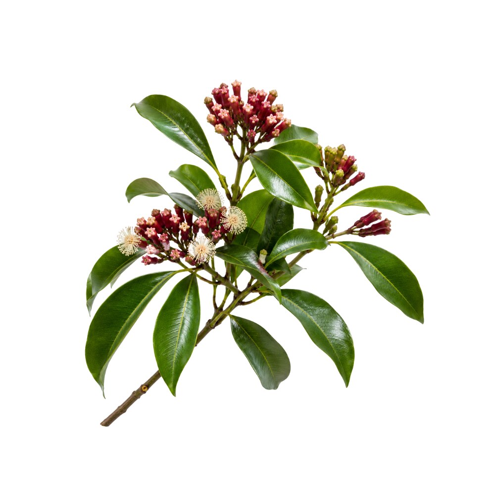
Casuarictin is a naturally occurring hydrolysable tannin (monomeric ellagitannin), found in Strawberry, Clove, Sea buckthorn leaf and 8 other sources. It is well tolerated orally (low toxicity).
Concentration
Content by Source
Concentrations not reported. No percent-dry-weight or mg/g figures have been published for this compound in its source plants.
Casuarictin at a Glance
Casuarictin is a monomeric ellagitannin — C₄₁H₂₈O₂₆, 936.6 Da, a single β-D-glucopyranose core carrying a galloyl group at O-1 and two hexahydroxydiphenoyl (HHDP) bridges spanning O-2/O-3 and O-4/O-6 1,
Its importance is mostly structural. Casuarictin is the repeating unit of the Rubus ellagitannin series: join two of them and you get sanguiin H-6, the dominant polyphenol of raspberry; join three and you get lambertianin C, the trimer 5Reference 5In vitroAntimicrobial activities of ellagitannins against Clostridium perfringens, Escherichia coli, Lactobacillus plantarum and Staphylococcus aureus — in vitroView study →. It is also the point at which the whole hydrolysable-tannin pathway forks — open its glucose ring and you get the C-glucosidic branch that leads to casuarinin, castalagin and vescalagin instead 4Reference 4ReviewStructural revisions in natural ellagitannins — reviewView study →. Almost every large ellagitannin in a berry is downstream of this molecule.
It also has a real, if modest, pharmacology of its own — anti-inflammatory activity at genuinely low micromolar concentrations in several independent laboratories, potent inhibition of digestive enzymes, and a well-characterised radical-scavenging profile 12,
Like every ellagitannin of this size, what reaches human tissue is not casuarictin. It hydrolyses to ellagic acid in the gut and is then converted by colonic bacteria into urolithins — urolithin A, urolithin B and their relatives — in amounts that vary enormously between people 7,
The Building Block: One Molecule, Three Compounds
The Rubus ellagitannins are an oligomer series, and casuarictin is what they are made of. Two units couple through a sanguisorboyl (galloyl-O-galloyl) bridge, losing two hydrogens in the oxidative C–C coupling; a third unit adds the same way.
| Compound | Form | Formula | Mass | Arithmetic |
|---|---|---|---|---|
| Casuarictin | monomer | C₄₁H₂₈O₂₆ | 936.6 Da | — |
| Sanguiin H-6 | dimer | C₈₂H₅₄O₅₂ | 1,871.3 Da | 2 × 936.6 − 2 H |
| Lambertianin C | trimer | C₁₂₃H₈₀O₇₈ | 2,805.9 Da | 3 × 936.6 − 4 H |
The masses are not approximate — each rung is exactly one casuarictin unit minus two hydrogens heavier than the one below it, which is the mass signature of an oxidative galloyl–galloyl coupling. The relationship is stated directly in the primary literature: sanguiin H-6 “consists of two casuarictin monomers” and lambertianin C is described in the same paper both as a “casuarictin trimer” and, more precisely, as two casuarictin units plus one potentillin unit 5Reference 5In vitroAntimicrobial activities of ellagitannins against Clostridium perfringens, Escherichia coli, Lactobacillus plantarum and Staphylococcus aureus — in vitroView study →. Our sanguiin H-6 page’s account of the dimer as two casuarictin-type units joined by a sanguisorboyl ester is correct and independently confirmed here.
That ladder is why casuarictin matters out of proportion to its own concentration. It is rarely the abundant molecule in a berry — it is the thing the abundant molecules are polymers of. It is also the thing they turn back into: strong acid hydrolysis of the oligomers regenerates 936 Da monomer, and casuarictin has been observed increasing in stored strawberry juice as larger ellagitannins depolymerise 6,
Casuarictin & Potentillin — Same Molecule, Different Anomer
This is the single most important caveat for reading anything written about casuarictin, and it is not a technicality.
Potentillin is the α-anomer of casuarictin. Same formula, same mass, same galloyl and HHDP substitution pattern; the only difference is the orientation of the glucose C-1 substituent. Casuarictin’s β-configuration was established by NMR coupling constant (³JH-1–H-2 = 8.5 Hz, ⁴C₁ chair) in Okuda’s original structural work 1,
The distinction has consequences:
- The two anomers build different dimers. Casuarictin dimerises to sanguiin H-6, the Rubus ellagitannin; potentillin dimerises to agrimoniin, the dominant ellagitannin of strawberry and of Agrimonia 3,
6, . Two dimers of the same mass and formula, built from two different anomers of the same monomer.41Reference 3Tannins of rosaceous medicinal plantsView study →Reference 6Transformation of oligomeric ellagitannins, typical for Rubus and Fragaria genus, during strong acid hydrolysisView study →Reference 41Agrimoniin and potentillin, an ellagitannin dimer and monomer having an α-glucose coreView study → - Chromatography frequently cannot separate them. Blackberry ellagitannin surveys report the peak as “casuarictin/potentillin” and leave it there 29Reference 29Ellagitannin composition of blackberry as determined by HPLC-ESI-MS and MALDI-TOF-MSView study →; a feijoa study did the same 40Reference 40In vitroPolyphenolic profile and biological activities in HT29 intestinal epithelial cells of Feijoa sellowiana fruit extracts — in vitroView study →. When a paper writes “casuarictin” on the strength of an m/z 935 peak alone, it may mean either.
- The conflation runs into structural claims. Acid-hydrolysis work on agrimoniin describes its 936 Da breakdown product as “casuarictin” in one sentence and correctly identifies the agrimoniin monomer as potentillin in the next 6Reference 6Transformation of oligomeric ellagitannins, typical for Rubus and Fragaria genus, during strong acid hydrolysisView study →. Agrimoniin’s monomer is potentillin; a 936 Da fragment released from agrimoniin should be potentillin, not casuarictin.
- PubChem does not resolve them either. Casuarictin is CID 73644 with full stereochemistry defined (InChIKey
SWRFKGRMQVLMKA-JIZJWZDPSA-N); potentillin is CID 452242, the same connectivity with the stereochemistry layer undefined (SWRFKGRMQVLMKA-UHFFFAOYSA-N). The database records the anomeric distinction as an absence, not as a difference.
Where the two are separated, they behave as distinct compounds with very different abundances. In Sibbaldianthe bifurca herb, run against authentic standards for both, casuarictin reached 5.28 mg/g at flowering while potentillin sat at 0.09 mg/g — a nearly sixty-fold difference in the same extract 28Reference 28Metabolites of Geum aleppicum and Sibbaldianthe bifurca — diversity and α-glucosidase inhibitory potentialView study →. A study that reports only one of them has probably not looked for the other.
What It's Used For
Forty-four PubMed records mention this molecule; perhaps a dozen tested the purified compound. What that produces is a set of consistent low-micromolar in-vitro findings from independent groups, no animal study of the isolate, and no human data at all.
Support is an experimental score — weighted by study type (human > animal > in vitro) and volume. A beta, not a validated metric. Scores are capped hard: there is no human study and no animal study of the isolated molecule, so no row can rise far. Each application links to its write-up.
The best-supported application, and the one where four unrelated groups converge. In LPS/IFN-γ-activated RAW264.7 macrophages, casuarictin isolated from Melastoma dodecandrum suppressed nitric oxide production with an ID₅₀ between 2.0 and 5.1 µM (shared with casuarinin, pedunculagin and nobotannin B) and reduced iNOS protein induction; against macrophages already activated for 16 hours, potency fell to roughly 30–130 µM 12Reference 12In vitroInhibitory effects of hydrolyzable tannins from Melastoma dodecandrum Lour. on nitric oxide production by RAW264.7 macrophages — in vitroView study →.
In human gastric epithelium, strawberry ellagitannins — agrimoniin and casuarictin, tested as isolates — inhibited IL-8 and IL-6 release in both TNF-α-challenged and Helicobacter pylori-infected GES-1 cells, an effect attributed at least in part to impaired NF-κB signalling 13Reference 13In vitroInvestigating ellagitannins from strawberry (Fragaria × ananassa Duch.) as a new strategy to counteract H. pylori infection and inflammation — in vitroView study →.
The earlier study in the same model separated the two mechanistically: casuarictin behaved as a clean NF-κB inhibitor, while agrimoniin inhibited IL-8 through NF-κB and additional targets 14Reference 14In vitroStrawberry tannins inhibit IL-8 secretion in a cell model of gastric inflammation — in vitroView study → — a rare case of an isolate comparison actually distinguishing two ellagitannins. In normal human epidermal keratinocytes, purified casuarictin and strictinin from Rosa roxburghii fruit inhibited poly(I:C)-induced IL-8 mRNA and release — a TLR-3 model of UV-damage inflammation — and the authors concluded the two ellagitannins accounted for the extract’s activity 15Reference 15In vitroEllagitannins from Rosa roxburghii suppress poly(I:C)-induced IL-8 production in human keratinocytes — in vitroView study →.
And in the closest thing to a human system anyone has used, casuarictin purified to 94% from sea buckthorn leaf strongly inhibited LPS-induced TNF-α synthesis in human whole blood ex vivo 16Reference 16In vitroInhibitory potential of sea buckthorn leaves extracts and its component casuarictin on pancreatic enzymes and LPS-induced TNF-α synthesis in human whole blood — in vitro / ex vivoView study →.
A separate line points the other way. Miyamoto found that casuarictin, alongside tellimagrandins I and II and rugosin A, increased IL-1β production from human peripheral macrophages about two-fold over unstimulated baseline 23Reference 23In vitroAntitumor activity and interleukin-1 induction by tannins — in vitro and mouseView study →. Immunomodulation is not the same as immunosuppression, and this molecule appears to do both depending on the cell and the stimulus.
Gap: every result above is a cell or blood culture. There is no animal inflammation model using the isolate, no human study, and no demonstration that any of it survives the gut. The one in-vivo anti-inflammatory result in the corpus — abolished oedema and myonecrosis — was obtained by co-injecting casuarictin with a snake venom enzyme, not by feeding it 17Reference 17In vitroEvaluation of the inhibitory potential of casuarictin, an ellagitannin isolated from white mangrove (Laguncularia racemosa) leaves, on snake venom secretory phospholipase A2 — in vitro and mouseView study →.
The most interesting application pharmacokinetically, because it is the one where non-absorption does not matter: α-glucosidase, α-amylase and pancreatic lipase all sit in the gut lumen, exactly where an unabsorbed 936 Da tannin stays.
The evidence is genuinely contradictory. Casuarictin was first isolated as a rat intestinal maltase inhibitor from clove in 2000, alongside eugeniin (tellimagrandin II) 10Reference 10In vitroToda M, Kawabata J, Kasai T (2000). α-Glucosidase inhibitors from clove (Syzygium aromaticum) — in vitro. Bioscience, Biotechnology, and Biochemistry. https://pubmed.ncbi.nlm.nih.gov/10737184/View study →.
The follow-up structure–activity study concluded that inhibitory potency tracks the number of galloyl groups, that HHDP units “had little effect,” and that pentagalloylglucose — five galloyls, no HHDP — was the strongest of the series 11Reference 11In vitroInhibitory effects of ellagi- and gallotannins on rat intestinal α-glucosidase complexes — in vitroView study →.
Casuarictin has one galloyl and two HHDP groups, so on that logic it should be weak. Yet a 2022 bioassay-guided study of Melastoma dodecandrum fruit identified casuarictin as the most potent α-glucosidase inhibitor present, IC₅₀ 0.21 µg/mL (~0.22 µM), binding competitively at the catalytic site, with the parent extract lowering postprandial glucose in maltose-loaded mice at 100 mg/kg 9Reference 9In vitroIdentification of a novel α-glucosidase inhibitor from Melastoma dodecandrum Lour. fruits and its effect on regulating postprandial blood glucose — in vitro and mouse studyView study →.
That is four to five orders of magnitude apart from Toda’s picture. The most likely explanation is the enzyme: yeast α-glucosidase, the standard cheap screening enzyme, is far more sensitive to polyphenols than mammalian intestinal maltase-glucoamylase, and the discrepancy is a recurring artefact in this literature. It is not resolved here, and the mouse experiment used the extract, not the molecule.
Two adjacent findings are cleaner. Purified casuarictin from sea buckthorn leaf inhibited pancreatic amylase and — described as a novel effect — pancreatic lipase 16Reference 16In vitroInhibitory potential of sea buckthorn leaves extracts and its component casuarictin on pancreatic enzymes and LPS-induced TNF-α synthesis in human whole blood — in vitro / ex vivoView study →.
And among six black raspberry seed ellagitannins, casuarictin was one of four that induced GLP-1 secretion in STC-1 enteroendocrine cells and upregulated colon-specific bitter taste receptors (mTAS2R131, mTAS2R140), with docking implicating the HHDP and glucose moieties in receptor binding 21Reference 21In vitroAnti-inflammatory activities of black raspberry seed ellagitannins and their structural effects on the stimulation of GLP-1 secretion and intestinal bitter taste receptors — in vitro and mouseView study →.
Gap: no isolate has been given to any animal for a glucose endpoint. The α-glucosidase potency figure that gets quoted 9Reference 9In vitroIdentification of a novel α-glucosidase inhibitor from Melastoma dodecandrum Lour. fruits and its effect on regulating postprandial blood glucose — in vitro and mouse studyView study → rests on an enzyme preparation that is not specified in the abstract and is contradicted by the only mammalian-enzyme study of the same molecule 11Reference 11In vitroInhibitory effects of ellagi- and gallotannins on rat intestinal α-glucosidase complexes — in vitroView study →. Treat the low-micromolar number with suspicion until someone repeats it on rat or human brush-border enzyme.
Straightforward chemistry, honestly quantified. Among ten polyphenols isolated from Syzygium guineense leaves, casuarictin was the most potent DPPH radical scavenger at IC₅₀ 3.9 ± 0.1 µM, narrowly ahead of casuarinin (4.5 µM) and pentagalloylglucose (5 µM); pentagalloylglucose, however, was much the better enzyme inhibitor 20Reference 20In vitroFlavonoids, gallotannins and ellagitannins in Syzygium guineense and the traditional use among Malian healers — in vitroView study →.
Casuarictin was also identified among the minor ellagitannins of red raspberry in the fractionation work that showed berry ellagitannins to be highly effective radical scavengers and protective of human LDL and lipid emulsions 30Reference 30In vitroAntioxidant activity of isolated ellagitannins from red raspberries and cloudberries — in vitroView study →.
It is one of the compounds abundant in Combretum racemosum leaf, whose extracts were active in FRAP and DPPH 37Reference 37Pharmacological property and cytotoxic effect showing antiproliferative potency in human melanoma cell lines (A375) of Combretum racemosum leaf and root extracts used in BeninView study →.
Gap: cell-free assays on a molecule with twelve phenolic hydroxyls will always come out well, and rank order in DPPH tracks hydroxyl count more than it tracks any distinctive pharmacology. No cellular antioxidant assay, no in-vivo oxidative-stress endpoint, and no plasma exposure to make a systemic antioxidant role plausible.
Real but generic. In a systematic comparison of 25 purified ellagitannins, casuarictin at 0.5 mM slowed growth and reduced total yield of Staphylococcus aureus, and inhibited Escherichia coli and Clostridium perfringens; the same study found sanguiin H-6 and lambertianin C — its own dimer and trimer — to be more effective than casuarictin, consistent with a size-dependent protein-binding mechanism rather than a specific target 5Reference 5In vitroAntimicrobial activities of ellagitannins against Clostridium perfringens, Escherichia coli, Lactobacillus plantarum and Staphylococcus aureus — in vitroView study →.
Strawberry ellagitannins including casuarictin showed antibacterial activity against a cagA⁺ H. pylori strain, with less effect on cagA⁻ 13Reference 13In vitroInvestigating ellagitannins from strawberry (Fragaria × ananassa Duch.) as a new strategy to counteract H. pylori infection and inflammation — in vitroView study →, and the Laguncularia racemosa isolate was reported to have “great antimicrobial potential” against a plant bacterial strain 17Reference 17In vitroEvaluation of the inhibitory potential of casuarictin, an ellagitannin isolated from white mangrove (Laguncularia racemosa) leaves, on snake venom secretory phospholipase A2 — in vitro and mouseView study →.
Gap: 0.5 mM is roughly 470 µg/mL — a topical or luminal concentration, not a systemic one. That the oligomers outperform the monomer, and that a size trend exists across the whole class, both point to non-specific tannin chemistry. No MIC, no MBC, and no infection model for this molecule.
A single self-contained paper, included because it is the only application where anyone has proposed a use. Casuarictin isolated from clove extract inhibited melanogenesis in B16F10 mouse melanoma cells at 30 µM without toxicity to those cells or to HaCaT keratinocytes, inhibited both intracellular and mushroom tyrosinase, showed copper-chelating ability comparable to 500 µM kojic acid, and downregulated MITF protein 18Reference 18In vitroAnti-melanogenic activity of ellagitannin casuarictin in B16F10 mouse melanoma cells — in vitroView study →.
Copper chelation is a coherent mechanism for a polyphenol with this many catechol groups.
Gap: one laboratory, one publication, one immortalised mouse melanoma line. No human skin model, no reconstructed epidermis, no in-vivo pigmentation study, and no formulation or penetration data — a 936 Da polyphenol is not an obvious candidate for crossing the stratum corneum. Treat as a hypothesis.
Mechanisms
| Target / pathway | Effect | Compartment | Relevant to |
|---|---|---|---|
| iNOS induction and NO production | ID₅₀ 2.0–5.1 µM in LPS/IFN-γ-activated macrophages; ~30–130 µM once already activated 12Reference 12In vitroInhibitory effects of hydrolyzable tannins from Melastoma dodecandrum Lour. on nitric oxide production by RAW264.7 macrophages — in vitroView study → | cell culture | anti-inflammatory |
| NF-κB → IL-8 / IL-6 | release suppressed in gastric epithelium (TNF-α and H. pylori) and in poly(I:C)-stimulated keratinocytes 13, | mucosal / epidermal surface | anti-inflammatory |
| TNF-α synthesis | strongly inhibited in LPS-stimulated human whole blood ex vivo 16Reference 16In vitroInhibitory potential of sea buckthorn leaves extracts and its component casuarictin on pancreatic enzymes and LPS-induced TNF-α synthesis in human whole blood — in vitro / ex vivoView study → | whole blood ex vivo | anti-inflammatory |
| IL-1β production | increased ~2-fold from human peripheral macrophages 23Reference 23In vitroAntitumor activity and interleukin-1 induction by tannins — in vitro and mouseView study → | cell culture | immunomodulation (opposite direction) |
| Secretory phospholipase A₂ | stable 1:1 complex (+937 Da by MALDI-TOF); enzymatic activity, oedema and myonecrosis abolished 17Reference 17In vitroEvaluation of the inhibitory potential of casuarictin, an ellagitannin isolated from white mangrove (Laguncularia racemosa) leaves, on snake venom secretory phospholipase A2 — in vitro and mouseView study → | co-injection | anti-inflammatoryantivenom |
| α-Glucosidase / maltase | IC₅₀ 0.21 µg/mL competitive in one system 9Reference 9In vitroIdentification of a novel α-glucosidase inhibitor from Melastoma dodecandrum Lour. fruits and its effect on regulating postprandial blood glucose — in vitro and mouse studyView study →; weak on rat intestinal maltase, where galloyl count governs 10, | gut lumen | hypoglycemic — figures conflict |
| Pancreatic α-amylase and lipase | both inhibited by the 94%-pure isolate 16Reference 16In vitroInhibitory potential of sea buckthorn leaves extracts and its component casuarictin on pancreatic enzymes and LPS-induced TNF-α synthesis in human whole blood — in vitro / ex vivoView study → | gut lumen | hypoglycemichypolipidemic |
| GLP-1 secretion via colonic TAS2Rs | GLP-1 released from STC-1 cells; mTAS2R131/140 upregulated 21Reference 21In vitroAnti-inflammatory activities of black raspberry seed ellagitannins and their structural effects on the stimulation of GLP-1 secretion and intestinal bitter taste receptors — in vitro and mouseView study → | gut lumen | metabolic signalling |
| Tyrosinase; Cu²⁺ chelation; MITF | melanin synthesis inhibited at 30 µM in B16F10 18Reference 18In vitroAnti-melanogenic activity of ellagitannin casuarictin in B16F10 mouse melanoma cells — in vitroView study → | cell culture | depigmenting |
| Phenolic radical scavenging | DPPH IC₅₀ 3.9 µM — most potent of ten S. guineense polyphenols 20Reference 20In vitroFlavonoids, gallotannins and ellagitannins in Syzygium guineense and the traditional use among Malian healers — in vitroView study → | cell-free | antioxidant |
| Non-specific protein binding | growth inhibition of S. aureus, E. coli, C. perfringens at 0.5 mM; astringency 5Reference 5In vitroAntimicrobial activities of ellagitannins against Clostridium perfringens, Escherichia coli, Lactobacillus plantarum and Staphylococcus aureus — in vitroView study → | luminal / topical | antimicrobialastringent |
| Gut hydrolysis → ellagic acid → urolithins | the actual in-vivo species; measured directly for this molecule 7, | systemic | the ceiling on every row above |
The last row governs the rest. The gut-lumen rows are the only ones an oral dose could plausibly reach; everything above them describes chemistry in a dish.
Pharmacokinetics
There is no pharmacokinetic study of casuarictin — no Cₘₐₓ, no half-life, no bioavailability figure — but unusually for a molecule this obscure, its gut fate has been measured directly, twice.
In 1991 Daniel and colleagues incubated purified casuarictin under gut conditions. It was hydrolysed to yield high quantities of ellagic acid at pH 7 and 8, and within two hours on incubation with rat caecal contents; small intestinal contents had no significant effect, and none of the purified digestive enzymes tested (β-glucosidase, esterases, α-amylase) catalysed the hydrolysis 8Reference 8In vitroThe effects of pH and rat intestinal contents on the liberation of ellagic acid from purified and crude ellagitannins — in vitroView study →. In other words the molecule survives the stomach and small intestine largely intact, then breaks down chemically and microbially in the colon — the classic ellagitannin route.
Twenty-five years later Piwowarski included casuarictin and potentillin as separate entries among fifteen ellagitannins incubated with ex vivo human gut microbiota cultures. Every ellagitannin bearing HHDP groups was metabolised to urolithins, and the amounts produced differed markedly between microbiota donors, leading the authors to conclude that both individual microbiota composition and the type of ellagitannin ingested determine the rate of urolithin production 7Reference 7Differences in metabolism of ellagitannins by human gut microbiota ex vivo culturesView study →. That is the metabotype story that governs this entire compound class: broadly, some people make urolithin A, some make urolithin B and isourolithin A, and some make effectively neither, so the same dietary dose is not the same exposure in two different people 38Reference 38Ellagic acid metabolism by human gut microbiota — consistent observation of three urolithin phenotypes in intervention trialsView study →.
Practically: casuarictin’s distribution volume is the gut lumen. Its pharmacologically relevant descendants are ellagic acid locally and the urolithins systemically, if your microbiota makes them. Any in-vitro result above 10 µM should be read as luminal, and nothing here should be read as a blood-level effect.
Clinical Trials
None. Casuarictin has never been administered to a human being as a purified compound in any registered or published trial, and there is no animal study of the isolate either. The nearest human-adjacent datum is ex vivo: LPS-stimulated whole blood drawn from human donors, treated in the tube 16Reference 16In vitroInhibitory potential of sea buckthorn leaves extracts and its component casuarictin on pancreatic enzymes and LPS-induced TNF-α synthesis in human whole blood — in vitro / ex vivoView study →.
| Completed | Planned | Terminated | Preclinical |
|---|---|---|---|
| None | — | — | In-vitro anti-inflammatory, enzyme-inhibition, antioxidant and antimicrobial assays; ex vivo human whole blood and gut microbiota |
Last checked: August 2026.
Isolate vs. Plant Studies
The split here is unusually favourable on one side and empty on the other. The isolate work is genuinely isolate work — Ishii’s NO assays, Toda’s maltase inhibitors, Nguyen’s DPPH panel, Rodrigues’s sPLA₂ complex, Goenka’s melanogenesis study, Puljula’s 25-compound antimicrobial comparison, Sokolova’s 94%-pure sea buckthorn preparation, Martinelli’s strawberry tannins and Takayama’s Rosa roxburghii compounds all used purified casuarictin, and several confirmed structure by NMR 5,
The plant side is where nothing joins up. No animal has ever been given casuarictin. The two in-vivo results in the corpus both used something else: the postprandial glucose experiment dosed a Melastoma fruit extract 9Reference 9In vitroIdentification of a novel α-glucosidase inhibitor from Melastoma dodecandrum Lour. fruits and its effect on regulating postprandial blood glucose — in vitro and mouse studyView study →, and the oedema/myonecrosis result co-injected the compound with a purified snake enzyme rather than administering it 17Reference 17In vitroEvaluation of the inhibitory potential of casuarictin, an ellagitannin isolated from white mangrove (Laguncularia racemosa) leaves, on snake venom secretory phospholipase A2 — in vitro and mouseView study →. Where whole plant materials rich in casuarictin have been fed — blackberry press cake to rats — the compounds recovered downstream were nasutin A and urolithin A, not the parent tannin 31Reference 31AnimalChemical composition of blackberry press cake, polyphenolic extract and defatted seeds, and their effects on cecal fermentation, bacterial metabolites and blood lipid profile — rat studyView study →.
One further isolate thread is real but too thin for a section of its own. Bioassay-guided separation of Rosae Rugosae Flos against a pseudovirus displaying the hepatitis C envelope proteins E1 and E2 identified casuarictin, tellimagrandin I and eugeniin as potent inhibitors of viral invasion, with the structure–activity follow-up pursued on tellimagrandin I 42Reference 42In vitroTellimagrandin I, HCV invasion inhibitor from Rosae Rugosae Flos — in vitroView study →. In the clove syncytia-formation work, casuarictin was again a co-isolate but tellimagrandin I was the compound with the reported IC₅₀ 35Reference 35In vitroIsolation of virus–cell fusion inhibitory components from Eugenia caryophyllata — in vitroView study →. There is no potency figure for casuarictin in either paper, so it is recorded here rather than scored.
Two structural cautions travel with any attribution to this molecule. First, the anomer problem above: a paper reporting “casuarictin” from an m/z 935 peak may have measured potentillin 29,
Its Role in Plants
Casuarictin sits at the branch point of the hydrolysable-tannin pathway, and this is the clearest statement of why it matters. Gallic acid esterified onto glucose gives β-pentagalloylglucose; oxidative coupling of adjacent galloyl pairs converts them into HHDP groups, giving tellimagrandin II and then casuarictin 1,
- The ester-linked branch. One further oxidative coupling between molecules gives the sanguisorboyl bridge and starts the oligomer series — sanguiin H-6, lambertianin C — that dominates berry chemistry 5Reference 5In vitroAntimicrobial activities of ellagitannins against Clostridium perfringens, Escherichia coli, Lactobacillus plantarum and Staphylococcus aureus — in vitroView study →. Via the α-anomer potentillin, the same step gives agrimoniin.
- The C-glucosidic branch. Casuarictin’s glucopyranose ring opens, and one HHDP unit forms a carbon–carbon bond to C-1, producing casuarinin and its epimer stachyurin; further coupling closes that unit into a nonahydroxytriphenoyl group and yields castalagin and vescalagin 4Reference 4ReviewStructural revisions in natural ellagitannins — reviewView study →.
Both of casuarictin’s own type-source companions — casuarinin from the 1981 paper, and the oligomers of the berry literature — therefore sit downstream of it.
Functionally these are defence polyphenols, and the clearest demonstration of that is in wood rather than fruit. In Eucalyptus nitens, casuarictin was one of eight hydrolysable tannins identified unequivocally in the reaction zone — the tissue a tree forms as an antimicrobial barrier around decay — where ellagitannins were considerably more abundant than in healthy sapwood 32Reference 32Identification of hydrolysable tannins in the reaction zone of Eucalyptus nitens wood by HPLC-ESI-MSView study →. The same logic explains its presence in galls on Carpinus tschonoskii buds 19Reference 19In vitroInhibitory effect of tannins from galls of Carpinus tschonoskii on the degranulation of RBL-2H3 cells — in vitroView study → and in leaves generally.
Its distribution within a fruit is equally telling. MALDI mass-spectrometry imaging of ripe strawberry located casuarictin in the pith, while strictinin sat in and around the achene seed coats — the first direct visualisation of ellagitannin distribution in plant tissue, and evidence that these compounds are placed, not merely present 26Reference 26Unique distribution of ellagitannins in ripe strawberry fruit revealed by mass spectrometry imagingView study →. In blackberry, most ellagitannins including the casuarictin/potentillin peak were detectable only in seed tissue 29Reference 29Ellagitannin composition of blackberry as determined by HPLC-ESI-MS and MALDI-TOF-MSView study →. Content also falls sharply as fruit ripens: across six Fragaria × ananassa cultivars and two F. vesca accessions, ellagitannins dropped significantly from the green stage to over-ripeness 25Reference 25Evolution of ellagitannin content and profile during fruit ripening in Fragaria sppView study →, and in Sibbaldianthe bifurca herb casuarictin peaked at flowering (1.60 → 5.28 → 4.16 mg/g across growth, flowering and fruiting) 28Reference 28Metabolites of Geum aleppicum and Sibbaldianthe bifurca — diversity and α-glucosidase inhibitory potentialView study →.
Prevalence in Nature
Casuarictin is taxonomically wide and quantitatively modest — reported from at least eight plant families, almost always as a minor component alongside larger relatives.
Rosaceae is where it matters most. It is genuinely abundant in strawberry: Gasperotti’s group isolated and structurally characterised it precisely because it was found to be abundant in Fragaria spp. alongside agrimoniin 25Reference 25Evolution of ellagitannin content and profile during fruit ripening in Fragaria sppView study →, MALDI imaging places it in the pith of ripe fruit 26Reference 26Unique distribution of ellagitannins in ripe strawberry fruit revealed by mass spectrometry imagingView study →, and in a strawberry fruit tannin preparation it reached 1,081 mg/100 g — about 5% of the preparation’s total ellagitannins, behind agrimoniin at 51% 27Reference 27In vitroAntilisterial activity of tannin-rich preparations isolated from raspberry (Rubus idaeus L.) and strawberry (Fragaria × ananassa Duch.) fruit — in vitroView study →. It is also reported from Sibbaldianthe bifurca 28Reference 28Metabolites of Geum aleppicum and Sibbaldianthe bifurca — diversity and α-glucosidase inhibitory potentialView study →, Geum 28Reference 28Metabolites of Geum aleppicum and Sibbaldianthe bifurca — diversity and α-glucosidase inhibitory potentialView study →, Rosa roxburghii fruit 15Reference 15In vitroEllagitannins from Rosa roxburghii suppress poly(I:C)-induced IL-8 production in human keratinocytes — in vitroView study → and Sanguisorba.
In Rubus, be careful. It is present — Kähkönen identified casuarictin among the minor ellagitannins of red raspberry 30Reference 30In vitroAntioxidant activity of isolated ellagitannins from red raspberries and cloudberries — in vitroView study →, a “casuarictin isomer” is among the monomers of blackberry press cake and defatted seed 31Reference 31AnimalChemical composition of blackberry press cake, polyphenolic extract and defatted seeds, and their effects on cecal fermentation, bacterial metabolites and blood lipid profile — rat studyView study →, the casuarictin/potentillin peak appears in blackberry seed 29Reference 29Ellagitannin composition of blackberry as determined by HPLC-ESI-MS and MALDI-TOF-MSView study →, and it is one of six ellagitannins isolated from black raspberry seed 21Reference 21In vitroAnti-inflammatory activities of black raspberry seed ellagitannins and their structural effects on the stimulation of GLP-1 secretion and intestinal bitter taste receptors — in vitro and mouseView study →. But it is not always detectable in raspberry fruit at all: the same 2025 study that measured 1,081 mg/100 g of casuarictin in a strawberry tannin preparation found no casuarictin peak in the matched raspberry preparation, whose 65 g/100 g of ellagitannins were 54% lambertianin C and 35% sanguiin H-6 27Reference 27In vitroAntilisterial activity of tannin-rich preparations isolated from raspberry (Rubus idaeus L.) and strawberry (Fragaria × ananassa Duch.) fruit — in vitroView study →. The monomer is consumed by the oligomers. Our sanguiin H-6 page’s description of casuarictin as a minor raspberry constituent is supported by 30Reference 30In vitroAntioxidant activity of isolated ellagitannins from red raspberries and cloudberries — in vitroView study → but should be read with that in mind.
Myrtaceae is the other stronghold: clove (Syzygium aromaticum), from which it was isolated as a maltase inhibitor and as a virus–cell fusion assay constituent 10,
Elsewhere: Casuarina stricta (Casuarinaceae) — the type source 1Reference 1Casuarictin and casuarinin, two new ellagitannins from Casuarina strictaView study →; Melastoma dodecandrum and Miconia rubiginosa (Melastomataceae) 9,
Major Natural Sources
Basis differs by row and is stated explicitly. These are not comparable figures and should not be read as a ranking.
| Source | Part | Reported level | Basis |
|---|---|---|---|
| Sibbaldianthe bifurca | herb | 1.60–5.28 mg/g 28Reference 28Metabolites of Geum aleppicum and Sibbaldianthe bifurca — diversity and α-glucosidase inhibitory potentialView study → | dried herb; standard-verified, potentillin resolved separately at 0.09 mg/g |
| Strawberry (Fragaria × ananassa) | fruit tannin preparation | 1,081 ± 71 mg/100 g 27Reference 27In vitroAntilisterial activity of tannin-rich preparations isolated from raspberry (Rubus idaeus L.) and strawberry (Fragaria × ananassa Duch.) fruit — in vitroView study → | preparation, not fruit — ~5% of its ellagitannins |
| Feijoa (Acca sellowiana) | pulp | 0.028 mg/g freeze-dried; 0.005 mg/g fresh 40Reference 40In vitroPolyphenolic profile and biological activities in HT29 intestinal epithelial cells of Feijoa sellowiana fruit extracts — in vitroView study → | unresolved casuarictin/potentillin peak |
| Clove (Syzygium aromaticum) | flower bud | isolated, not quantified 10, | — |
| Sea buckthorn (Hippophae rhamnoides) | leaf | isolated to 94% purity, not quantified 16Reference 16In vitroInhibitory potential of sea buckthorn leaves extracts and its component casuarictin on pancreatic enzymes and LPS-induced TNF-α synthesis in human whole blood — in vitro / ex vivoView study → | — |
| Black raspberry / blackberry | seed, press cake | present, not quantified 21, | — |
| Red raspberry (Rubus idaeus) | fruit | minor 30Reference 30In vitroAntioxidant activity of isolated ellagitannins from red raspberries and cloudberries — in vitroView study →; below detection in one quantified preparation 27Reference 27In vitroAntilisterial activity of tannin-rich preparations isolated from raspberry (Rubus idaeus L.) and strawberry (Fragaria × ananassa Duch.) fruit — in vitroView study → | see caveat above |
| Guava (Psidium guajava) | leaf | isolated, not quantified 34Reference 34n-Butyl alcohol-soluble chemical constituents of Psidium guajava leavesView study → | — |
| Eucalyptus nitens | reaction-zone wood | identified, enriched vs. healthy sapwood 32Reference 32Identification of hydrolysable tannins in the reaction zone of Eucalyptus nitens wood by HPLC-ESI-MSView study → | — |
Discovery & Synthesis
Casuarictin was isolated and named in 1981 by Takuo Okuda, Takashi Yoshida and Mitsuko Ashida at Okayama University, from the Australian she-oak Casuarina stricta — hence the name — and reported together with its C-glucosidic isomer casuarinin in Heterocycles 1Reference 1Casuarictin and casuarinin, two new ellagitannins from Casuarina strictaView study →. The full structural account, covering pedunculagin, casuarictin, strictinin, casuarinin, casuariin and stachyurin across Casuarina and Stachyurus, followed in 1983 2Reference 2Tannins of Casuarina and Stachyurus speciesView study →. The β-anomeric configuration and ⁴C₁ chair were settled by ¹H-NMR coupling constant, and the S,S axial chiralities of the two HHDP groups by circular dichroism — assignments so reliable that later ellagitannin structures were determined by comparison to them 4Reference 4ReviewStructural revisions in natural ellagitannins — reviewView study →.
A retrieval note worth having. Searching PubMed for casuarictin AND Casuarina returns zero records. The molecule is named for the genus, was first isolated from it, and has no PubMed-indexed paper linking the two — because both foundational papers appeared in journals outside PubMed’s coverage. Nothing about the attribution is doubtful; the index simply cannot see it. The same trap catches any pre-1985 natural-product chemistry.
Its α-anomer potentillin was announced separately in 1982, by Okuda’s group again, alongside agrimoniin — “an ellagitannin dimer and monomer having an α-glucose core” 41Reference 41Agrimoniin and potentillin, an ellagitannin dimer and monomer having an α-glucose coreView study → — with full structures following in 1984 3Reference 3Tannins of rosaceous medicinal plantsView study →. That split across two series of papers, one on Casuarina and one on rosaceous medicinal plants, is the origin of the confusion that has followed both molecules ever since.
No total synthesis of casuarictin has been reported. Ellagitannin total synthesis is an active field and molecules of this class have been made, but casuarictin specifically has not, and supply is extraction only — typically solvent partition followed by preparative HPLC, as in the 94%-pure sea buckthorn leaf preparation whose structure was confirmed by ¹H-NMR 16Reference 16In vitroInhibitory potential of sea buckthorn leaves extracts and its component casuarictin on pancreatic enzymes and LPS-induced TNF-α synthesis in human whole blood — in vitro / ex vivoView study →. Authentic standards are commercially available as research reagents 28Reference 28Metabolites of Geum aleppicum and Sibbaldianthe bifurca — diversity and α-glucosidase inhibitory potentialView study →, which is why the recent literature can resolve casuarictin from potentillin when it chooses to.
Patents: not yet researched.
Toxicity & Safety
Casuarictin carries a low flag, and the reasoning should be stated plainly: it is a normal constituent of strawberries, clove, guava and berry seeds with a long dietary history, it is not absorbed intact, and the handful of cell studies that measured viability found none. That is a class-and-route inference, not a toxicology dataset — no acute, repeat-dose, genotoxicity, carcinogenicity or reproductive study of the isolated molecule exists.
- Ordinary dietary compound. Present in strawberry, clove, guava, walnut and berry seeds; eaten by everyone who eats those foods, with no signal of harm attached to it.
- Not absorbed intact. At 936 Da it stays in the gut lumen and is hydrolysed to ellagic acid, which caps systemic exposure — and systemic risk.
- No cytotoxicity in the cell work that looked. Non-toxic to B16F10 and HaCaT cells at 30 µM 18Reference 18In vitroAnti-melanogenic activity of ellagitannin casuarictin in B16F10 mouse melanoma cells — in vitroView study →, and no cytotoxic effect after 24 hours in a 13-tannin panel 19Reference 19In vitroInhibitory effect of tannins from galls of Carpinus tschonoskii on the degranulation of RBL-2H3 cells — in vitroView study →.
- Class-level genotoxicity caution. Purified Rubus ellagitannins — the dimer and trimer casuarictin builds — caused DNA damage in Caco-2 colon cells at food-relevant concentrations. Casuarictin itself was not tested, so this is an inherited flag, not a measured one.
- Tannin astringency. Protein binding is its mechanism; concentrated tannin intake is drying and can hinder non-haem iron, mineral and protein absorption.
- No toxicology and no interaction data of any kind. Zero studies. Silence here is absence of data, not evidence of safety.
Full Safety & Interactions Detail
What has actually been measured. Viability controls in two cell studies: casuarictin at 30 µM was non-toxic to B16F10 mouse melanoma and HaCaT human keratinocyte cells over the melanogenesis assay 18Reference 18In vitroAnti-melanogenic activity of ellagitannin casuarictin in B16F10 mouse melanoma cells — in vitroView study →, and none of thirteen tannins from Carpinus galls, casuarictin included, showed cytotoxicity to RBL-2H3 cells after 24 hours’ incubation 19Reference 19In vitroInhibitory effect of tannins from galls of Carpinus tschonoskii on the degranulation of RBL-2H3 cells — in vitroView study →. In a 57-compound tannin cytotoxicity screen, casuarictin does not appear among the compounds with selective melanoma cytotoxicity — casuarinin and sanguiin H-6 do — and all tannins tested were inactive above 10 µg/mL against four other tumour lines 36Reference 36In vitroAntitumor agents, 129View study →. This is a molecule that has repeatedly failed to kill cells.
The inherited genotoxicity flag. Our sanguiin H-6 and lambertianin C pages both carry an in-vitro Caco-2 genotoxicity finding at concentrations the authors described as food-relevant. Those are casuarictin oligomers, and casuarictin was not among the compounds tested. It is recorded here as a class caution because a reader comparing the pages will otherwise wonder why it is missing — but it should not be reported as a finding about this molecule.
Tannin-class effects. Casuarictin binds and precipitates proteins; that is the mechanism behind its astringency, its antimicrobial activity at millimolar concentrations 5Reference 5In vitroAntimicrobial activities of ellagitannins against Clostridium perfringens, Escherichia coli, Lactobacillus plantarum and Staphylococcus aureus — in vitroView study → and its digestive-enzyme inhibition 11,
Interactions. No drug-interaction study exists — nothing on CYP isoforms, transporters or P-glycoprotein. Because the molecule does not reach plasma, a systemic pharmacokinetic interaction is implausible; a luminal one is not, and is entirely untested. Two mechanistic considerations deserve a mention without overstatement: casuarictin inhibits α-glucosidase and pancreatic lipase in vitro 9,
Reproductive note. There is no reproductive or developmental toxicity dataset for isolated casuarictin, and none should be inferred from the dietary sources. Pregnancy and lactation decisions belong on the herb pages where intake is actually decided — raspberry leaf, clove — not to a molecule page.
Dosage
There is no dose. Casuarictin is not sold as a supplement, has never been given to a human as a purified compound, and has no established or studied dose. What exists is dietary and research exposure:
| Context | Form | Amount |
|---|---|---|
| Strawberry tannin preparation | fruit tannin concentrate | ~1,081 mg per 100 g of preparation — about 5% of its ellagitannins 27Reference 27In vitroAntilisterial activity of tannin-rich preparations isolated from raspberry (Rubus idaeus L.) and strawberry (Fragaria × ananassa Duch.) fruit — in vitroView study → |
| Strawberry, fresh fruit | whole fruit | present and abundant among the ellagitannins; no clean per-100 g figure 25, |
| Sibbaldianthe bifurca herb | dried herb | 1.60–5.28 mg/g depending on growth stage 28Reference 28Metabolites of Geum aleppicum and Sibbaldianthe bifurca — diversity and α-glucosidase inhibitory potentialView study → |
| Clove, guava leaf, sea buckthorn leaf | culinary / infusion | isolated from all three; never quantified 10, |
| Red raspberry | fruit | minor to undetectable — the monomer is consumed by its own oligomers 27, |
| Cell culture | purified isolate | typically 2–200 µM — luminal, not plasma, concentrations 9, |
| Antimicrobial assays | purified isolate | 0.5 mM (~470 µg/mL) 5Reference 5In vitroAntimicrobial activities of ellagitannins against Clostridium perfringens, Escherichia coli, Lactobacillus plantarum and Staphylococcus aureus — in vitroView study → |
Content varies severalfold with species, cultivar, tissue, ripeness and processing 25,
References
- Okuda T, Yoshida T, Ashida M (1981). Casuarictin and casuarinin, two new ellagitannins from Casuarina stricta. Heterocycles, 16(10), 1681–1685. https://doi.org/10.3987/R-1981-10-1681
- Okuda T, Yoshida T, Ashida M, Yazaki K (1983). Tannins of Casuarina and Stachyurus species. Part 1. Structures of pedunculagin, casuarictin, strictinin, casuarinin, casuariin and stachyurin. Journal of the Chemical Society, Perkin Transactions 1, 1765–1772. https://doi.org/10.1039/P19830001765
- Okuda T, Yoshida T, Kuwahara M, Memon MU, Shingu T (1984). Tannins of rosaceous medicinal plants. I. Structures of potentillin, agrimonic acids A and B, and agrimoniin, a dimeric ellagitannin. Chemical and Pharmaceutical Bulletin, 32(6), 2165–2173. https://doi.org/10.1248/cpb.32.2165
- Yamada H, Wakamori S, Hirokane T, Ikeuchi K, Matsumoto S (2018). Structural revisions in natural ellagitannins — review. Molecules. https://pubmed.ncbi.nlm.nih.gov/30061530/
- Puljula E, Walton G, Woodward MJ, Karonen M (2020). Antimicrobial activities of ellagitannins against Clostridium perfringens, Escherichia coli, Lactobacillus plantarum and Staphylococcus aureus — in vitro. Molecules. https://pubmed.ncbi.nlm.nih.gov/32824081/
- Macierzyński J, Sójka M, Kosmala M, Karlińska E (2020). Transformation of oligomeric ellagitannins, typical for Rubus and Fragaria genus, during strong acid hydrolysis. Journal of Agricultural and Food Chemistry. https://pubmed.ncbi.nlm.nih.gov/32648752/
- Piwowarski JP, Granica S, Stefańska J, Kiss AK (2016). Differences in metabolism of ellagitannins by human gut microbiota ex vivo cultures. Journal of Natural Products. https://pubmed.ncbi.nlm.nih.gov/28006907/
- Daniel EM, Ratnayake S, Kinstle T, Stoner GD (1991). The effects of pH and rat intestinal contents on the liberation of ellagic acid from purified and crude ellagitannins — in vitro. Journal of Natural Products. https://pubmed.ncbi.nlm.nih.gov/1791480/
- Xu Y, Rashwan AK, Ge Z, et al. (2023). Identification of a novel α-glucosidase inhibitor from Melastoma dodecandrum Lour. fruits and its effect on regulating postprandial blood glucose — in vitro and mouse study. Food Chemistry. https://pubmed.ncbi.nlm.nih.gov/36037688/
- Toda M, Kawabata J, Kasai T (2000). α-Glucosidase inhibitors from clove (Syzygium aromaticum) — in vitro. Bioscience, Biotechnology, and Biochemistry. https://pubmed.ncbi.nlm.nih.gov/10737184/
- Toda M, Kawabata J, Kasai T (2001). Inhibitory effects of ellagi- and gallotannins on rat intestinal α-glucosidase complexes — in vitro. Bioscience, Biotechnology, and Biochemistry. https://pubmed.ncbi.nlm.nih.gov/11330666/
- Ishii R, Saito K, Horie M, Shibano T, Kitanaka S, Amano F (1999). Inhibitory effects of hydrolyzable tannins from Melastoma dodecandrum Lour. on nitric oxide production by RAW264.7 macrophages — in vitro. Biological & Pharmaceutical Bulletin. https://pubmed.ncbi.nlm.nih.gov/10408242/
- Martinelli G, Piazza S, Fumagalli M, et al. (2025). Investigating ellagitannins from strawberry (Fragaria × ananassa Duch.) as a new strategy to counteract H. pylori infection and inflammation — in vitro. Food & Function. https://pubmed.ncbi.nlm.nih.gov/40452546/
- Fumagalli M, Sangiovanni E, Vrhovsek U, et al. (2016). Strawberry tannins inhibit IL-8 secretion in a cell model of gastric inflammation — in vitro. Pharmacological Research. https://pubmed.ncbi.nlm.nih.gov/27473819/
- Takayama S, Kawanishi M, Yamauchi K, et al. (2021). Ellagitannins from Rosa roxburghii suppress poly(I:C)-induced IL-8 production in human keratinocytes — in vitro. Journal of Natural Medicines. https://pubmed.ncbi.nlm.nih.gov/33830449/
- Sokolova E, Adamov G, Krol’ T, Sheychenko O, Baleev D, Sidelnikov N (2025). Inhibitory potential of sea buckthorn leaves extracts and its component casuarictin on pancreatic enzymes and LPS-induced TNF-α synthesis in human whole blood — in vitro / ex vivo. Natural Product Research. https://pubmed.ncbi.nlm.nih.gov/40680206/
- Rodrigues CFB, Ferreira MJP, Belchor MN, et al. (2019). Evaluation of the inhibitory potential of casuarictin, an ellagitannin isolated from white mangrove (Laguncularia racemosa) leaves, on snake venom secretory phospholipase A2 — in vitro and mouse. Marine Drugs. https://pubmed.ncbi.nlm.nih.gov/31288445/
- Goenka S, Ceccoli J, Simon SR (2021). Anti-melanogenic activity of ellagitannin casuarictin in B16F10 mouse melanoma cells — in vitro. Natural Product Research. https://pubmed.ncbi.nlm.nih.gov/31274002/
- Yamada P, Ono T, Shigemori H, Han J, Isoda H (2012). Inhibitory effect of tannins from galls of Carpinus tschonoskii on the degranulation of RBL-2H3 cells — in vitro. Cytotechnology. https://pubmed.ncbi.nlm.nih.gov/22669603/
- Nguyen TL, Rusten A, Bugge MS, Malterud KE, Diallo D, Paulsen BS, Wangensteen H (2016). Flavonoids, gallotannins and ellagitannins in Syzygium guineense and the traditional use among Malian healers — in vitro. Journal of Ethnopharmacology. https://pubmed.ncbi.nlm.nih.gov/27647014/
- Kim RH, Lee GE, Lee K, Hwang KT, Park J, Lim T (2023). Anti-inflammatory activities of black raspberry seed ellagitannins and their structural effects on the stimulation of GLP-1 secretion and intestinal bitter taste receptors — in vitro and mouse. Food & Function. https://pubmed.ncbi.nlm.nih.gov/37073737/
- Miyamoto K, Nomura M, Murayama T, et al. (1993). Antitumor activities of ellagitannins against sarcoma-180 in mice. Biological & Pharmaceutical Bulletin. https://pubmed.ncbi.nlm.nih.gov/8358389/
- Miyamoto K, Murayama T, Nomura M, et al. (1993). Antitumor activity and interleukin-1 induction by tannins — in vitro and mouse. Anticancer Research. https://pubmed.ncbi.nlm.nih.gov/8476228/
- Nobushi Y, Wada T, Koike Y, et al. (2022). Inhibitory effects of hydrolysable tannins on lipid accumulation in 3T3-L1 cells — in vitro. Biological & Pharmaceutical Bulletin. https://pubmed.ncbi.nlm.nih.gov/36184503/
- Gasperotti M, Masuero D, Guella G, et al. (2013). Evolution of ellagitannin content and profile during fruit ripening in Fragaria spp. Journal of Agricultural and Food Chemistry. https://pubmed.ncbi.nlm.nih.gov/23992396/
- Enomoto H (2021). Unique distribution of ellagitannins in ripe strawberry fruit revealed by mass spectrometry imaging. Current Research in Food Science. https://pubmed.ncbi.nlm.nih.gov/34841268/
- Sójka M, Hejduk A, Klewicki R, et al. (2025). Antilisterial activity of tannin-rich preparations isolated from raspberry (Rubus idaeus L.) and strawberry (Fragaria × ananassa Duch.) fruit — in vitro. Scientific Reports. https://pubmed.ncbi.nlm.nih.gov/40133376/
- Kashchenko NI, Olennikov DN, Chirikova NK (2023). Metabolites of Geum aleppicum and Sibbaldianthe bifurca — diversity and α-glucosidase inhibitory potential. Metabolites. https://pubmed.ncbi.nlm.nih.gov/37367847/
- Hager TJ, Howard LR, Liyanage R, Lay JO, Prior RL (2008). Ellagitannin composition of blackberry as determined by HPLC-ESI-MS and MALDI-TOF-MS. Journal of Agricultural and Food Chemistry. https://pubmed.ncbi.nlm.nih.gov/18211030/
- Kähkönen M, Kylli P, Ollilainen V, Salminen JP, Heinonen M (2012). Antioxidant activity of isolated ellagitannins from red raspberries and cloudberries — in vitro. Journal of Agricultural and Food Chemistry. https://pubmed.ncbi.nlm.nih.gov/22229937/
- Kosmala M, Jurgoński A, Juśkiewicz J, et al. (2017). Chemical composition of blackberry press cake, polyphenolic extract and defatted seeds, and their effects on cecal fermentation, bacterial metabolites and blood lipid profile — rat study. Journal of Agricultural and Food Chemistry. https://pubmed.ncbi.nlm.nih.gov/28631469/
- Barry KM, Davies NW, Mohammed CL (2001). Identification of hydrolysable tannins in the reaction zone of Eucalyptus nitens wood by HPLC-ESI-MS. Phytochemical Analysis. https://pubmed.ncbi.nlm.nih.gov/11705240/
- Takayoshi J, Huang YL, Matsuo Y, Saito Y, Li DP, Tanaka T (2021). Ellagitannin digestion in moth larvae and a new dimeric ellagitannin from the leaves of Platycarya strobilacea. Molecules. https://pubmed.ncbi.nlm.nih.gov/34299409/
- Chen G, Wan KH, Fu HZ, Yan QW (2015). n-Butyl alcohol-soluble chemical constituents of Psidium guajava leaves. Zhong Yao Cai. https://pubmed.ncbi.nlm.nih.gov/26495653/
- Kim HJ, Lee JS, Woo ER, et al. (2001). Isolation of virus–cell fusion inhibitory components from Eugenia caryophyllata — in vitro. Planta Medica. https://pubmed.ncbi.nlm.nih.gov/11345703/
- Kashiwada Y, Nonaka G, Nishioka I, Chang JJ, Lee KH (1992). Antitumor agents, 129. Tannins and related compounds as selective cytotoxic agents — in vitro. Journal of Natural Products. https://pubmed.ncbi.nlm.nih.gov/1431932/
- Dah-Nouvlessounon D, Chokki M, Hoteyi IMS, et al. (2023). Pharmacological property and cytotoxic effect showing antiproliferative potency in human melanoma cell lines (A375) of Combretum racemosum leaf and root extracts used in Benin. Antioxidants. https://pubmed.ncbi.nlm.nih.gov/38247456/
- Tomás-Barberán FA, García-Villalba R, González-Sarrías A, Selma MV, Espín JC (2014). Ellagic acid metabolism by human gut microbiota — consistent observation of three urolithin phenotypes in intervention trials. Journal of Agricultural and Food Chemistry. https://pubmed.ncbi.nlm.nih.gov/24976365/
- Hejduk A, Sójka M, Klewicki R (2023). Stability of ellagitannins in processing products of selected Fragaria fruit during 12 months of storage. Food Science & Nutrition. https://pubmed.ncbi.nlm.nih.gov/36911835/
- Faraoni P, Campo M, Gnerucci A, et al. (2025). Polyphenolic profile and biological activities in HT29 intestinal epithelial cells of Feijoa sellowiana fruit extracts — in vitro. International Journal of Molecular Sciences. https://pubmed.ncbi.nlm.nih.gov/40869171/
- Okuda T, Yoshida T, Kuwahara M, Memon MU, Shingu T (1982). Agrimoniin and potentillin, an ellagitannin dimer and monomer having an α-glucose core. Journal of the Chemical Society, Chemical Communications, 163–164. https://doi.org/10.1039/C39820000163
- Tamura S, Yang GM, Yasueda N, Matsuura Y, Komoda Y, Murakami N (2010). Tellimagrandin I, HCV invasion inhibitor from Rosae Rugosae Flos — in vitro. Bioorganic & Medicinal Chemistry Letters. https://pubmed.ncbi.nlm.nih.gov/20144544/