

Overview
Hepatoprotective at a Glance
Hepatoprotective herbs help protect the liver from injury and preserve the function of its cells.
Some act directly on the hepatocyte itself.
Milk thistle is the classic example. Its silymarin complex can protect liver cells from toxins, oxidative stress, and inflammatory damage.
Schisandra, abuta, and bupleurum are used in much the same way.
Other herbs work by changing what is injuring the liver.
In fatty liver disease, for example, the problem is often excess fat accumulation and insulin resistance.
Herbs such as globe artichoke, barberry, and chrysanthemum may improve liver markers mainly by correcting those underlying metabolic problems rather than by directly shielding liver cells.
A smaller group acts on fibrosis — the process that turns repeated liver injury into scar tissue.
Milk thistle, bupleurum, dan shen, astragalus, and wormwood all have some evidence here, although much of it remains preclinical.
This is also different from simply being a “liver herb.”
Bitters, choleretics, and cholagogues may support digestion or increase bile flow without necessarily protecting liver tissue from injury.
Liver remedies have a long history in herbal medicine.
Culpeper recommended milk thistle for “obstructions of the liver” and jaundice, while andrographis and schisandra have long-standing roles in Ayurvedic and Chinese medicine for liver complaints.
Modern research is uneven.
Milk thistle has the clearest reputation as a direct hepatoprotective, while coffee has the strongest human association with lower rates of chronic liver disease despite its exact mechanism still being unclear.
Many other traditional liver herbs have good animal data but little or no controlled human evidence.
Caution
Changes drug clearance — schisandra induces CYP3A4, and milk thistle and the berberine herbs inhibit CYP3A4 and P-glycoprotein.
Some injure at high doses — several liver herbs protect at ordinary doses and become hepatotoxic at higher ones, so more is not better.
Liver symptoms need a clinician — jaundice, persistently raised liver enzymes or known liver disease should be properly assessed rather than self-treated.
Mechanisms
How It Works
01
Toxin Uptake and Bioactivation
Blocking a toxin before it reaches the liver cell, or before the cell converts it into its damaging form, prevents injury rather than limiting it.
Silybin competitively blocks the OATP transporters that carry amatoxins from Amanita phalloides into the hepatocyte and interrupts their enterohepatic recirculation, so the toxin never gets in.
This is the basis of intravenous silibinin as a death-cap antidote, and it is a transporter-level action rather than an antioxidant one.
Catnip’s essential oil works the other side of the problem. It inhibits CYP2E1, the enzyme that converts paracetamol into its toxic intermediate, while inducing the Phase II enzymes (UGTs, SULTs) that clear the drug safely.
Milk thistle’s protection does not depend on CYP2E1 inhibition, so these are two distinct routes.
The antidote evidence is for intravenous silibinin, not the oral herb, and the catnip finding is one rodent study of a supercritical-CO₂ oil.
Herbs: CatnipMilk Thistle
Key compounds: Silybin
02
Nrf2 Induction
Nrf2 is the transcription factor that switches on the cell’s own antioxidant programme, including glutathione synthesis and heme oxygenase-1.
Activating it is catalytic rather than one-for-one scavenging, which is why it tends to outperform simple antioxidancy.
Schisandra shows it most directly: schisandrol B protects against paracetamol injury by activating the Nrf2/ARE pathway that governs glutathione synthesis.
Forsythia’s forsythoside A reaches the same switch through two routes, Nrf2/HO-1 and AMPK/Nrf2, in two independent injury models, and turmeric, nigella, peony, carqueja, enoki and catnip engage it alongside their other actions.
This is the most frequently recurring route among the liver herbs.
All of it is animal and cell work; no human study has measured Nrf2 activation from any of these herbs.
Herbs: CarquejaCatnipEnokiForsythiaNigellaPeonySchisandraTurmeric
Key compounds: Schisandrol BSchisandrin ACurcumin
03
Antioxidant Scavenging and Enzyme Restoration
These herbs scavenge free radicals directly and restore the liver’s antioxidant enzymes (superoxide dismutase, catalase, glutathione peroxidase) after a toxic challenge, which shows up as normalised ALT and AST.
Milk thistle’s silymarin complex is the reference case.
Abuta produced the same profile with an effect size the literature compares to silymarin, and artichoke, yarrow, reishi, graviola, oyster mushroom and lion’s mane all report it. Greater celandine shows it in rodents as well.
It is the most common and least discriminating liver mechanism.
Almost any polyphenol-rich plant scores well on the assays that measure it, so a high antioxidant score alone says little about whether a herb is a useful liver remedy.
Herbs: AbutaAndrographisBauhinia ForficataBoldoCarquejaChicken Of The WoodsGraviolaGreater CelandineLion’s ManeMilk ThistleOyster MushroomPeonyReishiShiitakeSweetgrassYarrow
Key compounds: SilymarinChlorogenic acid
04
Inflammatory Signalling
Liver injury is amplified by inflammatory signalling — Kupffer-cell activation, TNF-α and IL-6 release, and inflammasome assembly — and suppressing that cascade limits how far the initial insult spreads.
Bupleurum’s saikosaponins block NF-κB activation, with downstream falls in TNF-α and IL-6. Rhubarb’s emodin works one step further along, inhibiting the NLRP3 inflammasome and macrophage pyroptosis.
Forsythia, reishi, schisandra and andrographis converge on the same NF-κB node, and gotu kola’s madecassoside protected mice in an LPS/D-galactosamine acute-failure model.
This is the mechanism most shared with the anti-inflammatory action, and the herbs strong here are usually strong there too.
It limits the spread of an injury rather than preventing it.
Herbs: AndrographisBupleurumCamu-camuCoptisEnokiForsythiaGotu KolaGrape Seed ExtractGynostemmaNigellaPoriaReishiRhubarbSchisandra
Key compounds: Saikosaponin DCurcuminEmodin
05
Stellate-Cell Suppression
Hepatic stellate cells are quiescent until injury activates them into collagen-secreting myofibroblasts; herbs that keep them quiet, or push activated ones into apoptosis, stop damage from becoming permanent scar.
Four unrelated chemistries reach the same cell. Milk thistle’s silybin suppresses stellate activation directly, bupleurum’s saikosaponin-d drives activated stellate cells into apoptosis, dan shen’s salvianolic acid A downregulates PI3K/AKT/mTOR to the same end, and wormwood returns them to quiescence through TGF-β/SMAD.
Astragalus’s cycloastragenol reduces collagen deposition in the same models.
Fibrosis, not the acute enzyme spike, is what becomes cirrhosis, so this is the mechanism that matters most in chronic liver disease.
The evidence is rodent apart from one small salvianolic acid B trial in hepatitis B fibrosis.
Herbs: AstragalusBupleurumDan ShenMilk ThistleWormwood
Key compounds: SilybinSaikosaponin D
06
Metabolic Correction
In fatty liver disease the liver is damaged by the fat accumulating in it, so shifting lipid handling and insulin sensitivity removes the cause, and the enzymes fall as a consequence.
Chrysanthemum’s water extract is a pan-PPAR (α/γ/δ) agonist, the same nuclear-receptor class as the glitazone and fibrate drugs, and it also remodels the gut microbiota.
Artichoke and barberry both lower transaminases in people with fatty liver, plausibly downstream of their lipid and insulin effects.
Camu-camu, maitake and turkey tail act through the gut microbiota, turmeric modulates bile-acid signalling through TGR5/GLP-1, and bupleurum overlaps here through PPARα/SREBP-1c.
The transaminase improvement looks identical on paper to direct protection. These herbs treat the cause of the injury rather than the injured cell.
Herbs: AbutaBarberryBoldoBupleurumCamu-camuCarquejaChrysanthemumCoptisGlobe ArtichokeGynostemmaLabrador TeaMaitakePeonyTurkey TailTurmeric
07
Unresolved Routes
Coffee has the largest human dataset of any liver herb and no established mechanism.
Coffee drinking is associated with a 35% lower risk of hepatocellular carcinoma per two cups a day across large prospective cohorts, and the association survives adjustment for alcohol, BMI, diabetes, smoking and hepatitis status.
The protection survives decaffeination, which argues for chlorogenic acid rather than caffeine; coffee’s Parkinson’s protection does not survive it, and so is caffeine-specific.
No pathway has been demonstrated in humans, and coffee drinking is confounded with a great deal else.
It is the mirror image of the other liver herbs, where the mechanism is mapped in animals and the human evidence is thin.
Herbs: Coffee
Key compounds: Chlorogenic acidCaffeine
The Molecules
Pharmacology
What produces a hepatoprotective effect at the molecular level. The herbs carry these molecules in different combinations and amounts, which is most of why their potency differs. Roles describe how central a molecule is to the action across plants generally, not how strong any one herb is.
Chiefly Phenolic Acids3Diterpenes2Saponins2Indole Alkaloids1Sesquiterpene Lactones1Tannins1
Carries the action largely on its own — the effect is attributed to the molecule, not just the extract it came from.
Adds to a plant's effect and is often present in quantity, but rarely accounts for it alone.
Real but narrow — one pathway, one tissue, or evidence too thin to lean on.
Salvianolic Acid A Phenolic Acids Primary mover Targets PI3K/AKT/mTOR ↓ and SIRT1→HSF1 → hepatic stellate-cell suppression
Salvianolic acid A suppresses hepatic stellate cells across different fibrosis models by different mechanisms.
In carbon-tetrachloride rat fibrosis it reduced collagen deposition and fibrosis indices by inhibiting PI3K/AKT/mTOR, blocking stellate-cell activation and extracellular-matrix synthesis, and separately regulated Bcl-2/Bax and caspase-3 to limit hepatocyte apoptosis 20Reference 20Salvianolic acid A attenuates CCl4-induced liver fibrosis by regulating the PI3K/AKT/mTOR, Bcl-2/Bax and caspase-3/cleaved caspase-3 signaling pathwaysView study →.
In bile-duct-ligation (cholestatic) fibrosis it worked through SIRT1-dependent deacetylation of HSF1, attenuating endoplasmic-reticulum stress; in human hepatic stellate LX2 cells SIRT1 siRNA abolished the effect and HSF1 overexpression blocked PDGF-BB-driven fibrosis 21Reference 21Salvianolic acid A attenuates endoplasmic reticulum stress and protects against cholestasis-induced liver fibrosis via the SIRT1/HSF1 pathwayView study →.
It also suppressed collagen accumulation, MMP/TIMP balance and TGF-β/Smad signalling in an areca-nut model of oral submucous fibrosis 22Reference 22In vitroInhibition of tanshinone IIA, salvianolic acid A and salvianolic acid B on areca nut extract-induced oral submucous fibrosis in vitroView study →, and improved kidney function while suppressing PDGF-C/PDGFR-α signalling, apoptosis and ER stress in renal interstitial fibrosis 23Reference 23Effects of salvianolic acid A and salvianolic acid B in renal interstitial fibrosis via PDGF-C/PDGFR-α signaling pathwayView study →, so the antifibrotic behaviour spans liver, kidney and oral mucosa.
The renal study is the one head-to-head: Sal A, Sal B and Sal A + B all worked, and whether the two differ is still unclear 23Reference 23Effects of salvianolic acid A and salvianolic acid B in renal interstitial fibrosis via PDGF-C/PDGFR-α signaling pathwayView study →.
All of it is animal and cell work.
Salvianolic Acid B Phenolic Acids Primary mover Targets TGF-β1/TβRI/Smad3 suppression + PDGFRβ antagonism in hepatic stellate cells
Salvianolic acid B is the one hepatoprotective constituent with an isolate-level human trial.
Sixty patients with chronic hepatitis B and biopsy-diagnosed fibrosis were randomised double-blind to oral Sal B tablets or intramuscular IFN-γ for six months with paired liver biopsies.
Fibrotic stage reversed in 36.7% on Sal B against 30.0% on IFN-γ, inflammation improved in 40.0% against 36.7%, serum hyaluronic acid and type-IV collagen fell significantly, and Sal B beat IFN-γ on the pooled four-marker serological panel and the ultrasound score, with no reported adverse effects against a 50% fever rate in the comparator arm 25Reference 25RCTClinical observation of salvianolic acid B in treatment of liver fibrosis in chronic hepatitis B — randomised double-blind trial with paired liver biopsiesView study →.
The mechanism is replicated in rodents: Sal B lowers TGF-β1 and TβRI expression, inhibits TβRI kinase activity and blocks Smad3 nuclear translocation, reducing α-SMA, collagen deposition and hydroxyproline in fibrotic liver 26Reference 26In vitroSalvianolic acid B inhibits hepatic stellate cell activation through transforming growth factor beta-1 signal transduction pathway in vivo and in vitroView study →, and it antagonises PDGFRβ directly, reversing PDGF-BB-driven stellate-cell proliferation, restoring apoptosis and improving CCl₄ fibrosis in mice 27Reference 27Salvianolic acid B inhibits hepatic stellate cell activation and liver fibrosis by targeting PDGFRβView study →.
The trial was small, single-centre, from 2002, against an active comparator rather than placebo, published no milligram dose, and has not been replicated.
Salvianolic Acids Phenolic Acids Primary mover Targets Hepatic stellate-cell activation ↓ via PI3K/AKT/mTOR
The family’s human evidence is congener-specific.
The trial is salvianolic acid B, oral, six months, double-blind against interferon-γ, with fibrotic stage reversing in 36.7% against 30.0% on the comparator and no side effects on Sal B against fever in half the interferon arm 25Reference 25RCTClinical observation of salvianolic acid B in treatment of liver fibrosis in chronic hepatitis B — randomised double-blind trial with paired liver biopsiesView study →.
The rodent mechanism belongs to salvianolic acid A, which attenuated CCl₄-induced fibrosis in rats by suppressing hepatic stellate-cell activation through PI3K/AKT/mTOR, with Bcl-2/Bax and caspase-3 modulation 20Reference 20Salvianolic acid A attenuates CCl4-induced liver fibrosis by regulating the PI3K/AKT/mTOR, Bcl-2/Bax and caspase-3/cleaved caspase-3 signaling pathwaysView study →.
The pathway has not been shown for the trialled molecule, and the trial’s English abstract does not state the dose, so it supports oral Sal B having been tried rather than any dosing.
Dan shen itself potentiates warfarin 24Reference 24Systematic overview of warfarin and its drug and food interactionsView study →.
8-Deoxylactucin Sesquiterpene Lactones Contributor Targets NRF2 induction; NF-κB repression
In LPS/D-galactosamine-induced acute hepatitis in mice, 8-deoxylactucin reduced injury markers through NF-κB repression plus NRF2 induction and nuclear translocation, the antioxidant-response arm that protects hepatocytes from oxidative injury 1Reference 1Sesquiterpene lactones from Cichorium intybus exhibit potent anti-inflammatory and hepatoprotective effects by repression of NF-κB and enhancement of NRF2View study →.
NRF2 activation is the expected consequence of Keap1 cysteine alkylation by an α-methylene-γ-lactone, so this is the class mechanism landing on a liver endpoint.
It is the only whole-animal result for the isolate.
Carnosic acid Diterpenes Contributor Targets Nrf2/SIRT1 + inflammasome suppression + lipogenic gene repression
In a mouse alcoholic-liver model, isolated carnosic acid reduced hepatic lipid accumulation, immune-cell infiltration, P2X7R–NLRP3 activation and NET formation, and in hepatocytes suppressed ethanol-driven lipid accumulation via SREBP1 2Reference 2AnimalCarnosic acid suppressed the formation of NETs in alcoholic hepatosteatosis based on the P2X7R–NLRP3 axis — miceView study →.
In a 16-week high-fat-diet study, a carnosic-acid-enriched rosemary extract (extract, not isolate) lowered liver weight, liver triglyceride, free fatty acids, plasma ALT and AST, hepatic malondialdehyde and advanced glycation end-products, and raised the liver GSH/GSSG ratio 3Reference 3AnimalCarnosic acid as a major bioactive component in rosemary extract ameliorates high-fat-diet-induced obesity and metabolic syndrome in miceView study →.
Carnosic acid also protects normal mouse hepatocytes against H₂O₂ cytotoxicity through SIRT1-mediated signalling 4Reference 4In vitroCarnosic acid protects normal mouse hepatocytes against H₂O₂-induced cytotoxicity via sirtuin 1-mediated signaling — in vitroView study →.
In primary human hepatocytes it is itself hepatotoxic, with an EC₅₀ of 94.8 µM by cellular ATP without caspase-3/7 activation 5Reference 5In vitroIn vitro hepatotoxicity and cytochrome P450 induction and inhibition characteristics of carnosic acid, a dietary supplement with antiadipogenic properties — human hepatocytes and microsomesView study →: protective below, toxic above, a biphasic profile.
Found in Holy Basil, Rosemary, Sage
Full Carnosic acid profile →Carnosol Diterpenes Contributor Targets PRDX3 stabilization + AMPK activation
In high-fat-diet mice and palmitate-treated hepatocytes, carnosol reduced steatosis, mitochondrial dysfunction and hepatocyte apoptosis by binding and stabilising the mitochondrial peroxidase PRDX3, an effect abolished by PRDX3 siRNA 6Reference 6Carnosol alleviates nonalcoholic fatty liver disease by inhibiting mitochondrial dysfunction and apoptosis through targeting of PRDX3View study →.
Upstream of that, carnosol and carnosic acid, but not rosmarinic acid, phosphorylate AMPK and its downstream ACC1 in HepG2 cells, suppress the gluconeogenic genes G6PC and PCK1, and cut lipogenic FAS and SREBP-1c 7Reference 7Carnosic acid and carnosol activate AMPK, suppress expressions of gluconeogenic and lipogenic genes, and inhibit proliferation of HepG2 cellsView study →.
The two diterpenes, and no other rosemary compound tested, also inhibited differentiation of mouse 3T3-L1 preadipocytes through ARE activation, phase-2 induction and raised intracellular glutathione 8Reference 8AnimalCarnosic acid and carnosol inhibit adipocyte differentiation in mouse 3T3-L1 cells through induction of phase 2 enzymes and activation of glutathione metabolismView study →.
All of it is rodent and cell work with no human metabolic or hepatic data, and every finding except PRDX3 is shared with carnosic acid.
Casuarinin Tannins Contributor Targets TGF-β/SMAD signaling in hepatic stellate cells
Casuarinin was one of three compounds (with pedunculagin and gallic acid) isolated from Osbeckia octandra leaf by an LX-2 hepatic-stellate-cell antifibrotic screen; all three ameliorated the fibrotic state, and the mechanism was traced to inhibition of TGF-β/SMAD signalling 9Reference 9Identification and mechanistic investigation of ellagitannins from Osbeckia octandra that attenuate liver fibrosis via the TGF-β/SMAD signaling pathwayView study →.
The parent animal work used a boiled leaf extract rather than the isolate, so the in-vivo evidence is extract-level and the molecular attribution is cell-culture only.
Found in Raspberry Leaf
Full Casuarinin profile →Dioscin Saponins Contributor Targets CYP2E1; NF-κB/TLR4-MyD88; mitochondrial apoptosis (Bcl-2/Bax, cytochrome c, caspase-3/-8/-9)
Dioscin suppresses induction and activity of CYP2E1, the isoform that bioactivates both ethanol and paracetamol into the reactive species that cause the injury, and simultaneously blunts NF-κB-driven TNF-α and IL-6 output and stabilises the mitochondrial apoptotic switch 10,
The mechanism is the same across five chemically distinct rodent injury models (CCl₄, paracetamol, ethanol, thioacetamide, endotoxin), and in one head-to-head 100 mg/kg matched silymarin at 200 mg/kg 13Reference 13AnimalApplication of proteomic and bioinformatic techniques for studying the hepatoprotective effect of dioscin against CCl4-induced liver damage in miceView study →.
The corpus is dominated by one laboratory, and several constituent papers carry published corrigenda.
At the cell level dioscin was cytotoxic to HepG2 at 10–50 µM and was excluded from the only wild-yam saponin hepatoprotection assay on that basis 14Reference 14Hepatoprotective effect of steroidal glycosides from Dioscorea villosa on hydrogen peroxide-induced hepatotoxicity in HepG2 cellsView study →, and how much of the in-vivo effect belongs to the diosgenin liberated from it is unresolved.
Found in Fenugreek, Tribulus, Wild Yam
Full Dioscin profile →Vincamine Indole Alkaloids Contributor Targets S100A4/farnesoid-X-receptor; NLRP3; AMPK/mTOR autophagy
Vincamine inhibits S100A4-mediated farnesoid-X-receptor activation to reduce hepatic fibrosis 28Reference 28AnimalVincamine ameliorates hepatic fibrosis via inhibiting S100A4-mediated farnesoid X receptor activation — animal modelView study → and induces cytoprotective autophagy through AMPK/mTOR in gentamicin-induced liver and kidney injury 29Reference 29AnimalVincamine induces cytoprotective autophagy via regulation of AMPK/mTOR signalling in gentamicin-induced hepatotoxicity and nephrotoxicity in rats — animal modelView study →.
Five independently modelled injuries are covered across separate laboratories: fibrosis 28Reference 28AnimalVincamine ameliorates hepatic fibrosis via inhibiting S100A4-mediated farnesoid X receptor activation — animal modelView study →, alcoholic liver injury via a CDK1–glycolysis–NLRP3 axis 30Reference 30AnimalVincamine attenuates alcoholic liver injury through modulation of a CDK1-glycolysis-NLRP3 immunometabolic axis — animal modelView study →, sepsis 31Reference 31AnimalVincamine exerts hepato-protective activity during colon-ligation-puncture-induced sepsis by modulating oxidative stress, apoptosis and TNFα/Nrf-2/Keap-1 — animal modelView study →, intrahepatic cholestasis via NF-κB/PDGF/KLF6/PPARγ and PI3K/Akt 32Reference 32AnimalVincamine alleviates intrahepatic cholestasis in rats through modulation of NF-κB/PDGF/KLF6/PPARγ and PI3K/Akt pathways — animal modelView study →, and methotrexate fibrosis 33Reference 33AnimalVincamine mitigates a methotrexate-induced liver fibrosis model — animal modelView study →.
There are no human liver data of any kind: no trial, no dose-ranging, no biomarker study.
The hepatoprotection is the general Nrf2/NF-κB/autophagy phenotype expressed in liver rather than a liver-specific molecular identity.
Gracillin Saponins Minor role Targets Akt/GSK3β → Bcl-2/Bax
Gracillin pretreatment protected mice against hepatic ischaemia–reperfusion injury, with lower serum ALT, AST, ALP and LDH, less histological damage, raised Bcl-2 and lowered Bax, and blunted H₂O₂-driven oxidative stress and hepatocyte apoptosis in vitro; the mechanism was pinned on Akt/GSK3β activation, with the Akt inhibitor LY294002 reversing the protection 15Reference 15Gracillin protects liver ischemia-reperfusion injury from oxidative stress-induced apoptosisView study →.
Paris saponins including gracillin distribute preferentially to liver and lung 16,
The oncology literature reports the opposite effect in liver cells: gracillin is cytotoxic to HepG2 at about 1.2 µM 18Reference 18Diosgenin glycosides from Paris vietnamensis (Takht) H.Li with cytotoxic and inhibitory NO production activitiesView study →, and a 2026 Dracaena study found dioscin and gracillin significantly toxic to HepG2 while their rearranged degradation products were not 19Reference 19Dracaenogenins C and D, two new 12(13→14)-abeo-spirostanols from the red resin of Dracaena cochinchinensisView study →.
Protection in an intact mouse liver and toxicity to hepatoma cells in a dish have not been reconciled, and the protective finding is one group, one model, with no dose-ranging.
What It's For
Indications
- Support for a strained or fatty liver
- Recovery from alcohol or drug load on the liver
- Sluggish digestion tied to poor liver function
- Ongoing exposure to environmental toxins
Materia Medica
Hepatoprotective Herbs by Potency
The hepatoprotective herbs, ordered by the relative strength of the effect rather than by how well studied it is. Each herb is described by the mechanism behind its effect and the evidence for it.
None of this is a substitute for professional care.
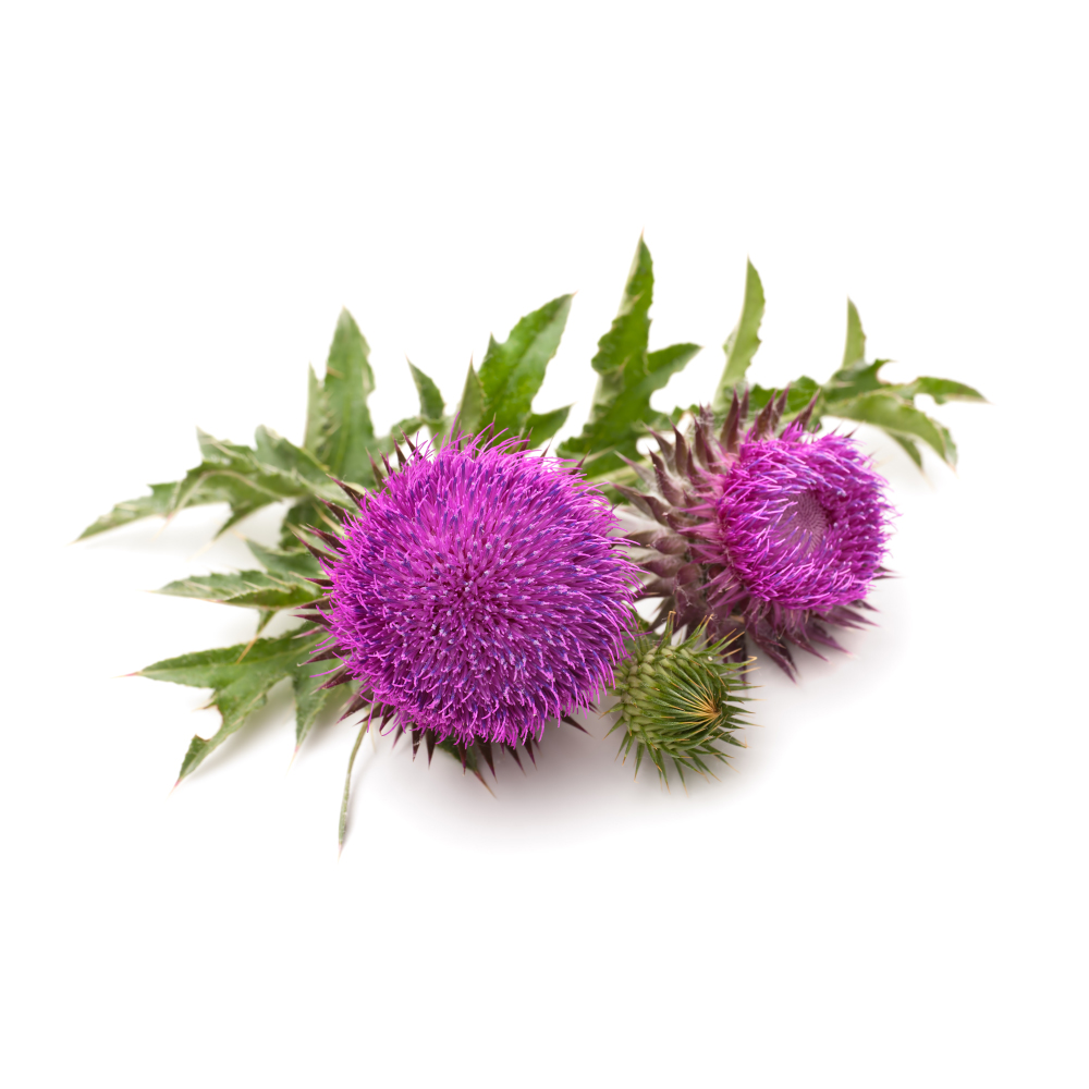 PotentPotentMilk ThistleSilybum marianumThe reference liver herb: silymarin lowers liver enzymes in fatty liver and prevents drug-induced liver injury, and intravenous silibinin is the antidote to death-cap poisoning.Mechanismradical scavenging; glutathione ↑; hepatic stellate-cell inhibition; OATP blockadeRead the evidenceShow lessMonograph →
A 2017 meta-analysis of randomised trials found silymarin significantly reduced ALT and AST in NAFLD, with modest effect sizes across heterogeneous trials 97Reference 97Meta-analysisThe therapeutic effect of silymarin in the treatment of nonalcoholic fatty disease: a meta-analysis (PRISMA) of randomized control trials — meta-analysisView study →, and the Realsil complex (silybin with phosphatidylcholine and vitamin E) improved liver enzymes, insulin resistance and biopsy-scored fibrosis over 12 months 98Reference 98RCTSilybin combined with phosphatidylcholine and vitamin E in patients with nonalcoholic fatty liver disease: a randomized controlled trial — randomised controlled trialView study →.
Silymarin also prevents injury. It reduced the incidence of anti-tuberculosis drug-induced liver injury in a double-blind RCT 99Reference 99RCTA double-blinded randomized controlled trial of silymarin for the prevention of antituberculosis drug-induced liver injury — randomised placebo-controlled trialView study →, confirmed by a 2019 meta-analysis 100Reference 100Meta-analysisProphylactic therapy of silymarin (milk thistle) on antituberculosis drug-induced liver injury: a meta-analysis of randomized controlled trials — meta-analysisView study →.
In Amanita phalloides poisoning, intravenous silibinin competitively blocks the OATP transporters that carry amatoxins into hepatocytes and interrupts their enterohepatic recirculation, and 40 years of registry data associate early administration with lower mortality 101Reference 101Clinical trialLegalon SIL: the antidote of choice in patients with acute hepatotoxicity from amatoxin poisoning — clinical review (case series)View study →.
The antidote is intravenous silibinin, not the oral herb. In chronic viral and alcoholic hepatitis the herb largely fails: a Cochrane review found no convincing effect on mortality or histology 102Reference 102Meta-analysisMilk thistle for alcoholic and/or hepatitis B or C virus liver diseases — Cochrane systematic review and meta-analysisView study →, and the 2012 NIH multicentre trial in hepatitis C found no ALT reduction even at 420–700 mg three times daily 103Reference 103RCTEffect of silymarin (milk thistle) on liver disease in patients with chronic hepatitis C unsuccessfully treated with interferon therapy: a randomized controlled trial — multicentre randomised controlled trialView study →.
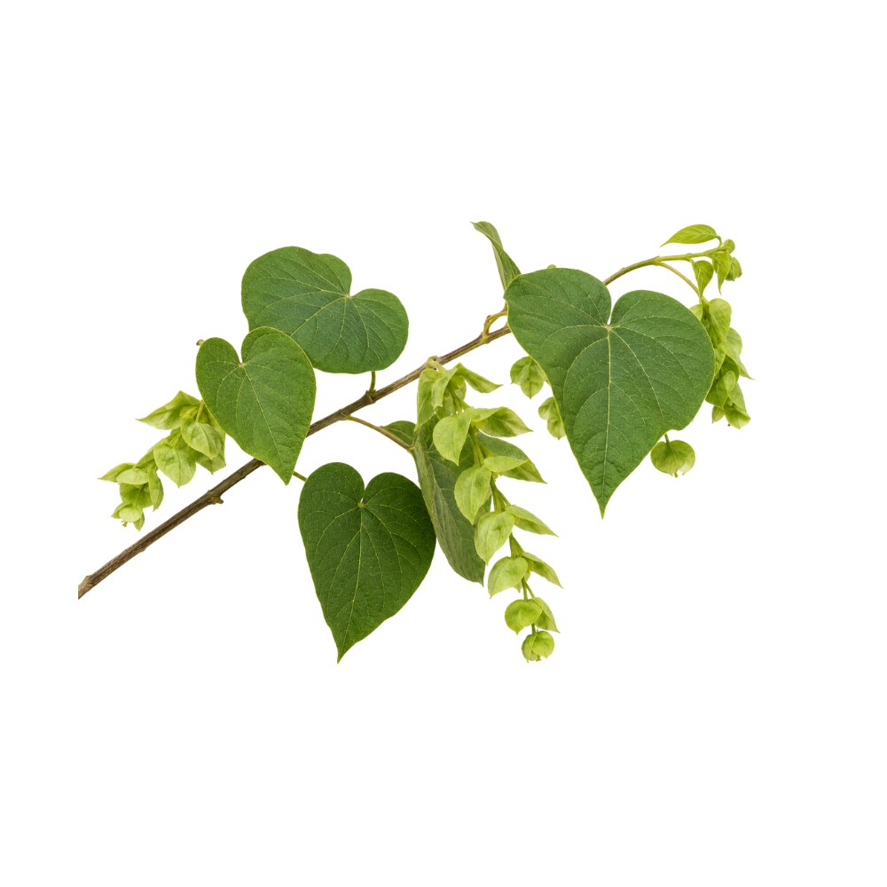 StrongStrongAbutaCissampelos pareiraRestores the liver's antioxidant enzymes and lowers raised liver enzymes after carbon-tetrachloride injury in rats, with an effect size compared to silymarin.Mechanismantioxidant-enzyme restoration (SOD/CAT/GSH); lipid-peroxidation ↓Read the evidenceShow lessMonograph →
Against carbon-tetrachloride injury, a hydroalcoholic root extract (100–400 mg/kg) lowered elevated AST, ALT, ALP and bilirubin toward normal, reduced lipid peroxidation, and raised SOD, catalase, GST, GPx and GSH, with parallel dose-dependent protection of freshly isolated rat hepatocytes and HepG2 cells in the same study 34Reference 34In vitroIn vitro and in vivo hepatoprotective activity of Cissampelos pareira against carbon-tetrachloride-induced hepatic damage — in vitro and animal model (rat)View study →.
The effect size compared favourably to silymarin, the relevant benchmark. The evidence is rodent and cell only, with no human data.
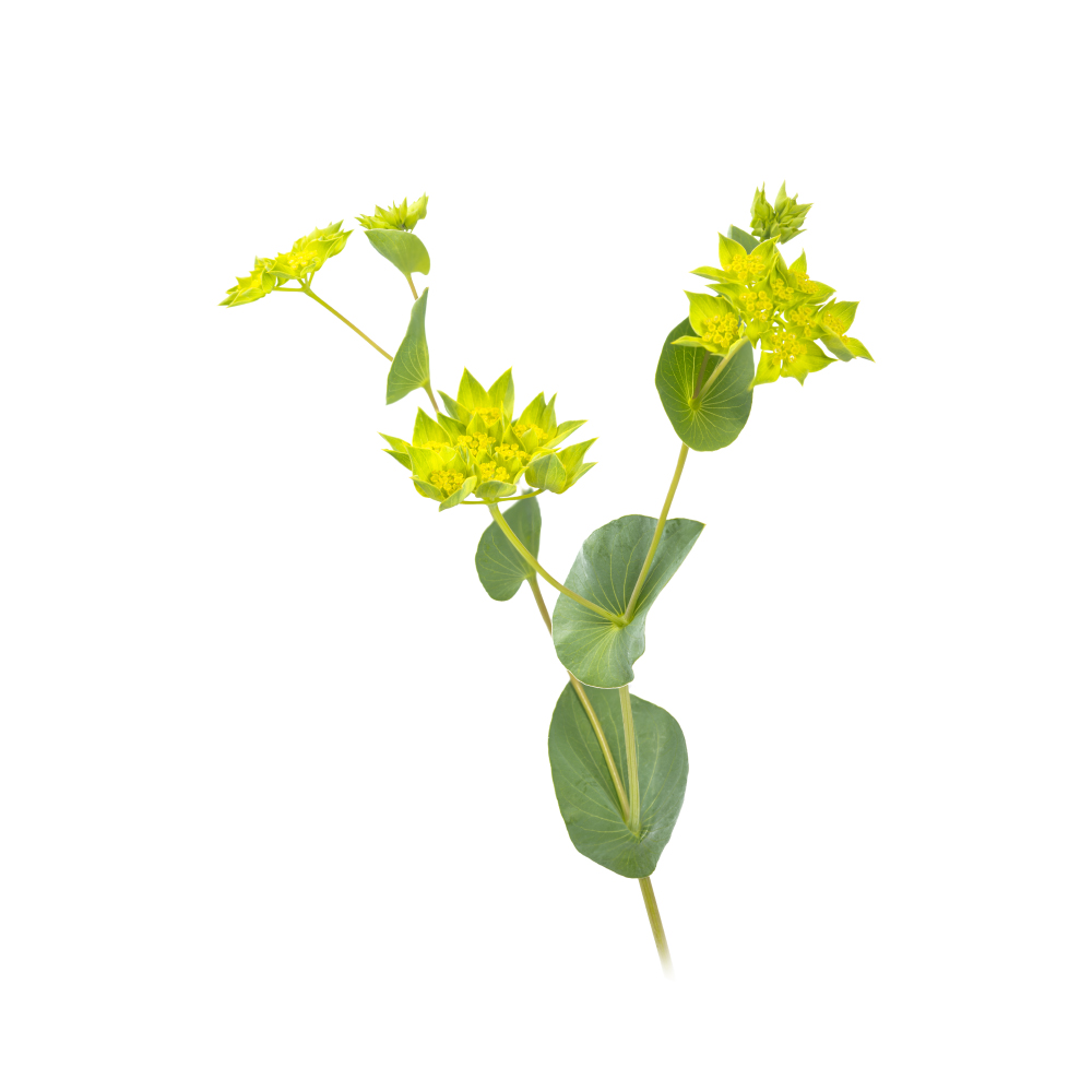 StrongStrongBupleurumBupleurum falcatumSaikosaponins drive activated hepatic stellate cells into apoptosis and damp NF-κB signalling, reducing fibrosis in rats; the same molecules injure the liver at higher doses.Mechanismhepatic stellate-cell apoptosis; NF-κB ↓; PPARα/SREBP-1cRead the evidenceShow lessMonograph →
In CCl₄-treated rats, saikosaponin-d (1.0–2.0 mg/kg/day) reduced hepatic fibrogenesis and downregulated NF-κB, TNF-α and IL-6 62Reference 62AnimalInhibitory effects of saikosaponin-d on CCl₄-induced hepatic fibrogenesis in rats — animal modelView study →; dietary saikosaponin-a improved hepatic antioxidant capacity and lowered ALT/AST over eight weeks 63Reference 63AnimalCurcumin or saikosaponin a improves hepatic antioxidant capacity and protects against CCl₄-induced liver injury in rats — animal modelView study →.
The antifibrotic mechanism runs through apoptosis of activated hepatic stellate cells, a targeted route shared with milk thistle’s silybin and dan shen’s salvianolic acids.
Saikosaponin-d is hepatotoxic at higher doses, so the action is dose-dependent in both directions. Human evidence is formula-level only, from the sho-saiko-to trials 64Reference 64RCTProspective study of chemoprevention of hepatocellular carcinoma with Sho-saiko-to (TJ-9) — randomized controlled trialView study →.
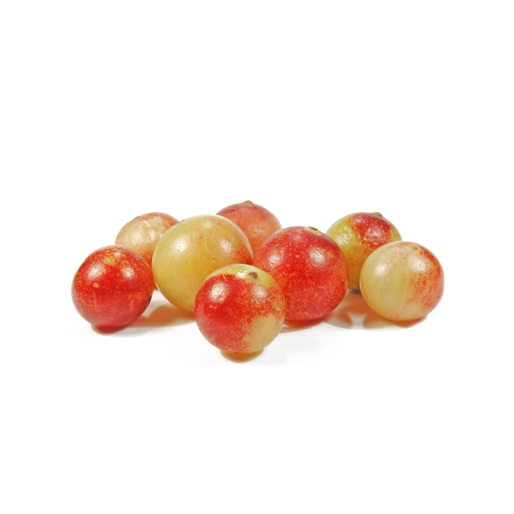 StrongStrongCamu-camuMyrciaria dubiaWhole-fruit powder cut liver fat and lowered AST and ALT over 12 weeks in a placebo-controlled crossover trial, an effect that mouse work traces to the gut microbiota.Mechanismhepatic steatosis ↓; AST/ALT ↓; gut-microbiota remodeling (gut–liver axis); hepatic NF-κB ↓Read the evidenceShow lessMonograph →
In a randomised, double-blind, placebo-controlled crossover trial, 30 overweight, hypertriglyceridaemic adults took 1.5 g of whole-fruit powder daily for 12 weeks. Liver fat fell 7.43% on camu-camu while rising 8.42% on placebo, a net 15.85% difference, with reduced plasma AST and ALT and measurable shifts in gut-microbiota composition 65Reference 65RCTCamu-camu decreases hepatic steatosis and liver injury markers in overweight, hypertriglyceridemic individuals: a randomized crossover trialView study →.
A camu-camu extract fully protected high-fat/high-sucrose-fed mice from hepatic steatosis, and the protection transferred by faecal microbiota transplant 66Reference 66AnimalTreatment with camu camu (Myrciaria dubia) prevents obesity by altering the gut microbiota and increasing energy expenditure in diet-induced obese miceView study →, which places the mechanism in the gut–liver axis rather than in direct hepatocyte antioxidancy. 1-Methylmalate isolated from the juice suppressed D-galactosamine liver injury in rats where malate, citrate and tartrate did not 67Reference 67AnimalAkachi T, et al. 1-Methylmalate from camu-camu (Myrciaria dubia) suppressed D-galactosamine-induced liver injury in rats. Bioscience, Biotechnology, and Biochemistry. 2010;74(3):573–578. https://pubmed.ncbi.nlm.nih.gov/20208347/View study →, and a chick-embryo gut–liver-axis model showed reduced hepatic NF-κB, lipid droplets and inflammatory infiltrate 68Reference 68An extract from camu-camu (Myrciaria dubia) attenuates DSS-induced gut–liver axis alterations in ovo (Gallus gallus)View study →.
The human evidence is a single 12-week trial in one metabolic phenotype, with no dose-ranging, no histology, and nothing beyond 12 weeks. The trial used whole-fruit powder, not juice and not a standardised extract.
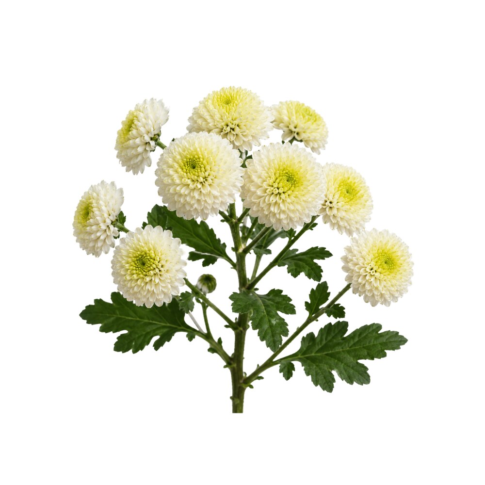 StrongStrongChrysanthemumChrysanthemum morifoliumThe water extract is a pan-PPAR agonist, the nuclear-receptor class of the glitazone and fibrate drugs, and it clears fatty liver in mice by that route.Mechanismpan-PPAR (α/γ/δ) agonism; gut-microbiota modulationRead the evidenceShow lessMonograph →
The water extract prevents and treats metabolic-associated fatty liver disease through pan-PPAR (α/γ/δ) agonism, cutting hepatic lipid accumulation and enzyme elevation in mice 70Reference 70AnimalChrysanthemum morifolium water extract prevents and treats MAFLD via pan-PPAR agonism (mouse model)View study →, and attenuates metabolic- and alcohol-associated liver disease through gut-microbiota modulation plus PPAR-α/γ activation 71Reference 71AnimalChrysanthemum morifolium attenuates metabolic- and alcohol-associated liver disease via gut microbiota and PPAR-α/γ (mouse model)View study →. Germacrane sesquiterpenoids separately protect against paracetamol injury.
Nuclear-receptor agonism is a drug-like mechanism not shared by the other liver herbs. All of it is mouse work, with no human data.
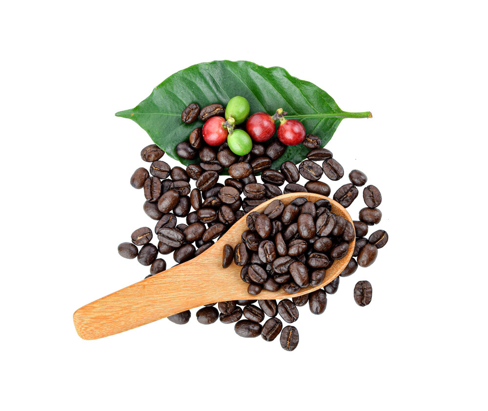 StrongStrongCoffeeCoffea arabicaEach extra two cups a day is associated with a 35% lower risk of liver cancer across large cohorts, an effect that survives decaffeination; the mechanism is not established.Mechanismnot established (chlorogenic acid + caffeine candidates)Read the evidenceShow lessMonograph →
A dose-response meta-analysis found each extra two cups a day associated with a 35% lower risk of hepatocellular carcinoma (RR 0.65), an association not significantly altered by stage of liver disease, alcohol, BMI, diabetes, smoking or hepatitis B/C, and present, though weaker, for decaffeinated coffee 72Reference 72Meta-analysisCoffee, including caffeinated and decaffeinated coffee, and the risk of hepatocellular carcinoma: a systematic review and dose-response meta-analysisView study →.
The decaffeinated signal argues that the protection is not simply caffeine, and points at chlorogenic acid.
The data are observational cohorts, not trials. Coffee drinking is confounded with a great deal else, and no mechanism has been demonstrated in humans.
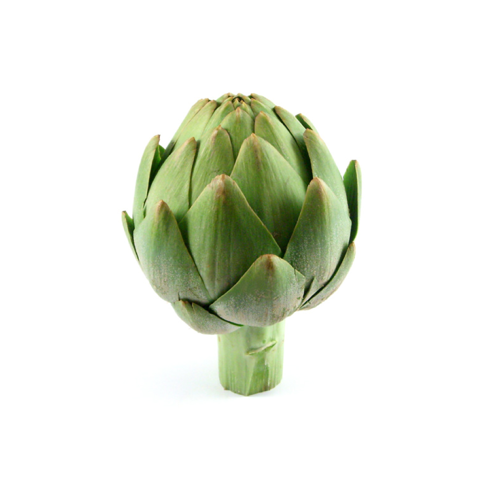 StrongStrongGlobe ArtichokeCynara scolymusLeaf extract lowers ALT and AST in fatty liver across meta-analyses of randomised trials and reduced liver size on ultrasound in one; the effect tracks its lipid action.Mechanismhepatic lipid mobilization; ROS scavengingRead the evidenceShow lessMonograph →
A meta-analysis of 5 RCTs (333 NAFLD patients) found extract significantly reduced ALT and AST alongside total cholesterol, LDL and triglycerides 37Reference 37Meta-analysisTherapeutic potential of artichoke in the treatment of fatty liver — systematic review and meta-analysis of randomised controlled trialsView study →, and a separate meta-analysis of 8 trials confirmed the enzyme fall, clearest in NAFLD and overweight subjects 38Reference 38Meta-analysisEffects of Cynara scolymus L. supplementation on liver enzymes — systematic review and meta-analysisView study →.
A pilot double-blind RCT in 100 NAFLD patients reported reduced liver size and improved hepatic blood flow on ultrasound plus lower ALT/AST after two months at 600 mg/day 39Reference 39RCTEfficacy of artichoke leaf extract in non-alcoholic fatty liver disease — pilot double-blind randomised controlled trialView study →.
The transaminase fall is plausibly downstream of the lipid action rather than a distinct hepatocyte-protective mechanism. Artichoke appears to treat the steatosis that drives the enzymes.
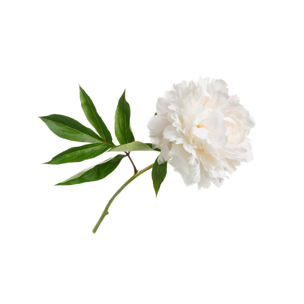 StrongStrongPeonyPaeonia lactifloraTotal glucosides of paeony reduced the liver toxicity of methotrexate and leflunomide in two multicentre randomised trials in rheumatoid arthritis.Mechanismdrug-induced hepatotoxicity ↓ (MTX/leflunomide); Nrf2 antioxidant response; gut-microbiota restoration (paeoniflorin, rodent)Read the evidenceShow lessMonograph →
Two multicentre randomised trials in active rheumatoid arthritis, in patients on methotrexate plus leflunomide, found that adding total glucosides of paeony (TGP) significantly reduced the incidence and severity of hepatotoxicity 112,
Paeoniflorin protects rodent liver against high-fat-diet steatosis through antioxidant and anti-inflammatory action and restoration of gut-microbiota balance 114,
General hepatoprotection in fatty liver or fibrosis is animal-only. The tested article is TGP, a purified extract of at least 40% paeoniflorin licensed as a drug in China, not a decoction or tincture of the raw root.
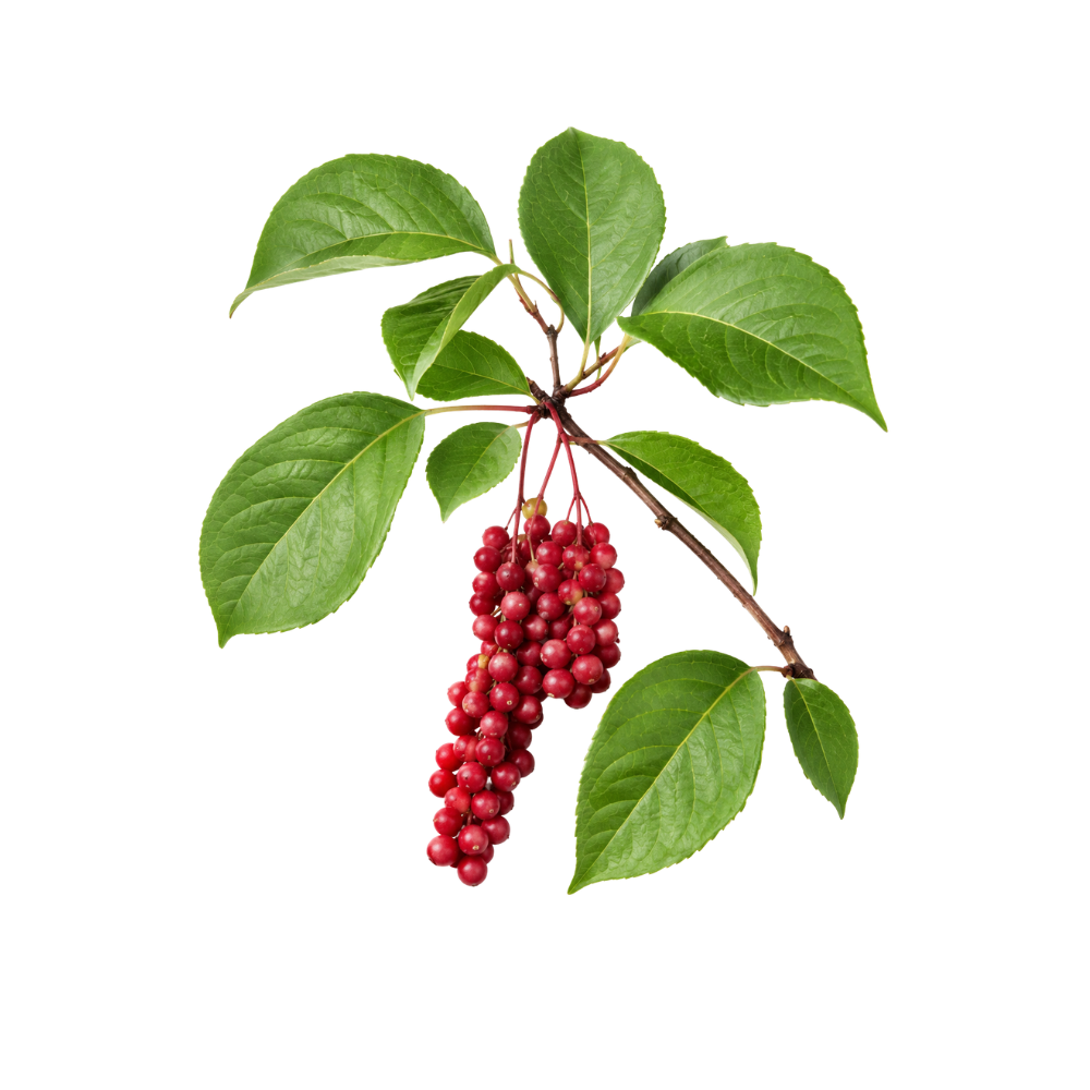 StrongStrongSchisandraSchisandra chinensisWu wei zi, the classical liver berry of Chinese medicine; its lignans switch on the liver's own glutathione defences, and a meta-analysis of 54 animal studies shows large falls in liver enzymes.MechanismNRF2/ARE activation; glutathione synthesis ↑; NF-κB ↓Read the evidenceShow lessMonograph →
A 2025 systematic review and meta-analysis pooling 54 animal studies reported large reductions in ALT (standardised mean difference about −4.7) and other injury markers, attributed to combined anti-inflammatory, antioxidant and anti-apoptotic mechanisms 124Reference 124Meta-analysisEfficacy of Schisandra chinensis in liver injury: a systematic review and preclinical meta-analysis — systematic review, meta-analysis of animal studiesView study →.
The lignan schisandrol B protects against paracetamol-induced injury by activating the NRF2/ARE pathway that governs glutathione synthesis and detoxification 125Reference 125AnimalSchisandrol B protects against acetaminophen-induced acute hepatotoxicity in mice via activation of the NRF2/ARE signaling pathway — animal (mouse) studyView study →. It upregulates the liver’s own defence rather than scavenging on its behalf.
The entire meta-analysis is animal work. There is no controlled human liver trial.
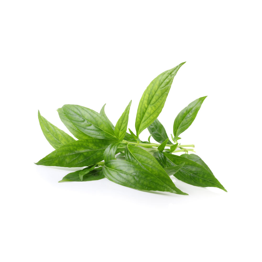 ModerateModerateAndrographisAndrographis paniculataKalmegh, the bitter liver herb of Ayurveda; andrographolide protects mouse liver against paracetamol, and no human trial exists.Mechanismantioxidant/anti-inflammatory hepatocyte protectionRead the evidenceShow lessMonograph →
Oral extract at 100–200 mg/kg gave dose-dependent protection against paracetamol-induced hepatotoxicity in mice, normalising serum transaminases and improving liver histology 35Reference 35AnimalHepatoprotective activity of Andrographis paniculata and Swertia chirayita — animal model (mouse)View study →. Reviews attribute this to andrographolide’s antioxidant and anti-inflammatory action on hepatocytes 36Reference 36ReviewOverview of pharmacological activities of Andrographis paniculata and its major compound andrographolide — reviewView study →.
The finding is coherent and replicated in rodents, and untested in people.
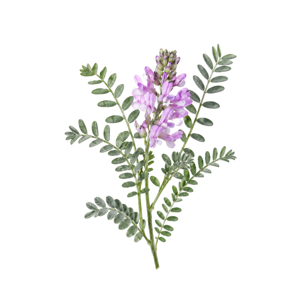 ModerateModerateAstragalusAstragalus membranaceusThree separate constituents protect rodent liver against alcohol, carbon tetrachloride and chronic chemical injury; the one human trial is a two-herb decoction.Mechanismhepatic oxidative stress ↓; inflammatory cytokines ↓; anti-fibrotic (collagen/ECM ↓)Read the evidenceShow lessMonograph →
Astragaloside IV attenuates alcoholic liver injury in rats, lowering ALT/AST, inflammatory cytokines and oxidative markers 40Reference 40AnimalMetabolomics combined with network pharmacology reveals the protective effect of astragaloside IV on alcoholic liver disease (rat model)View study →; cycloastragenol reduces carbon-tetrachloride-induced fibrosis in mice 41Reference 41AnimalHepatoprotective efficacy of cycloastragenol alleviated the progression of liver fibrosis in carbon-tetrachloride-treated miceView study →; and astragalus polysaccharide is synergistically protective with schisandra lignans against chronic chemical liver injury in rats 42Reference 42AnimalSynergistic hepatoprotective effect of Schisandra lignans with Astragalus polysaccharides on chronic liver injury in ratsView study →.
The one human study is a 48-week RCT (n=92) in chronic hepatitis B liver fibrosis using an astragalus–Polygonum anti-fibrosis decoction, which improved serum fibrosis markers (HA, LN, PCIII, CIV) and liver function more than the comparator 43Reference 43RCTComparison of efficacy in treating liver fibrosis of chronic hepatitis B between Astragalus-Polygonum anti-fibrosis decoction and Jinshuibao capsule — a randomized controlled trialView study →.
That trial tested a two-herb formula against an active comparator rather than placebo. Standalone-root efficacy in liver disease is untested.
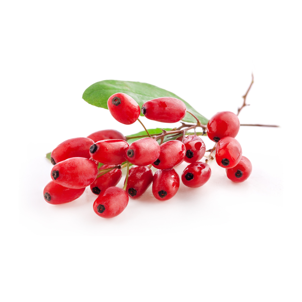 ModerateModerateBarberryBerberis spp.Berberine-bearing extract lowered ALT and AST in fatty liver in randomised trials, most likely through its effect on insulin and lipids.Mechanisminsulin sensitization; hepatic lipid handling (berberine)Read the evidenceShow lessMonograph →
Two RCTs of Berberis vulgaris extract in non-alcoholic fatty liver disease reported significant falls in transaminases, with an 80-patient trial at 750 mg/day for three months lowering ALT from 49 to 27 U/L and AST from 48 to 30 U/L against placebo 50Reference 50Clinical trialThe effect of Berberis vulgaris extract on transaminase activities in non-alcoholic fatty liver disease — clinical trialView study →.
NAFLD tracks with dyslipidaemia and insulin resistance, so the transaminase benefit is plausibly downstream of barberry’s metabolic effects rather than a direct hepatocyte-protective action, the same pattern as artichoke.
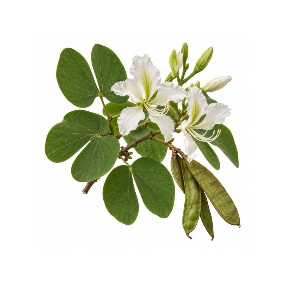 ModerateModerateBauhinia ForficataBauhinia forficataProtects the liver of diabetic mice through antioxidant defence, an extension of its blood-sugar action.Mechanismantioxidant defence in diabetic-mouse liverRead the evidenceShow less
Hepatoprotection has been demonstrated in diabetic mouse liver through antioxidant defence 51,
The effect is coherent with the antidiabetic action rather than independent of it. It remains preclinical.
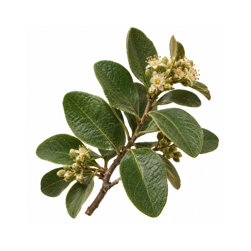 ModerateModerateBoldoPeumus boldusBoldine protects the liver against oxidative injury across animal and cell models using the infusion, the extract and the isolated alkaloid; the infusion has also injured the liver in case reports.Mechanismboldine → antioxidant defence, lipid-peroxidation suppressionRead the evidenceShow less
Protection is reproducible across several independent animal and cell models using the infusion, the hydroethanolic extract and isolated boldine 53,
At least three case reports link boldo infusion to reversible jaundice and raised liver enzymes 59,
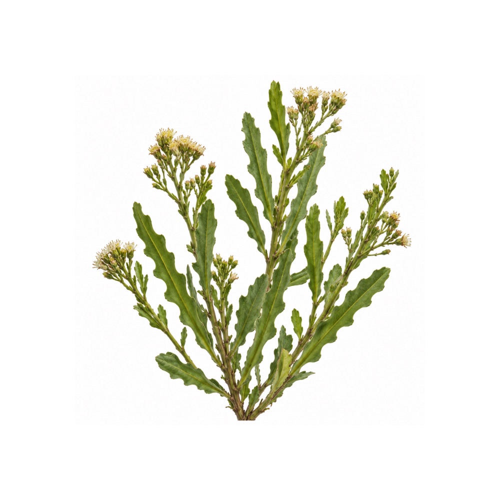 ModerateModerateCarquejaBaccharis trimeraProtects rat liver across three different injury models, carbon tetrachloride, ethanol and paracetamol, from independent research groups, through antioxidant defence.Mechanism↑SOD/catalase/glutathione; Nrf2 activation; lipid-peroxidation suppressionRead the evidenceShow less
Independent groups converge across three chemically distinct liver-injury models, carbon tetrachloride, ethanol and paracetamol 44,
Most preclinical liver herbs rest on a single model from a single group; three models from independent groups is a higher bar. There are no human data, and the efficacy work uses hydroethanolic extract rather than the tea.
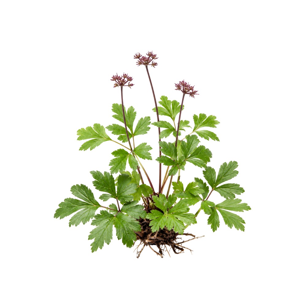 ModerateModerateCoptisCoptis chinensisBerberine, its main alkaloid, lowers liver enzymes in fatty liver across ten randomised trials; the rhizome itself has only been tested in mice.Mechanismhepatic steatosis ↓ (AMPK/SREBP-1c, PPARα/LXRα); ALT/AST/GGT ↓; IL-17RA/NF-κB ↓Read the evidenceShow lessMonograph →
A meta-analysis of 10 berberine RCTs (811 patients) found significant reductions in ALT, AST and GGT alongside improved triglycerides, LDL-C, insulin resistance and BMI, with only mild gastrointestinal side effects 73Reference 73Meta-analysisThe clinical efficacy and safety of berberine in the treatment of non-alcoholic fatty liver disease — meta-analysis and systematic reviewView study →.
Whole-herb work supplies the mechanism. Coptidis rhizoma ameliorated diabetes-associated steatohepatitis in mice by downregulating IL-17RA/NF-κB signalling 74Reference 74AnimalCoptidis rhizoma ameliorates type 2 diabetes-related metabolic-dysfunction-associated steatohepatitis by downregulating IL-17RA/NF-κB — mouse modelView study →, and Coptis total alkaloids reduced hepatic steatosis via AMPK/SREBP-1c and PPARα/LXRα 75Reference 75AnimalTotal alkaloids from Coptis chinensis Franch ameliorate hyperlipidaemia and hepatic steatosis via AMPK/SREBP-1c and PPARα/LXRα in mice — animal modelView study →.
No human trial has tested Coptis extract itself in NAFLD. The human support is berberine’s.
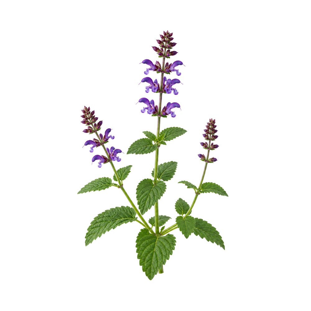 ModerateModerateDan ShenSalvia miltiorrhizaA classical Chinese liver herb; salvianolic acid A suppresses the stellate cells that lay down scar, and salvianolic acid B reversed fibrosis in a small hepatitis B trial.Mechanismhepatic stellate-cell suppression; PI3K/AKT/mTOR ↓Read the evidenceShow lessMonograph →
Salvianolic acid A attenuated CCl₄-induced liver fibrosis in rats by suppressing hepatic stellate-cell activation and downregulating PI3K/AKT/mTOR signalling 20Reference 20Salvianolic acid A attenuates CCl4-induced liver fibrosis by regulating the PI3K/AKT/mTOR, Bcl-2/Bax and caspase-3/cleaved caspase-3 signaling pathwaysView study →, consistent with the phenolic-acid fraction’s antioxidant and anti-inflammatory activity.
Stellate-cell suppression is the same antifibrotic route as milk thistle’s, reached by a different molecule. Oral salvianolic acid B reversed fibrotic stage in 36.7% of hepatitis B patients in a six-month trial against interferon-γ 25Reference 25RCTClinical observation of salvianolic acid B in treatment of liver fibrosis in chronic hepatitis B — randomised double-blind trial with paired liver biopsiesView study →.
Controlled human trials of dan shen monotherapy for liver disease are absent.
 ModerateModerateForsythiaForsythia suspensaForsythoside A protects mouse liver in two independent injury models, reaching Nrf2 by two routes.MechanismNrf2/HO-1 ↑; NF-κB/TNF-α ↓; AMPK/Nrf2Read the evidenceShow less
Forsythoside A reduced liver injury in LPS/D-galactosamine challenge, with lower ALT/AST/MDA through Nrf2/HO-1 up and NF-κB/TNF-α down 76Reference 76AnimalForsythiaside A attenuates LPS/D-galactosamine-induced liver injury in mice via NF-κB and Nrf2/HO-1. https://pubmed.ncbi.nlm.nih.gov/25797347/View study →, and in carbon tetrachloride injury on oral dosing, confirmed by biochemistry and histology through AMPK/Nrf2 77Reference 77AnimalForsythiaside A protects against CCl₄-induced liver injury via AMPK/Nrf2 (mouse model)View study →.
Two models and two mechanisms converge on Nrf2. The evidence is one isolated constituent in mice, with no whole-extract or human confirmation for this action.
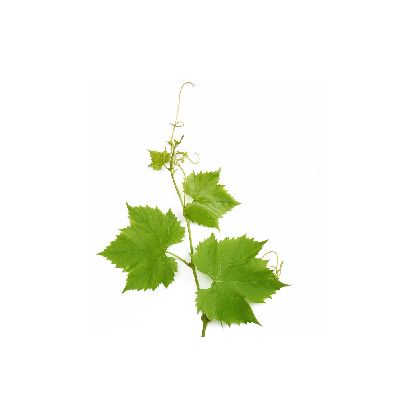 ModerateModerateGrape Seed ExtractVitis viniferaStandardised proanthocyanidin extract improved oxidative-stress markers and steatosis in two short placebo-controlled trials in fatty liver, after reversing steatosis in rats as well as metformin did.Mechanismhepatic steatosis ↓; oxidative-stress markers ↓ (MDA ↓, SOD/GPx ↑); NF-κB ↓ → IL-6 ↓Read the evidenceShow lessMonograph →
Proanthocyanidins reduced dimethylnitrosamine-induced liver damage 80Reference 80AnimalThe proanthocyanidins inhibit dimethylnitrosamine-induced liver damage in ratsView study →, attenuated hepatic injury in streptozotocin-diabetic rats 81Reference 81AnimalAntioxidant effects of proanthocyanidin from grape seed on hepatic tissue injury in diabetic ratsView study →, and reversed high-fat/fructose-diet steatosis comparably to metformin 82Reference 82AnimalGrape seed proanthocyanidin rescues rats from steatosis: a comparative and combination study with metforminView study →.
Two 2-month randomised placebo-controlled trials in NAFLD patients reported improved oxidative-stress markers and quality of life 83Reference 83Grape seed extract supplementation in non-alcoholic fatty liver diseaseView study → and improvements in metabolic factors and steatosis severity 84Reference 84Clinical trialThe effects of grape seed extract supplementation on cardiovascular risk factors, liver enzymes and hepatic steatosis in patients with non-alcoholic fatty liver diseaseView study →. The route is NF-κB suppression by the flavanol fraction, driving IL-6 and malondialdehyde down in the same cohort 83,
Both trials tested grape seed extract alone.
The trials are small and short (8 weeks), with surrogate endpoints and no histology. They use standardised OPC extracts at 100–600 mg/day (Leucoselect, Enovita, MegaNatural, IH636), and none of it transfers to grape juice, wine or whole seed 86Reference 86Masquelier’s grape seed extract: from basic flavonoid research to a well-characterized food supplement with health benefitsView study →.
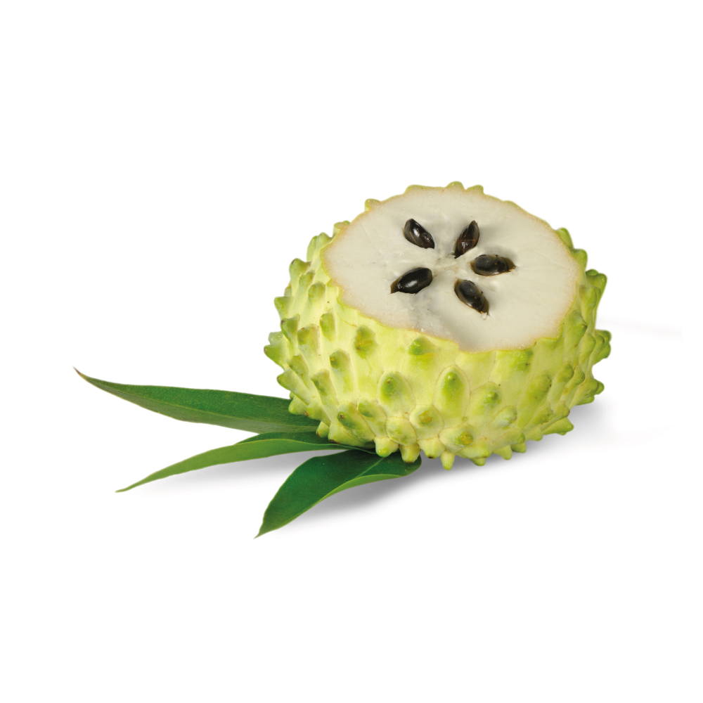 ModerateModerateGraviolaAnnona muricataLeaf, stem-bark and flower extracts lower raised liver enzymes across several rodent toxin models.Mechanismradical scavenging; antioxidant-enzyme shiftRead the evidenceShow lessMonograph →
Results are directionally consistent across streptozotocin-diabetic liver damage 87Reference 87AnimalAmeliorative effect of Annona muricata (Graviola) extract on hyperglycaemia-induced hepatic damage in STZ diabetic rats — animal modelView study →, stem-bark extract against chemically induced injury 88Reference 88AnimalEffect of ethanol extract of soursop (Annona muricata L.) stem bark on rat liver function — animal modelView study →, and paracetamol and MSG challenges, with lowered ALT/AST and improved histology.
The studies use different plant parts (leaf, stem bark, flower) and different injury models, so they agree in direction while measuring different things. No human data exist.
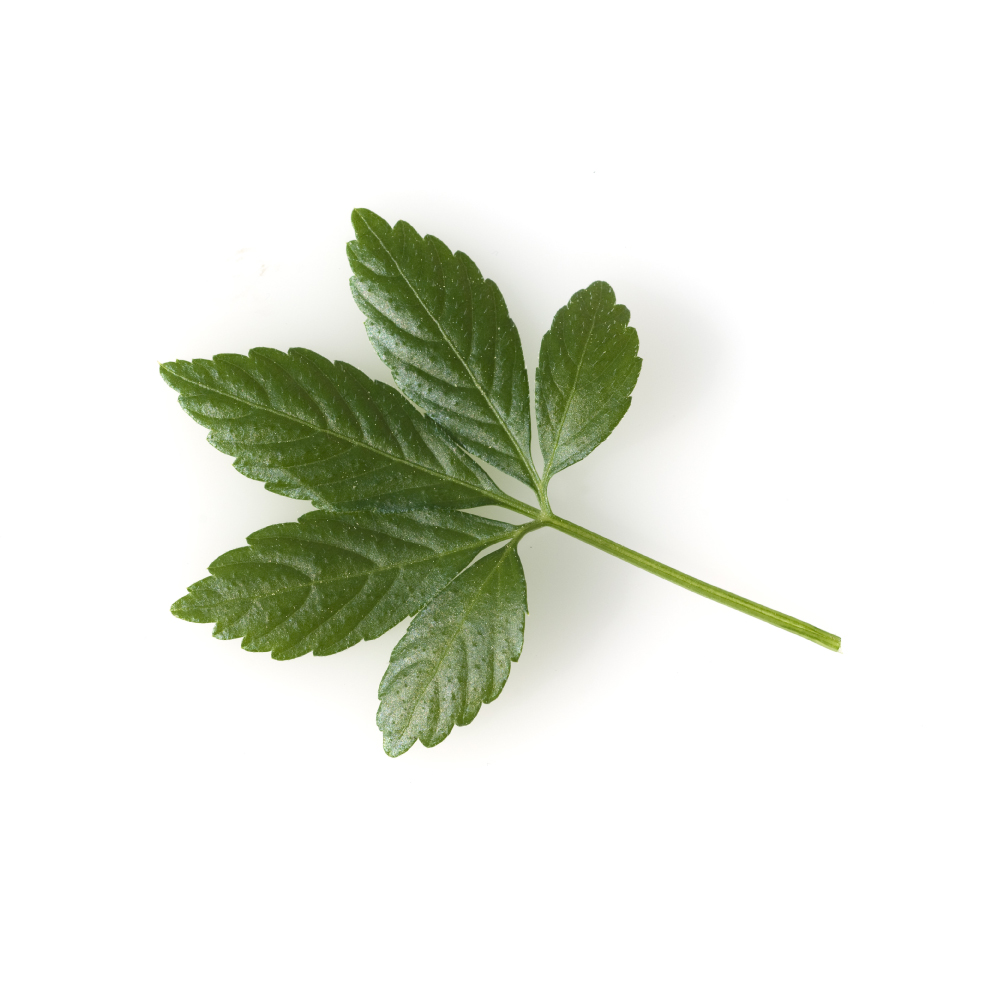 ModerateModerateGynostemmaGynostemma pentaphyllumAdded to a diet programme for four months, the extract improved AST, insulin and HOMA-IR in fatty liver beyond diet alone; the Cochrane review noted the benefit and the bias.Mechanismhepatic steatosis ↓ (AST, insulin, HOMA-IR); hepatic NF-κB and TLR4/MyD88 ↓; Akkermansia enrichmentRead the evidenceShow lessMonograph →
Adding 80 mL/day of extract to a diet programme for four months significantly improved BMI, AST, insulin and HOMA-IR in NAFLD patients beyond diet alone 89Reference 89RCTThe add-on effects of Gynostemma pentaphyllum on nonalcoholic fatty liver disease — randomised controlled trialView study →. The 2013 Cochrane review of herbal medicines for fatty liver singled out Gynostemma pentaphyllum among the single-herb products showing benefit on liver enzymes and imaging, while cautioning that the trials were at high risk of bias and used surrogate endpoints only 90Reference 90Systematic reviewHerbal medicines for fatty liver diseases — systematic reviewView study →.
In methionine/choline-deficient NASH mice the extract cut hepatic inflammation and lipid peroxidation, enriched Akkermansia and repressed hepatic NF-κB and TLR4/MyD88 signalling 91Reference 91AnimalGynostemma pentaphyllum extract alleviates NASH in mice: inflammation and gut microbiota — mouseView study →; older rat models showed protection against paracetamol- and CCl₄-induced injury 92Reference 92AnimalAntioxidant and hepatoprotective effects of Anoectochilus formosanus and Gynostemma pentaphyllum — ratView study →.
Every human result is enzymes, insulin markers and ultrasound, with no biopsy confirmation and no long-term outcome. The protective rat study was biphasic and dose-sensitive.
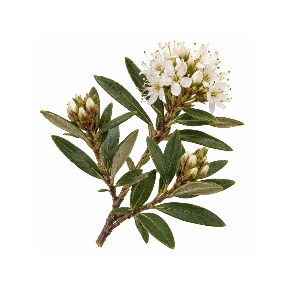 ModerateModerateLabrador TeaRhododendron groenlandicumReduces liver fat in diet-induced obese mice, an extension of its metabolic action.Mechanismreduced hepatic fat accumulation; anti-steatoticRead the evidenceShow less
The extract reduced hepatic fat in diet-induced obese mouse models 119,
The effect is an extension of the metabolic action rather than an independent hepatic one. It is mouse-only.
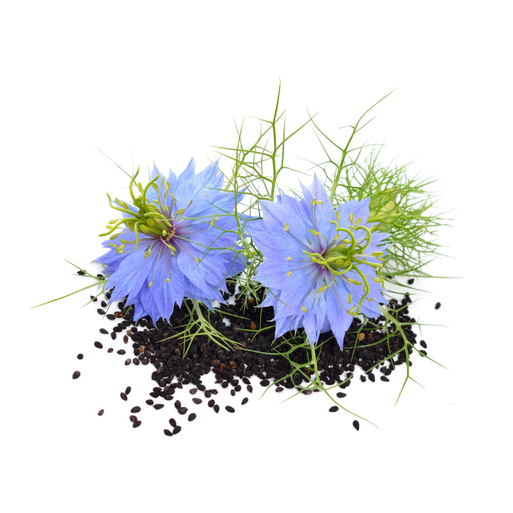 ModerateModerateNigellaNigella sativaBlack seed lowered liver enzymes and ultrasound steatosis grade across five randomised trials in fatty liver, without changing lipids or insulin in the same patients.MechanismALT/AST ↓; ultrasound steatosis grade ↓; Nrf2 activation and NF-κB suppression (rodent)Read the evidenceShow lessMonograph →
A meta-analysis of five NAFLD trials found N. sativa improved alanine and aspartate aminotransferases and ultrasound steatosis grade, but did not significantly change cholesterol, triglycerides, insulin or TNF-α in the same patients 104Reference 104Meta-analysisEffect of Nigella sativa in the treatment of nonalcoholic fatty liver disease — systematic review and meta-analysis of randomized controlled trialsView study →. The liver effect is not simply the lipid effect showing up downstream.
A standardised seed-oil RCT (2.5 mL twice daily for three months) reduced hepatic steatosis grade and aminotransferases 105Reference 105RCTStandardized Nigella sativa seed oil ameliorates hepatic steatosis, aminotransferase and lipid levels in NAFLD — a randomized, double-blind, placebo-controlled clinical trialView study →, with further RCTs reporting cardiometabolic and inflammatory improvements in fatty-liver patients 106Reference 106RCTNigella sativa and inflammatory biomarkers in patients with nonalcoholic fatty liver disease — a randomized, double-blind, placebo-controlled clinical trialView study →. Extensive rodent work supports protection against chemical and drug-induced injury through Nrf2 activation and NF-κB suppression 107Reference 107AnimalProtective effect of Nigella sativa against 5-fluorouracil-induced liver damage in rats — animal studyView study →.
Human data are enzyme and imaging surrogates, never biopsy-confirmed fibrosis or long-term progression. A dose-response meta-analysis of liver and kidney parameters found aminotransferases and alkaline phosphatase fell, with a small creatinine rise and higher blood urea nitrogen above 2000 mg/day 108Reference 108Meta-analysisThe effect of Nigella sativa on measures of liver and kidney parameters — systematic review and dose-response meta-analysis of randomized controlled trialsView study →.
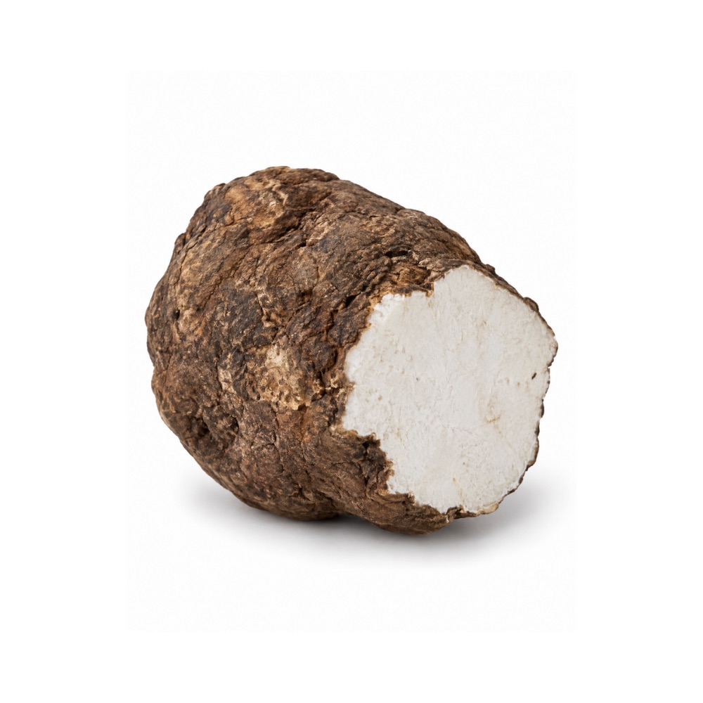 ModerateModeratePoriaWolfiporia extensaFu ling's polysaccharides protect mouse liver across paracetamol, alcohol and fatty-liver models.Mechanismpolysaccharide protection; NF-κB ↓Read the evidenceShow less
Polysaccharide fractions protected against paracetamol, alcohol and NASH injury in mice 143,
The evidence is animal-only.
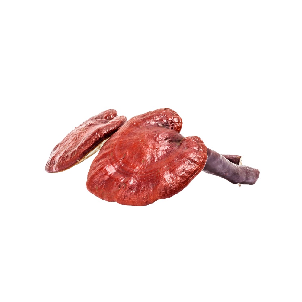 ModerateModerateReishiGanoderma lucidumEthanolic extract blunted liver-enzyme rises in rats as well as silymarin did in the same model; the only human data are a safety study.Mechanismantioxidant enzyme restoration; NF-κB/MAPK ↓Read the evidenceShow lessMonograph →
In rats, ethanolic extract dose-dependently blunted CCl₄-induced ALT/AST rises and restored antioxidant enzymes, performing comparably to silymarin in the same model 117Reference 117AnimalAmelioration of CCl₄-induced oxidative stress and hepatotoxicity by Ganoderma lucidum in Long Evans rats — animal modelView study →. Isolated ganoderic acid likewise protected mice against CCl₄- and BCG/LPS-induced injury 118Reference 118In vitroAnti-hepatitis B activities of ganoderic acid from Ganoderma lucidum — in vitro and mouse studyView study →.
The only human signal is a six-month healthy-volunteer study, which speaks to safety rather than treatment.
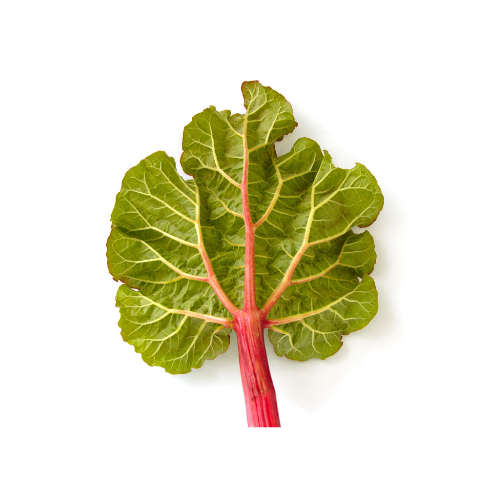 ModerateModerateRhubarbRheum palmatumRhubarb anthraquinones reduce liver inflammation in fatty-liver mice by inhibiting the NLRP3 inflammasome; the same extract injures the liver at higher doses.MechanismNLRP3 inflammasome ↓; NF-κB ↓ — dose-dependently two-edgedRead the evidenceShow lessMonograph →
In mice with non-alcoholic fatty liver disease, rhubarb free anthraquinones reduced hepatic inflammation by inhibiting the NLRP3 inflammasome 122Reference 122AnimalRhubarb free anthraquinones improved mice non-alcoholic fatty liver disease by inhibiting NLRP3 inflammasome — mouse, in vivoView study →.
A metabolomic study in rats found the same R. palmatum extract hepatoprotective at lower doses and hepatotoxic at higher ones; the paper is titled “Hepatotoxicity or hepatoprotection?” 123Reference 123AnimalHepatotoxicity or hepatoprotection? Pattern recognition for the paradoxical effect of the Chinese herb Rheum palmatum L. in treating rat liver injury — rat, in vivoView study →. The dose-dependence is the finding.
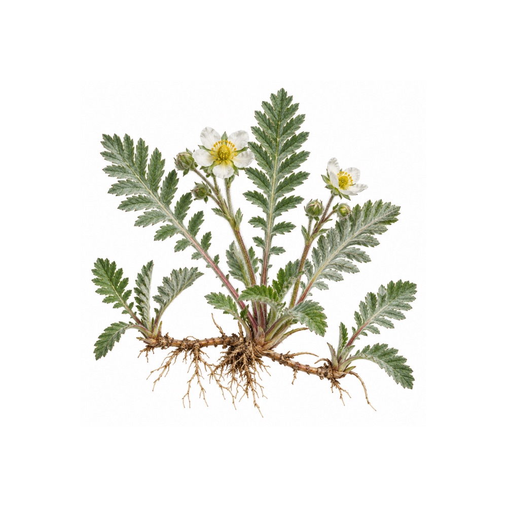 ModerateModerateSilverweedArgentina anserinaUrsane triterpenes protect the liver against oxidative injury in rodents, replicated across independent groups.Mechanismtriterpene-mediated protection against oxidative injuryRead the evidenceShow less
The protection recurs across independent groups and is carried by the ursane triterpenes rosamultin and kaji-ichigoside F1.
It is part of a wider organ-protection profile rather than an isolated finding, and it is preclinical.
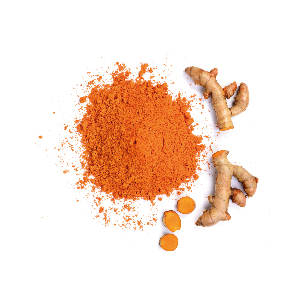 ModerateModerateTurmericCurcuma longaCurcumin reduced liver fat over 24 weeks in a randomised trial and lowers AST in fatty liver across trials, through Nrf2 and bile-acid signalling.MechanismNrf2/ARE cytoprotection; bile-acid/TGR5/GLP-1 modulation; hepatic-fat ↓Read the evidenceShow lessMonograph →
A 24-week RCT reduced hepatic fat alongside gut-microbiota-dependent bile-acid metabolism and TGR5/GLP-1 signalling, and a 4-RCT meta-analysis lowered AST, triglycerides, glucose and HOMA-IR in NAFLD 140,
Curcumin reaches the liver through the Nrf2/ARE antioxidant axis and a separate metabolic route. Trials are small and short, and histological benefit is untested.
Concentrated, high-bioavailability turmeric extracts have been linked to idiosyncratic liver injury.
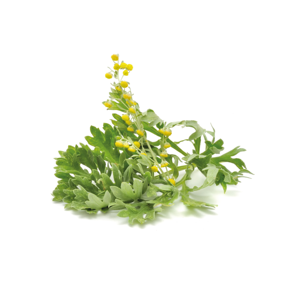 ModerateModerateWormwoodArtemisia absinthiumExtracts protect rodent liver against chemical injury and, in a 2026 mouse study, reverse fibrosis by returning hepatic stellate cells to quiescence.MechanismTGF-β/SMAD ↓; hepatic stellate-cell quiescenceRead the evidenceShow lessMonograph →
Aqueous extract protected mouse liver against chemically and immunologically induced injury 145Reference 145AnimalIn vivo hepatoprotective activity of the aqueous extract of Artemisia absinthium L. against chemically and immunologically induced liver injuries in mice — animal modelView study → and reversed CCl₄-induced damage in rats, normalising raised transaminases and lipid peroxidation 146Reference 146AnimalReversal of carbon tetrachloride-induced hepatic injury by aqueous extract of Artemisia absinthium in Sprague-Dawley rats — animal modelView study →.
A 2026 mouse study found methanol extract (400 mg/kg) attenuated CCl₄-induced fibrosis by suppressing the TGF-β/SMAD axis and returning hepatic stellate cells to quiescence 147Reference 147AnimalArtemisia absinthium attenuates hepatic fibrosis in mice by suppressing hepatic stellate cell activation and modulating inflammatory chemokine signalling — animal modelView study →, a third independent route to stellate-cell suppression alongside milk thistle and dan shen.
The evidence is animal-only.
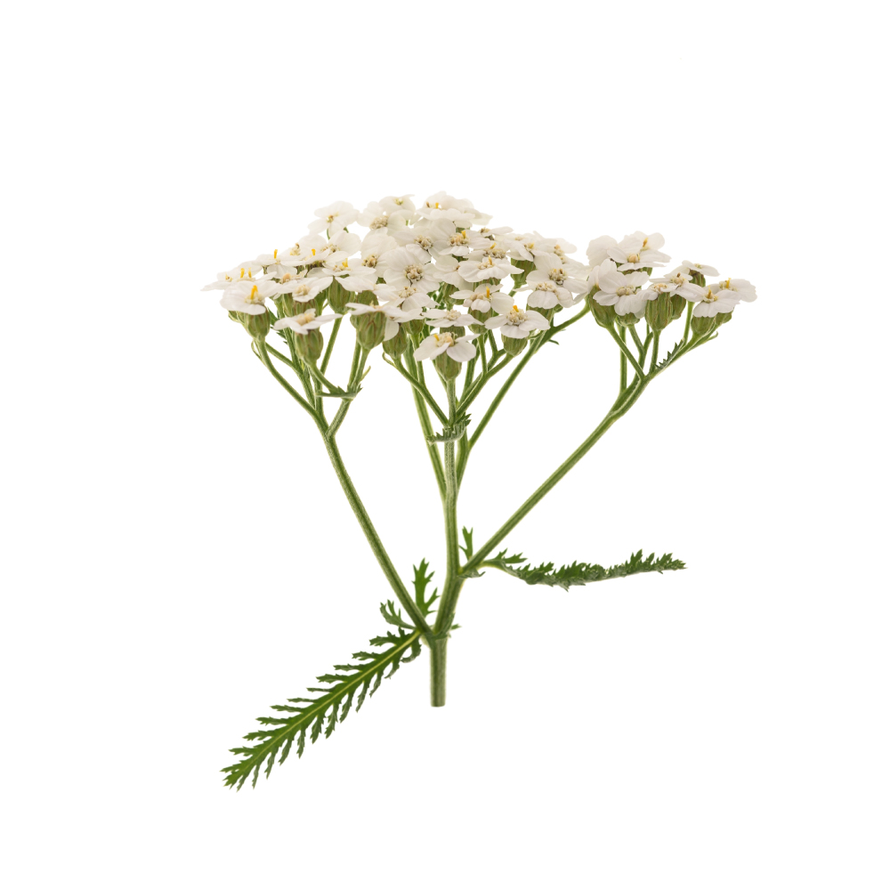 ModerateModerateYarrowAchillea millefoliumLong used for liver and gallbladder complaints; crude extract cut mortality and blunted the liver-enzyme rise in mice given a lethal liver toxin.Mechanismantioxidant-enzyme support; radical scavengingRead the evidenceShow lessMonograph →
Pre-treatment with crude extract (150–600 mg/kg) cut mortality and blunted the ALT/AST rise in mice given D-galactosamine plus LPS, with histology confirming preserved architecture 148Reference 148AnimalStudies on hepatoprotective, antispasmodic and calcium antagonist activities of the aqueous-methanol extract of Achillea millefolium — [mouse in vivo]View study →.
In streptozotocin-diabetic rats a hydroalcoholic extract lowered elevated liver enzymes alongside glucose and lipids, comparably to metformin on several markers 149Reference 149AnimalHydroalcoholic extract of Achillea millefolium improved blood glucose, liver enzymes and lipid profile compared to metformin in streptozotocin-induced diabetic rats — [rat in vivo]View study →. The evidence is animal-only.
Show 13 more herbs with a mild hepatoprotective action
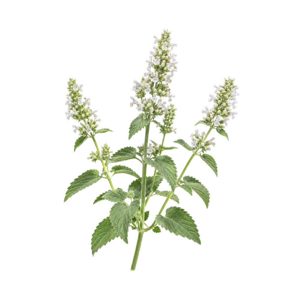 MildMildCatnipNepeta catariaThe essential oil protected rodent liver against paracetamol by inhibiting CYP2E1, the enzyme that makes the drug toxic, and inducing the enzymes that clear it.MechanismCYP2E1 inhibition; Phase II (UGT/SULT) induction; Nrf2Read the evidenceShow lessMonograph →
Supercritical-CO₂ essential oil of Nepeta cataria attenuated paracetamol-induced liver damage in rodents by up-regulating Phase II conjugating enzymes (UGTs and SULTs), inhibiting CYP2E1 and apparently activating Nrf-2, together shifting the drug toward safe conjugation 69Reference 69AnimalHepatoprotective effect of essential oils of Nepeta cataria L. on acetaminophen-induced liver dysfunction — [animal in vivo]View study →.
That is a specific detoxification-pathway mechanism rather than generic antioxidant protection, and the inverse of milk thistle’s, whose protection does not depend on CYP2E1 inhibition.
One study, one model, on a supercritical-CO₂ essential oil rather than the tea catnip is usually taken as.
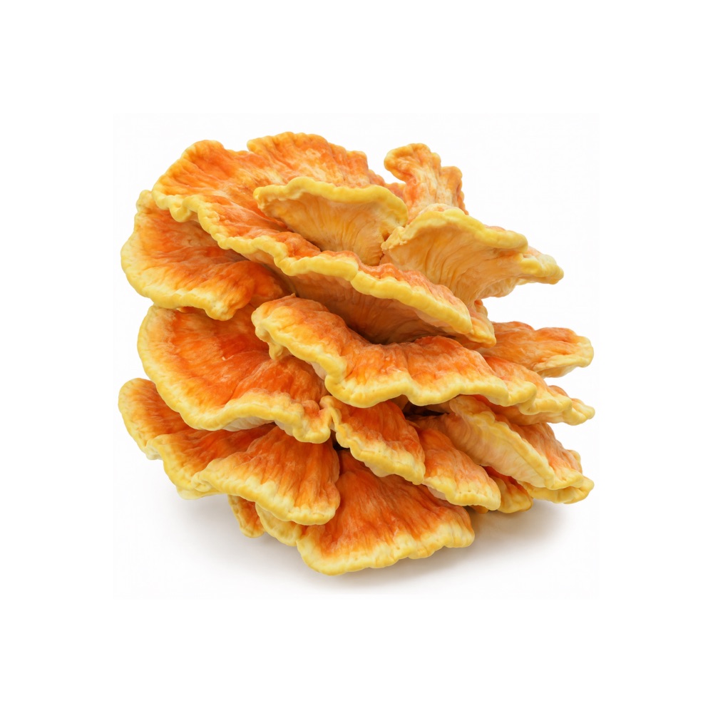 MildMildChicken Of The WoodsLaetiporus sulphureusPolysaccharides protected mouse liver against alcohol injury in a single study, coherent with the mushroom's antioxidant chemistry.Mechanismpolysaccharide antioxidant protectionRead the evidenceShow less
One alcohol-liver mouse study and one mechanistic cell study 93Reference 93AnimalAntioxidant and hepatoprotective activities of polysaccharides from spent mushroom substrates (Laetiporus sulphureus) in acute alcohol-induced mice — in vivo mouse studyView study →, each from a single research group.
The finding is coherent with the antioxidant activity and unreplicated independently.
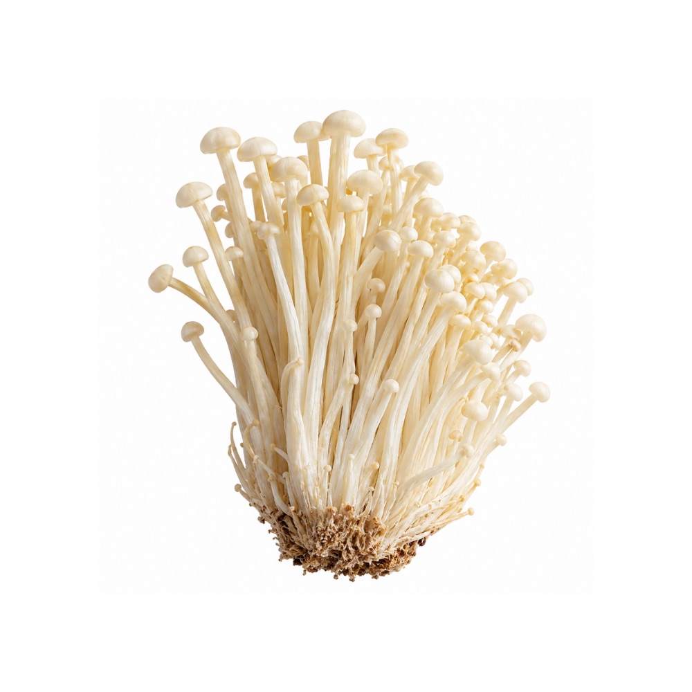 MildMildEnokiFlammulina velutipesPolysaccharide fractions protect rodent liver against chemical injury through Nrf2 activation and suppression of NLRP3-driven pyroptosis.MechanismKeap1/Nrf2 ↑, NLRP3 pyroptosis ↓Read the evidenceShow less
CCl₄ rat and mouse models and DSS-colitis work using polysaccharide fractions map the mechanism through Keap1/Nrf2 and NLRP3-driven pyroptosis.
The evidence is animal-only.
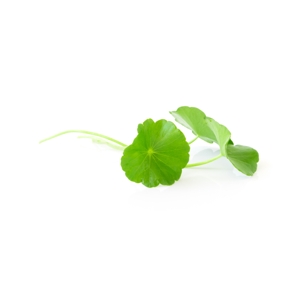 MildMildGotu KolaCentella asiaticaMadecassoside prevented acute liver failure in mice by suppressing inflammation; the herb is also among the more frequently reported causes of herbal liver injury.Mechanismanti-inflammatory (LPS/D-GalN liver-failure model)Read the evidenceShow lessMonograph →
Madecassoside prevented LPS/D-galactosamine-induced acute liver failure in mice by suppressing inflammation 78Reference 78AnimalMadecassoside prevents acute liver failure in LPS/D-GalN-induced mice — animal modelView study →.
Centella asiatica is among the more frequently reported herbal causes of drug-induced liver injury, hepatocellular and predominantly in women 79Reference 79Systematic reviewProfile of herbal and dietary supplement-induced liver injury in Latin America — systematic reviewView study →. The animal protection and the human case series point in opposite directions.
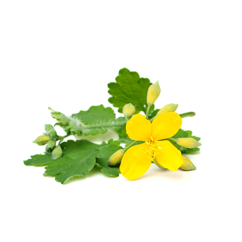 MildMildGreater CelandineChelidonium majusWhole-plant extract and its alkaloid chelidonine protect rodent liver against chemical toxins; in people the herb is a documented cause of idiosyncratic liver injury.Mechanismantioxidant-enzyme restoration (GSH/SOD/CAT); lipid-peroxidation ↓ (rodent)Read the evidenceShow lessMonograph →
Ethanolic whole-plant extract reduced p-dimethylaminoazobenzene-induced hepatocarcinogenesis in mice across cytogenetic, biochemical and histological endpoints 150Reference 150AnimalEfficacy of a plant extract (Chelidonium majus L.) in combating induced hepatocarcinogenesis in mice — animal modelView study →, and chelidonine protected mouse liver against cadmium-induced oxidative injury, restoring glutathione and normalising ALT, AST and ALP 151Reference 151AnimalPLGA nano-encapsulation of chelidonine enhances its potential against cadmium-induced oxidative stress and hepatic injury in mice — animal modelView study →.
Greater celandine is a documented cause of idiosyncratic hepatocellular liver injury in people, with jaundice and transaminases often above 1000 IU/L, reported even from tea 152,
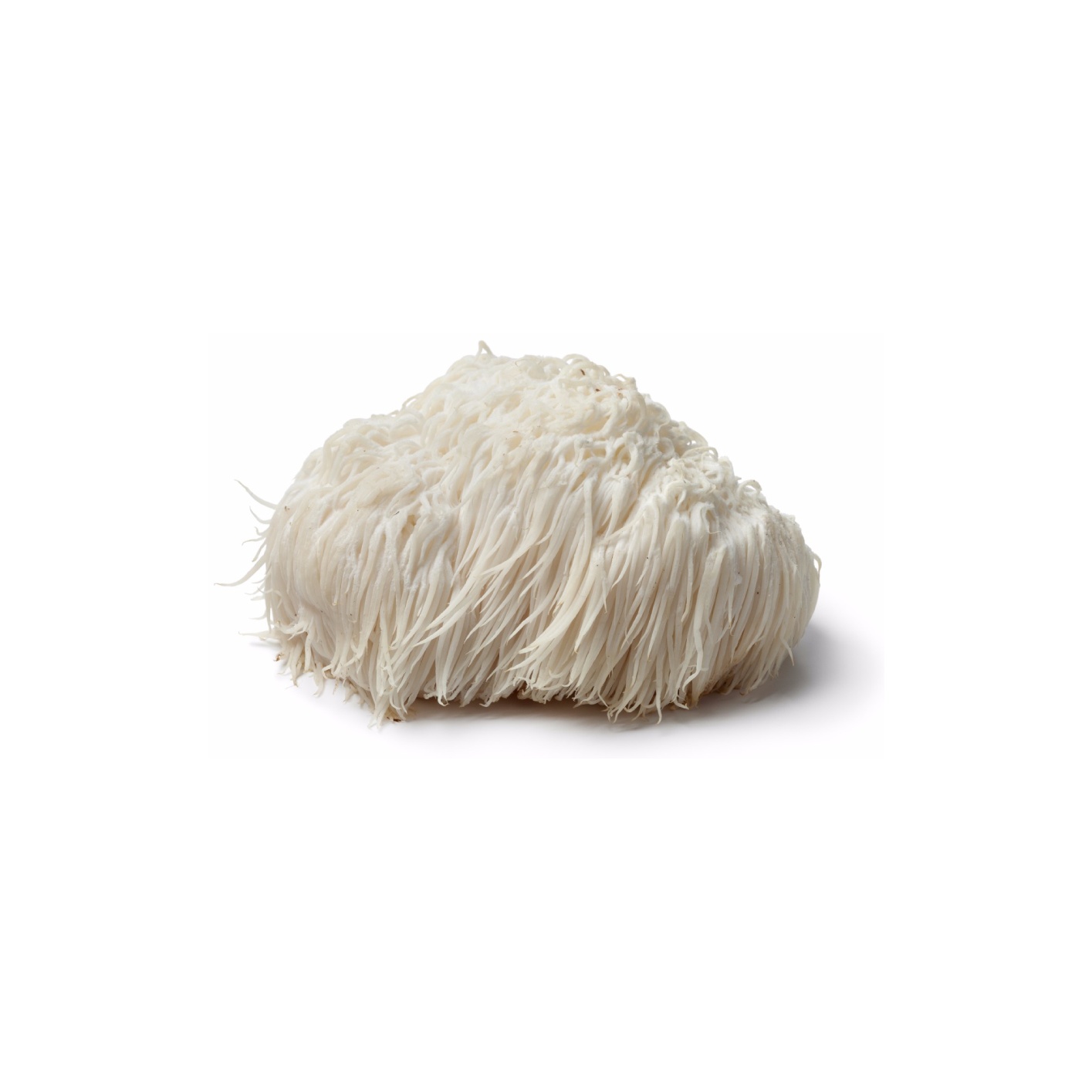 MildMildLion’s ManeHericium erinaceusMycelial polysaccharide fractions protected mouse liver, apparently through antioxidant capacity.Mechanismpolysaccharide antioxidant protectionRead the evidenceShow lessMonograph →
Mycelial endo-polysaccharide fractions showed a hepatoprotective effect in mice, linked to their antioxidant capacity 94Reference 94In vitroAntioxidant and hepatoprotective potential of endo-polysaccharides from Hericium erinaceus — in vitro and mouse studyView study →, with related work reporting protection against high-fat-diet fatty liver.
The evidence is single-lab and mouse-only, and tied to an isolated mycelial polysaccharide fraction rather than the fruiting body most products supply.
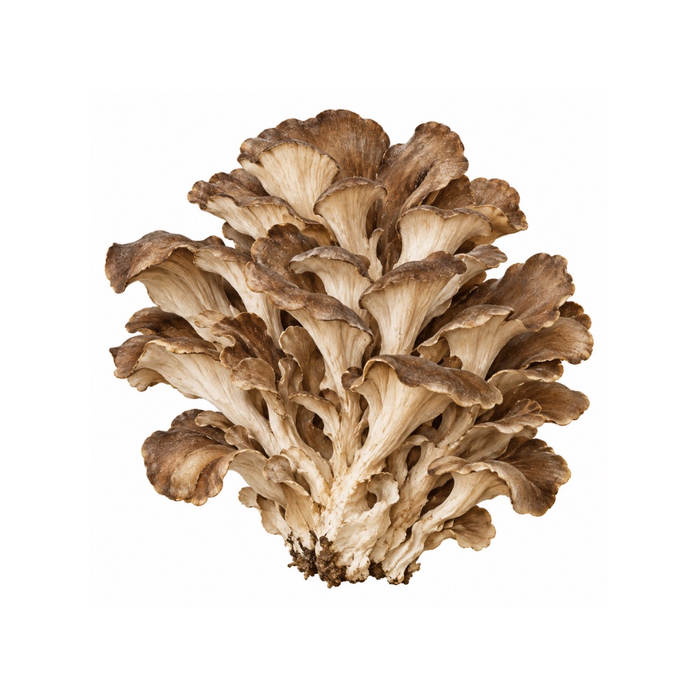 MildMildMaitakeGrifola frondosaA heteropolysaccharide reduced liver fat and inflammation in fatty-liver mice, apparently by remodelling the gut microbiota.Mechanismreduced hepatic fat accumulation via gut-microbiota remodellingRead the evidenceShow less
A heteropolysaccharide reduced hepatic fat and inflammation in NAFLD mice, with the benefit attributed to gut-microbiota remodelling 95Reference 95AnimalThe positive effects of Grifola frondosa heteropolysaccharide on NAFLD and regulation of the gut microbiota — mouse in vivoView study →, consistent with hepatic gene-expression work 96Reference 96AnimalThe mechanisms underlying the hypolipidaemic effects of Grifola frondosa in the liver of rats — animal in vivo (rat)View study →.
The result is mouse-only, and the microbiota mechanism is inferred from association.
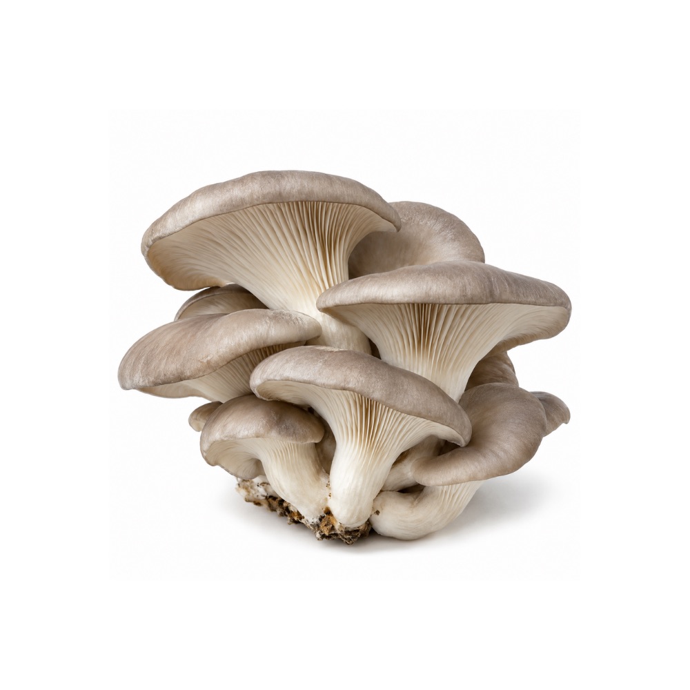 MildMildOyster MushroomPleurotus ostreatusPolysaccharides lower liver enzymes, restore antioxidant enzymes and reduce apoptosis in rats given carbon tetrachloride, reproduced by three preparations.Mechanismantioxidant-coupled protection; Bax/caspase ↓, Bcl-2 ↑Read the evidenceShow less
Polysaccharides lowered ALT/AST, raised antioxidant enzymes and reduced apoptosis markers in CCl₄-injured rats 109Reference 109AnimalThe hepatoprotective effect of polysaccharides from Pleurotus ostreatus on carbon tetrachloride-induced acute liver injury — rat modelView study →, with an exopolysaccharide-peptide complex 110Reference 110AnimalExopolysaccharide-peptide complex from oyster mushroom (Pleurotus ostreatus) protects against hepatotoxicity — rat modelView study → and a crude extract 111Reference 111AnimalAntioxidant activity of the oyster mushroom, Pleurotus ostreatus, on CCl4-induced liver injury — rat model in vivoView study → reproducing it.
Protection tracks restored redox balance rather than a distinct hepatic pathway, and all of it is rodent chemical-injury work.
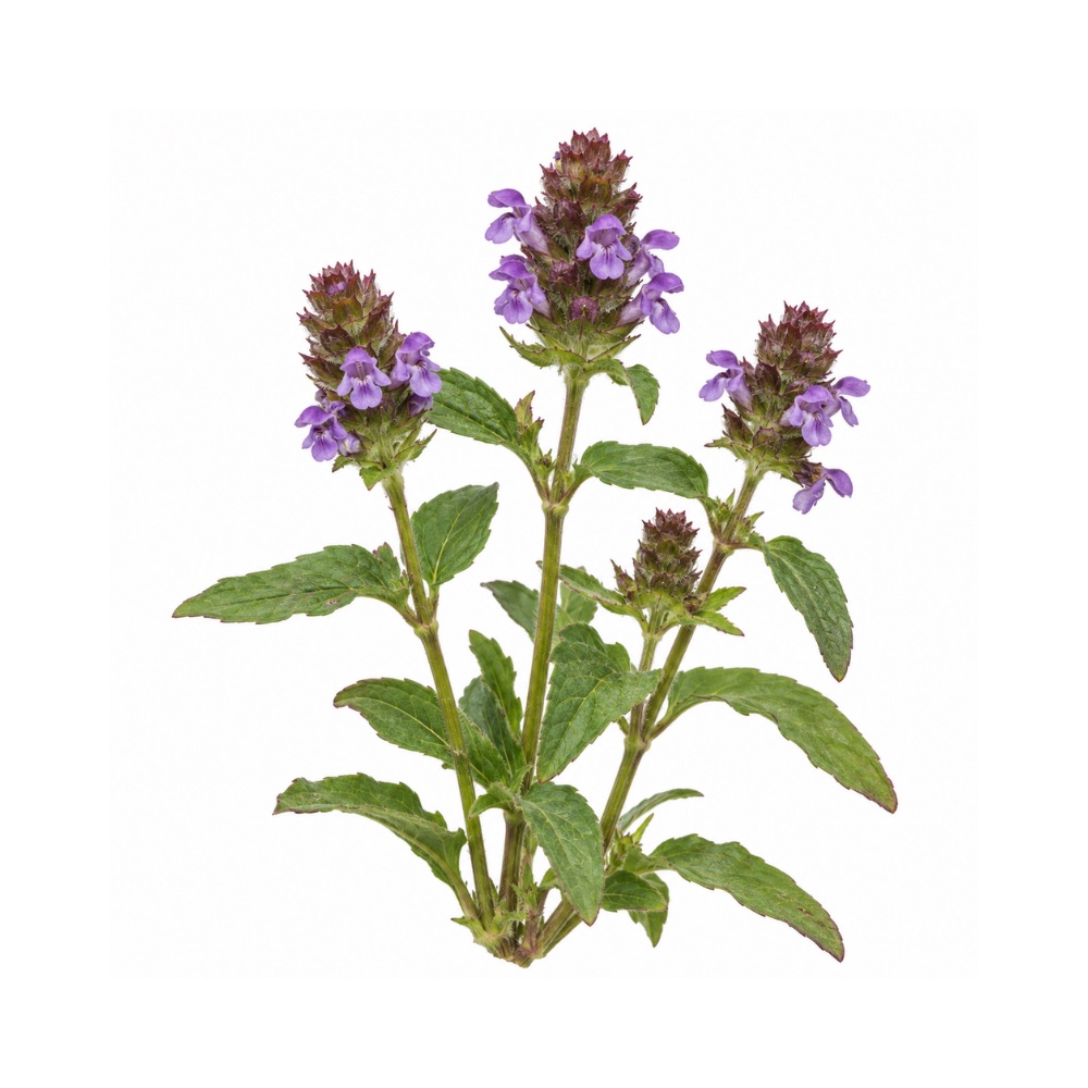 MildMildSelf-HealPrunella vulgarisTriterpenes and polyphenols protect the liver in rodent and cell models.Mechanismnot resolved; triterpene and polyphenol activityRead the evidenceShow less
Hepatoprotection has been shown in rodent and cell models.
The evidence is preclinical and the active fraction is not resolved.
MildShiitakeLentinula edodesThe mycelial extract LEM lowered liver-injury markers in rodents, and shiitake extract limited paracetamol damage to hepatocytes in vitro.Mechanismantioxidant/radical scavenging; glutathione supportRead the evidenceShow less
The mycelial extract LEM reduced liver-injury markers in rodents 126Reference 126AnimalLiver protective effect of Lentinula edodes mycelia (LEM) — animal model. https://pubmed.ncbi.nlm.nih.gov/22790046/View study → and shiitake extract limited paracetamol-induced hepatocyte damage in vitro 127Reference 127In vitroIn vitro antioxidant activity and hepatoprotective effects of Lentinula edodes against paracetamol-induced hepatotoxicity — in vitro. https://pubmed.ncbi.nlm.nih.gov/20657455/View study →, attributed to antioxidant activity with sterols and β-glucans as candidate actives.
There are no human trials, and the rodent data are for LEM rather than the mushroom.
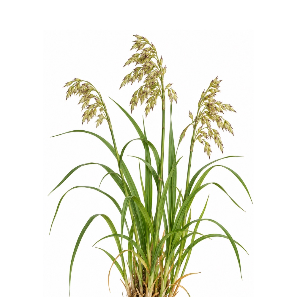 MildMildSweetgrassHierochloe odorataExtract protects rat liver against ethanol-induced oxidative stress, an expression of its antioxidant activity.Mechanismantioxidant protection in ethanol-induced liver stressRead the evidenceShow less
Three rat ethanol-stress models show liver protection 128,
The plant’s coumarin content is itself the reason coumarin-bearing plants carry hepatotoxicity restrictions.
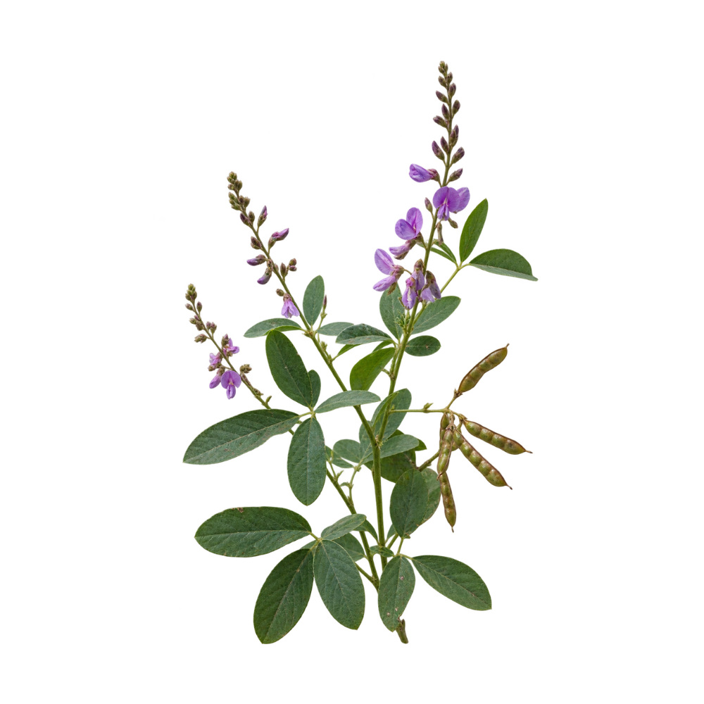 MildMildTick TrefoilDesmodium spp.Desmodium adscendens decoction protects, rather than repairs, the liver in injury models, with the effect tied to D-pinitol.MechanismD-pinitol-linked protection in liver-injury modelsRead the evidenceShow lessMonograph →
D. adscendens decoction is protective, not curative, in liver-injury models, with the effect tied to D-pinitol 134Reference 134AnimalAntihepatotoxic activity of a quantified Desmodium adscendens decoction and D-pinitol against chemically-induced liver damage — animal model (rat)View study →, and results are mixed across models.
The signal does not extend to antiviral activity, so it says nothing about viral hepatitis. The same species induced hepatic CYP enzymes on subchronic dosing 135Reference 135AnimalAcute and subchronic toxicity studies of aqueous extract of Desmodium adscendens — animal model (rat)View study → and was cytotoxic to hepatocytes at 100 mg/mL in vitro 136Reference 136In vitroSafety of Desmodium adscendens extract on hepatocytes and renal cells; protective effect against oxidative stress — in vitro studyView study →.
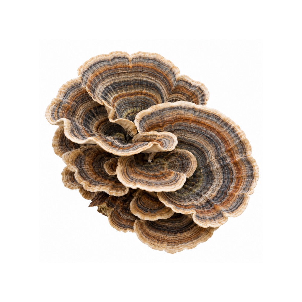 MildMildTurkey TailTrametes versicolorYun zhi, a traditional Chinese liver mushroom; its polysaccharides reduced liver fat in high-fat-diet mice through the gut microbiota.Mechanismgut-microbiota-mediated reduction in hepatic lipid accumulationRead the evidenceShow less
T. versicolor polysaccharides reduced hepatic lipid accumulation and steatosis in high-fat-diet mice, largely through the gut microbiota rather than by direct hepatocyte protection 137Reference 137AnimalPolysaccharides from Trametes versicolor as a potential prebiotic to improve the gut microbiota in high-fat-diet mice — in vivo (mouse)View study →.
The clinical liver claim traces to older Chinese-language, largely uncontrolled reports 138,
No herbs match these filters.
Safety
- No liver herb replaces assessment: jaundice, a persistent enzyme rise or known liver disease needs a clinician.
- Several liver herbs protect at ordinary doses and injure at high ones, so more is not better.
- Liver herbs change drug clearance: schisandra induces CYP3A4, and milk thistle and the berberine herbs inhibit CYP3A4 and P-glycoprotein.
- Stop any liver herb at the first sign of jaundice, dark urine, unexplained fatigue or right-upper-quadrant pain and have liver enzymes checked.
- Choleretic liver herbs are inappropriate with obstructing gallstones or bile-duct obstruction.
- The cautions attach to concentrated extracts rather than culinary amounts, and in pregnancy the group is an avoid unless specifically advised otherwise.
References
- Zhou Y, Wen T, Yang S, Meng B, Wei J, Zhang J, Wang L, Shen X (2025). Sesquiterpene lactones from Cichorium intybus exhibit potent anti-inflammatory and hepatoprotective effects by repression of NF-κB and enhancement of NRF2. Journal of Ethnopharmacology. https://pubmed.ncbi.nlm.nih.gov/39904423/
- Zuo RM, Jiao JY, Chen N, Jiang XL, Wu YL, Nan JX, Lian LH. (2023). Carnosic acid suppressed the formation of NETs in alcoholic hepatosteatosis based on the P2X7R–NLRP3 axis — mice. Phytomedicine. https://pubmed.ncbi.nlm.nih.gov/36577209/
- Zhao Y, Sedighi R, Wang P, Chen H, Zhu Y, Sang S. (2015). Carnosic acid as a major bioactive component in rosemary extract ameliorates high-fat-diet-induced obesity and metabolic syndrome in mice. Journal of Agricultural and Food Chemistry. https://pubmed.ncbi.nlm.nih.gov/25929334/
- Wang T, Takikawa Y (2016). Carnosic acid protects normal mouse hepatocytes against H₂O₂-induced cytotoxicity via sirtuin 1-mediated signaling — in vitro. Hepatology Research. https://pubmed.ncbi.nlm.nih.gov/26223904/
- Dickmann LJ, VandenBrink BM, Lin YS. (2012). In vitro hepatotoxicity and cytochrome P450 induction and inhibition characteristics of carnosic acid, a dietary supplement with antiadipogenic properties — human hepatocytes and microsomes. Drug Metabolism and Disposition. https://pubmed.ncbi.nlm.nih.gov/22531045/
- Geng Y, Wang Y, Sun R, et al. (2021). Carnosol alleviates nonalcoholic fatty liver disease by inhibiting mitochondrial dysfunction and apoptosis through targeting of PRDX3. Toxicology and Applied Pharmacology, 432, 115758. https://pubmed.ncbi.nlm.nih.gov/34678374/
- Hasei S, Yamamotoya T, Nakatsu Y, et al. (2021). Carnosic acid and carnosol activate AMPK, suppress expressions of gluconeogenic and lipogenic genes, and inhibit proliferation of HepG2 cells. International Journal of Molecular Sciences, 22(8), 4040. https://pubmed.ncbi.nlm.nih.gov/33919842/
- Takahashi T, Tabuchi T, Tamaki Y, Kosaka K, Takikawa Y, Satoh T. (2009). Carnosic acid and carnosol inhibit adipocyte differentiation in mouse 3T3-L1 cells through induction of phase 2 enzymes and activation of glutathione metabolism. Biochemical and Biophysical Research Communications, 382(3), 549–554. https://pubmed.ncbi.nlm.nih.gov/19289108/
- Bogahawaththa S, Kawamura T, Bandaranayake U, et al. (2023). Identification and mechanistic investigation of ellagitannins from Osbeckia octandra that attenuate liver fibrosis via the TGF-β/SMAD signaling pathway. Bioscience, Biotechnology, and Biochemistry, 87(11), 1295-1309. https://pubmed.ncbi.nlm.nih.gov/37580142/
- Lu B, Xu Y, Xu L, Cong X, Yin L, Li H, Peng J (2012). Mechanism investigation of dioscin against CCl4-induced acute liver damage in mice. Environmental Toxicology and Pharmacology. https://pubmed.ncbi.nlm.nih.gov/22516057/
- Zhao X, Cong X, Zheng L, Xu L, Yin L, Peng J (2012). Dioscin, a natural steroid saponin, shows remarkable protective effect against acetaminophen-induced liver damage in vitro and in vivo. Toxicology Letters. https://pubmed.ncbi.nlm.nih.gov/22939915/
- Xu T, Zheng L, Xu L, Yin L, Qi Y, Xu Y, Han X, Peng J (2014). Protective effects of dioscin against alcohol-induced liver injury. Archives of Toxicology. Correction published 2019. https://pubmed.ncbi.nlm.nih.gov/24146112/
- Lu B, Yin L, Xu L, Peng J (2011). Application of proteomic and bioinformatic techniques for studying the hepatoprotective effect of dioscin against CCl4-induced liver damage in mice. Planta Medica. https://pubmed.ncbi.nlm.nih.gov/20979020/
- Siddiqui MA, Ali Z, Chittiboyina AG, Khan IA (2018). Hepatoprotective effect of steroidal glycosides from Dioscorea villosa on hydrogen peroxide-induced hepatotoxicity in HepG2 cells. Frontiers in Pharmacology. https://pubmed.ncbi.nlm.nih.gov/30083104/
- Shen Y, Shi R, Wang G, Liu H, Li C, Liu F, Cai J (2025). Gracillin protects liver ischemia-reperfusion injury from oxidative stress-induced apoptosis. Drug Design, Development and Therapy. https://pubmed.ncbi.nlm.nih.gov/41262334/
- Lu W, Pan M, Zhang P, Zheng T, Huang L, Ye F, Lei P (2020). The pharmacokinetics and tissue distributions of nine steroidal saponins from Paris polyphylla in rats. European Journal of Drug Metabolism and Pharmacokinetics. https://pubmed.ncbi.nlm.nih.gov/32661907/
- Wang G, Yan M, Hao R, Lv P, Wang Y, Man S, Gao W (2022). Q-marker identification of Paris polyphylla var. yunnanensis in pulmonary metastasis of liver cancer mice. Journal of Ethnopharmacology. https://pubmed.ncbi.nlm.nih.gov/35461989/
- Hoang LTA, Pham VC, Ngo VD, et al. (2025). Diosgenin glycosides from Paris vietnamensis (Takht) H.Li with cytotoxic and inhibitory NO production activities. Natural Product Research. https://pubmed.ncbi.nlm.nih.gov/41021975/
- Dai B, Wang L, Su LH, Li HZ (2026). Dracaenogenins C and D, two new 12(13→14)-abeo-spirostanols from the red resin of Dracaena cochinchinensis. Molecules. https://pubmed.ncbi.nlm.nih.gov/41828837/
- Wang R, Song F, Li S, et al. (2019). Salvianolic acid A attenuates CCl4-induced liver fibrosis by regulating the PI3K/AKT/mTOR, Bcl-2/Bax and caspase-3/cleaved caspase-3 signaling pathways. Drug Design, Development and Therapy, 13, 1889–1900. https://pubmed.ncbi.nlm.nih.gov/31213776/
- Zhu J, Wang R, Xu T, et al. (2018). Salvianolic acid A attenuates endoplasmic reticulum stress and protects against cholestasis-induced liver fibrosis via the SIRT1/HSF1 pathway. Frontiers in Pharmacology, 9, 1277. https://pubmed.ncbi.nlm.nih.gov/30455644/
- Dai JP, Zhu DX, Sheng JT, et al. (2015). Inhibition of tanshinone IIA, salvianolic acid A and salvianolic acid B on areca nut extract-induced oral submucous fibrosis in vitro. Molecules, 20(4), 6794–6807. https://pubmed.ncbi.nlm.nih.gov/25884554/
- Yao L, Zhao R, He S, et al. (2022). Effects of salvianolic acid A and salvianolic acid B in renal interstitial fibrosis via PDGF-C/PDGFR-α signaling pathway. Phytomedicine, 106, 154414. https://pubmed.ncbi.nlm.nih.gov/36057144/
- Holbrook AM, Pereira JA, Labiris R, et al. (2005). Systematic overview of warfarin and its drug and food interactions. Archives of Internal Medicine, 165(10), 1095–1106. https://pubmed.ncbi.nlm.nih.gov/15911722/
- Liu P, Hu YY, Liu C, et al. (2002). Clinical observation of salvianolic acid B in treatment of liver fibrosis in chronic hepatitis B — randomised double-blind trial with paired liver biopsies. World Journal of Gastroenterology, 8(4), 679–685. https://pubmed.ncbi.nlm.nih.gov/12174378/
- Tao YY, Wang QL, Shen L, Fu WW, Liu CH. (2013). Salvianolic acid B inhibits hepatic stellate cell activation through transforming growth factor beta-1 signal transduction pathway in vivo and in vitro. Experimental Biology and Medicine, 238(11), 1284–1296. https://pubmed.ncbi.nlm.nih.gov/24006304/
- Liu F, et al. (2023). Salvianolic acid B inhibits hepatic stellate cell activation and liver fibrosis by targeting PDGFRβ. International Immunopharmacology, 122, 110550. https://pubmed.ncbi.nlm.nih.gov/37451016/
- Jiang, Y. C., et al. (2025). Vincamine ameliorates hepatic fibrosis via inhibiting S100A4-mediated farnesoid X receptor activation — animal model. British Journal of Pharmacology. https://pubmed.ncbi.nlm.nih.gov/39940076/
- Abouelhamd, A., et al. (2025). Vincamine induces cytoprotective autophagy via regulation of AMPK/mTOR signalling in gentamicin-induced hepatotoxicity and nephrotoxicity in rats — animal model. Scientific Reports. https://pubmed.ncbi.nlm.nih.gov/40542078/
- Xin, X., et al. (2026). Vincamine attenuates alcoholic liver injury through modulation of a CDK1-glycolysis-NLRP3 immunometabolic axis — animal model. International Immunopharmacology. https://pubmed.ncbi.nlm.nih.gov/42048736/
- Alaaeldin, R., et al. (2024). Vincamine exerts hepato-protective activity during colon-ligation-puncture-induced sepsis by modulating oxidative stress, apoptosis and TNFα/Nrf-2/Keap-1 — animal model. Scientific Reports. https://pubmed.ncbi.nlm.nih.gov/39174578/
- Alaaeldin, R., et al. (2024). Vincamine alleviates intrahepatic cholestasis in rats through modulation of NF-κB/PDGF/KLF6/PPARγ and PI3K/Akt pathways — animal model. Naunyn-Schmiedeberg’s Archives of Pharmacology. https://pubmed.ncbi.nlm.nih.gov/38761209/
- Ürün, Y. Y., et al. (2025). Vincamine mitigates a methotrexate-induced liver fibrosis model — animal model. Turkish Journal of Gastroenterology. https://pubmed.ncbi.nlm.nih.gov/41084779/
- Surendran S, Eswaran MB, Vijayakumar M, et al. (2011). In vitro and in vivo hepatoprotective activity of Cissampelos pareira against carbon-tetrachloride-induced hepatic damage — in vitro and animal model (rat). Indian Journal of Experimental Biology. https://pubmed.ncbi.nlm.nih.gov/22403868/
- Nagalekshmi, R., et al. (2011). Hepatoprotective activity of Andrographis paniculata and Swertia chirayita — animal model (mouse). Food and Chemical Toxicology. https://pubmed.ncbi.nlm.nih.gov/21983487/
- Dai, Y., et al. (2019). Overview of pharmacological activities of Andrographis paniculata and its major compound andrographolide — review. Critical Reviews in Food Science and Nutrition. https://pubmed.ncbi.nlm.nih.gov/30040451/
- Kamel AM, et al. (2022). Therapeutic potential of artichoke in the treatment of fatty liver — systematic review and meta-analysis of randomised controlled trials. Journal of Medicinal Food. https://pubmed.ncbi.nlm.nih.gov/35763310/
- Moradi S, et al. (2021). Effects of Cynara scolymus L. supplementation on liver enzymes — systematic review and meta-analysis. International Journal of Clinical Practice. https://pubmed.ncbi.nlm.nih.gov/34383355/
- Panahi Y, Kianpour P, Mohtashami R, et al. (2018). Efficacy of artichoke leaf extract in non-alcoholic fatty liver disease — pilot double-blind randomised controlled trial. Phytotherapy Research. https://pubmed.ncbi.nlm.nih.gov/29520889/
- Hao, J., et al. (2024). Metabolomics combined with network pharmacology reveals the protective effect of astragaloside IV on alcoholic liver disease (rat model). Phytomedicine. https://pubmed.ncbi.nlm.nih.gov/39270570/
- Luangmonkong, T., et al. (2023). Hepatoprotective efficacy of cycloastragenol alleviated the progression of liver fibrosis in carbon-tetrachloride-treated mice. Biomedicines. https://pubmed.ncbi.nlm.nih.gov/36672739/
- Yan, F., et al. (2009). Synergistic hepatoprotective effect of Schisandra lignans with Astragalus polysaccharides on chronic liver injury in rats. Phytomedicine. https://pubmed.ncbi.nlm.nih.gov/19345075/
- Chen, H., Weng, L., et al. (2000). Comparison of efficacy in treating liver fibrosis of chronic hepatitis B between Astragalus-Polygonum anti-fibrosis decoction and Jinshuibao capsule — a randomized controlled trial. Chinese Journal of Integrated Traditional and Western Medicine. https://pubmed.ncbi.nlm.nih.gov/11789260/
- Silveira Rabelo, A. C., & Caldeira Costa, D. (2018). A review of biological and pharmacological activities of Baccharis trimera — review. Chemico-Biological Interactions, 296, 65–75. https://pubmed.ncbi.nlm.nih.gov/30240600/
- Pádua, B. C., Silva, L. D., Rossoni, J. V., et al. (2010). Antioxidant properties of Baccharis trimera in the neutrophils of Fisher rats — in vitro and in vivo (rat). Journal of Ethnopharmacology, 129(3), 381–386. https://pubmed.ncbi.nlm.nih.gov/20430095/
- Pádua, B. C., Rossoni Júnior, J. V., de Brito Magalhães, C. L., et al. (2014). Protective effect of Baccharis trimera extract on acute hepatic injury in a model of inflammation induced by acetaminophen — in vivo (rat). Mediators of Inflammation, 2014, 196598. https://doi.org/10.1155/2014/196598
- Pádua, B. C., Rossoni Júnior, J. V., de Brito Magalhães, C. L., et al. (2013). Baccharis trimera improves the antioxidant defense system and inhibits iNOS and NADPH oxidase expression in a rat model of inflammation — in vivo (rat). Current Pharmaceutical Biotechnology, 14(11), 975–984. https://pubmed.ncbi.nlm.nih.gov/24372242/
- Chaves, P. F. P., Iacomini, M., & Cordeiro, L. M. C. (2020). Chemical characterization of polysaccharides from Baccharis trimera (Less.) DC. infusion and its hepatoprotective effects — in vitro and in vivo (mouse). Food Research International, 136, 109510. https://doi.org/10.1016/j.foodres.2020.109510
- Barbosa, R. J., Ratti da Silva, G., Cola, I. M., et al. (2020). Promising therapeutic use of Baccharis trimera (Less.) DC. as a natural hepatoprotective agent against hepatic lesions caused by multiple risk factors — in vivo (rat). Journal of Ethnopharmacology, 254, 112729. https://pubmed.ncbi.nlm.nih.gov/32145332/
- Iloon Kashkooli, R. et al. (2015). The effect of Berberis vulgaris extract on transaminase activities in non-alcoholic fatty liver disease — clinical trial. Hepatitis Monthly. https://pubmed.ncbi.nlm.nih.gov/25788958/
- Salgueiro, A. C., Folmer, V., da Silva, M. P., et al. (2016). Effects of Bauhinia forficata tea on oxidative stress and liver damage in diabetic mice — animal study. Oxidative Medicine and Cellular Longevity. https://pubmed.ncbi.nlm.nih.gov/26839634/
- Pinafo, M. S., Benedetti, P. R., Gaiotte, L. B., et al. (2019). Effects of Bauhinia forficata on glycaemia, lipid profile, hepatic glycogen content and oxidative stress in rats exposed to bisphenol A — animal study. Toxicology Reports. https://pubmed.ncbi.nlm.nih.gov/30911467/
- Lanhers, M. C., et al. (1991). Hepatoprotective and anti-inflammatory effects of a traditional medicinal plant of Chile, Peumus boldus — animal in vivo and in vitro. Planta Medica. https://pubmed.ncbi.nlm.nih.gov/1891491/
- Fernández, J., et al. (2009). Effect of boldo (Peumus boldus Molina) infusion on lipoperoxidation induced by cisplatin in mice liver — animal in vivo. Phytotherapy Research. https://pubmed.ncbi.nlm.nih.gov/19145575/
- Ezhilarasan, D. (2021). Boldine treatment protects acetaminophen-induced liver inflammation and acute hepatic necrosis in mice — animal in vivo. Journal of Biochemical and Molecular Toxicology. https://pubmed.ncbi.nlm.nih.gov/33393705/
- Shuker, E., et al. (2022). Potential effects of boldine on oxidative stress, apoptosis, and inflammatory changes induced by methylprednisolone hepatotoxicity in male Wistar rats — animal in vivo. Dose-Response. https://pubmed.ncbi.nlm.nih.gov/35360456/
- O’Brien, P., et al. (2006). Boldine and its antioxidant or health-promoting properties — review. Chemico-Biological Interactions. https://pubmed.ncbi.nlm.nih.gov/16221469/
- Bannach, R., et al. (1996). Cytoprotective and antioxidant effects of boldine on tert-butyl hydroperoxide-induced damage to isolated hepatocytes — in vitro. Cell Biology and Toxicology. https://pubmed.ncbi.nlm.nih.gov/8738478/
- Piscaglia, F., et al. (2005). Caution in the use of boldo in herbal laxatives: a case of hepatotoxicity — case report. Scandinavian Journal of Gastroenterology. https://pubmed.ncbi.nlm.nih.gov/15764158/
- Ribeiro, R. J., et al. (2017). Hidden risks of alternative medicines: a case of boldo-induced hepatotoxicity — case report. Journal of Dietary Supplements. https://pubmed.ncbi.nlm.nih.gov/27576017/
- Oliveira Sá, A., et al. (2020). Boldo-induced hepatotoxicity: a case of unexplained jaundice — case report. European Journal of Case Reports in Internal Medicine. https://doi.org/10.12890/2020_002116
- Dang, S. S., Wang, B. F., Cheng, Y. A., et al. (2007). Inhibitory effects of saikosaponin-d on CCl₄-induced hepatic fibrogenesis in rats — animal model. World Journal of Gastroenterology. https://pubmed.ncbi.nlm.nih.gov/17278221/
- Wu, S.-J., Lin, Y.-H., et al. (2008). Curcumin or saikosaponin a improves hepatic antioxidant capacity and protects against CCl₄-induced liver injury in rats — animal model. Journal of Medicinal Food. https://pubmed.ncbi.nlm.nih.gov/18598162/
- Oka, H., Yamamoto, S., Kuroki, T., et al. (1995). Prospective study of chemoprevention of hepatocellular carcinoma with Sho-saiko-to (TJ-9) — randomized controlled trial. Cancer. https://pubmed.ncbi.nlm.nih.gov/8625175/
- Agrinier AL, et al. Camu-camu decreases hepatic steatosis and liver injury markers in overweight, hypertriglyceridemic individuals: a randomized crossover trial. Cell Reports Medicine. 2024. https://pubmed.ncbi.nlm.nih.gov/39168095/
- Anhê FF, et al. Treatment with camu camu (Myrciaria dubia) prevents obesity by altering the gut microbiota and increasing energy expenditure in diet-induced obese mice. Gut. 2019. https://pubmed.ncbi.nlm.nih.gov/30064988/
- Akachi T, et al. 1-Methylmalate from camu-camu (Myrciaria dubia) suppressed D-galactosamine-induced liver injury in rats. Bioscience, Biotechnology, and Biochemistry. 2010;74(3):573–578. https://pubmed.ncbi.nlm.nih.gov/20208347/
- Pereira SMS, et al. An extract from camu-camu (Myrciaria dubia) attenuates DSS-induced gut–liver axis alterations in ovo (Gallus gallus). British Journal of Nutrition. 2026 (epub). https://pubmed.ncbi.nlm.nih.gov/42290501/
- Tan J, et al. (2019). Hepatoprotective effect of essential oils of Nepeta cataria L. on acetaminophen-induced liver dysfunction — [animal in vivo]. Bioscience Reports. https://pubmed.ncbi.nlm.nih.gov/31337687/
- Zhu et al. (2025). Chrysanthemum morifolium water extract prevents and treats MAFLD via pan-PPAR agonism (mouse model). Journal of Ethnopharmacology. https://pubmed.ncbi.nlm.nih.gov/40921229/
- Liu et al. (2024). Chrysanthemum morifolium attenuates metabolic- and alcohol-associated liver disease via gut microbiota and PPAR-α/γ (mouse model). Phytomedicine. https://pubmed.ncbi.nlm.nih.gov/38820659/
- Kennedy, O. J., Roderick, P., Buchanan, R., et al. (2017). Coffee, including caffeinated and decaffeinated coffee, and the risk of hepatocellular carcinoma: a systematic review and dose-response meta-analysis. BMJ Open. https://pubmed.ncbi.nlm.nih.gov/28490552/
- Nie, Q., Li, M., Huang, C., et al. (2024). The clinical efficacy and safety of berberine in the treatment of non-alcoholic fatty liver disease — meta-analysis and systematic review. Journal of Translational Medicine. https://pubmed.ncbi.nlm.nih.gov/38429794/
- Yao, N., Wang, X., Chen, Y., et al. (2025). Coptidis rhizoma ameliorates type 2 diabetes-related metabolic-dysfunction-associated steatohepatitis by downregulating IL-17RA/NF-κB — mouse model. Frontiers in Pharmacology. https://pubmed.ncbi.nlm.nih.gov/41089852/
- Li, X., Zhang, X., Wei, N., et al. (2025). Total alkaloids from Coptis chinensis Franch ameliorate hyperlipidaemia and hepatic steatosis via AMPK/SREBP-1c and PPARα/LXRα in mice — animal model. Frontiers in Pharmacology. https://pubmed.ncbi.nlm.nih.gov/41244823/
- Pan CW, Zhou GY, Chen WL, et al. (2015). Forsythiaside A attenuates LPS/D-galactosamine-induced liver injury in mice via NF-κB and Nrf2/HO-1. https://pubmed.ncbi.nlm.nih.gov/25797347/
- Kim YE, Bak SB, Kim MJ, et al. (2023). Forsythiaside A protects against CCl₄-induced liver injury via AMPK/Nrf2 (mouse model). International Journal of Molecular Sciences. https://www.ncbi.nlm.nih.gov/pmc/articles/PMC10707188/
- Wang, W., et al. (2018). Madecassoside prevents acute liver failure in LPS/D-GalN-induced mice — animal model. Biomed Pharmacother. https://pubmed.ncbi.nlm.nih.gov/29715757/
- Santos, G., et al. (2021). Profile of herbal and dietary supplement-induced liver injury in Latin America — systematic review. Phytotherapy Research. https://pubmed.ncbi.nlm.nih.gov/32525269/
- Shin, M. O., et al. (2010). The proanthocyanidins inhibit dimethylnitrosamine-induced liver damage in rats. Archives of Pharmacal Research. https://pubmed.ncbi.nlm.nih.gov/20191358/
- Mansouri, E., et al. (2014). Antioxidant effects of proanthocyanidin from grape seed on hepatic tissue injury in diabetic rats. Iranian Journal of Basic Medical Sciences. https://pubmed.ncbi.nlm.nih.gov/25140209/
- Yogalakshmi, B., et al. (2013). Grape seed proanthocyanidin rescues rats from steatosis: a comparative and combination study with metformin. Journal of Lipids. https://pubmed.ncbi.nlm.nih.gov/24307947/
- Ghanbari, P., et al. (2024). Grape seed extract supplementation in non-alcoholic fatty liver disease. International Journal for Vitamin and Nutrition Research. https://pubmed.ncbi.nlm.nih.gov/38419408/
- Ghanbari, P., et al. (2024). The effects of grape seed extract supplementation on cardiovascular risk factors, liver enzymes and hepatic steatosis in patients with non-alcoholic fatty liver disease. BMC Complementary Medicine and Therapies. https://pubmed.ncbi.nlm.nih.gov/38755622/
- Bagchi, D., et al. (2014). Free radical scavenging, antioxidant and cancer chemoprevention by grape seed proanthocyanidin: an overview. Mutation Research. https://pubmed.ncbi.nlm.nih.gov/24751946/
- Weseler, A. R., et al. (2017). Masquelier’s grape seed extract: from basic flavonoid research to a well-characterized food supplement with health benefits. Nutrition Journal. https://pubmed.ncbi.nlm.nih.gov/28103873/
- Son, Y., et al. (2021). Ameliorative effect of Annona muricata (Graviola) extract on hyperglycaemia-induced hepatic damage in STZ diabetic rats — animal model. Antioxidants, 10. https://pubmed.ncbi.nlm.nih.gov/34679681/
- Masykur, et al. (2023). Effect of ethanol extract of soursop (Annona muricata L.) stem bark on rat liver function — animal model. Pakistan Journal of Biological Sciences, 26. https://pubmed.ncbi.nlm.nih.gov/38110557/
- Chou, S. C., et al. (2006). The add-on effects of Gynostemma pentaphyllum on nonalcoholic fatty liver disease — randomised controlled trial. Alternative Therapies in Health and Medicine. https://pubmed.ncbi.nlm.nih.gov/16708768/
- Liu, Z. L., et al. (2013). Herbal medicines for fatty liver diseases — systematic review. The Cochrane Database of Systematic Reviews. https://pubmed.ncbi.nlm.nih.gov/23975682/
- Jiang, F. Y., et al. (2024). Gynostemma pentaphyllum extract alleviates NASH in mice: inflammation and gut microbiota — mouse. Nutrients. https://pubmed.ncbi.nlm.nih.gov/38892715/
- Lin, C. C., Huang, P. C., & Lin, J. M. (2000). Antioxidant and hepatoprotective effects of Anoectochilus formosanus and Gynostemma pentaphyllum — rat. The American Journal of Chinese Medicine. https://pubmed.ncbi.nlm.nih.gov/10794120/
- Zhao H et al. (2017). Antioxidant and hepatoprotective activities of polysaccharides from spent mushroom substrates (Laetiporus sulphureus) in acute alcohol-induced mice — in vivo mouse study. Oxid Med Cell Longev. https://pubmed.ncbi.nlm.nih.gov/29430281/
- Zhang, Z., et al. (2012). Antioxidant and hepatoprotective potential of endo-polysaccharides from Hericium erinaceus — in vitro and mouse study. Int J Biol Macromol. https://pubmed.ncbi.nlm.nih.gov/22982810/
- Li, X., et al. (2019). The positive effects of Grifola frondosa heteropolysaccharide on NAFLD and regulation of the gut microbiota — mouse in vivo. International Journal of Molecular Sciences. https://pubmed.ncbi.nlm.nih.gov/31653116/
- Ding, Y., et al. (2016). The mechanisms underlying the hypolipidaemic effects of Grifola frondosa in the liver of rats — animal in vivo (rat). Frontiers in Microbiology. https://pubmed.ncbi.nlm.nih.gov/27536279/
- Zhong S, Fan Y, Yan Q, et al. (2017). The therapeutic effect of silymarin in the treatment of nonalcoholic fatty disease: a meta-analysis (PRISMA) of randomized control trials — meta-analysis. Medicine (Baltimore). https://pubmed.ncbi.nlm.nih.gov/29245314/
- Loguercio C, Andreone P, Brisc C, et al. (2012). Silybin combined with phosphatidylcholine and vitamin E in patients with nonalcoholic fatty liver disease: a randomized controlled trial — randomised controlled trial. Free Radic Biol Med. https://pubmed.ncbi.nlm.nih.gov/22343419/
- Luangchosiri C, Thakkinstian A, Chitphuk S, et al. (2015). A double-blinded randomized controlled trial of silymarin for the prevention of antituberculosis drug-induced liver injury — randomised placebo-controlled trial. BMC Complement Altern Med. https://pubmed.ncbi.nlm.nih.gov/26400476/
- Tao L, Qu X, Zhang Y, et al. (2019). Prophylactic therapy of silymarin (milk thistle) on antituberculosis drug-induced liver injury: a meta-analysis of randomized controlled trials — meta-analysis. Can J Gastroenterol Hepatol. https://pubmed.ncbi.nlm.nih.gov/30733935/
- Mengs U, Pohl RT, Mitchell T. (2012). Legalon SIL: the antidote of choice in patients with acute hepatotoxicity from amatoxin poisoning — clinical review (case series). Curr Pharm Biotechnol. https://pubmed.ncbi.nlm.nih.gov/22352731/
- Rambaldi A, Jacobs BP, Gluud C. (2007). Milk thistle for alcoholic and/or hepatitis B or C virus liver diseases — Cochrane systematic review and meta-analysis. Cochrane Database Syst Rev. https://pubmed.ncbi.nlm.nih.gov/17943794/
- Fried MW, Navarro VJ, Afdhal N, et al. (2012). Effect of silymarin (milk thistle) on liver disease in patients with chronic hepatitis C unsuccessfully treated with interferon therapy: a randomized controlled trial — multicentre randomised controlled trial. JAMA. https://pubmed.ncbi.nlm.nih.gov/22797645/
- Tang G, Zhang L, et al. (2021). Effect of Nigella sativa in the treatment of nonalcoholic fatty liver disease — systematic review and meta-analysis of randomized controlled trials. Phytother Res. https://pubmed.ncbi.nlm.nih.gov/33728708/
- Khonche A, Huseini HF, et al. (2019). Standardized Nigella sativa seed oil ameliorates hepatic steatosis, aminotransferase and lipid levels in NAFLD — a randomized, double-blind, placebo-controlled clinical trial. J Ethnopharmacol. https://pubmed.ncbi.nlm.nih.gov/30639231/
- Darand M, Darabi Z, et al. (2019). Nigella sativa and inflammatory biomarkers in patients with nonalcoholic fatty liver disease — a randomized, double-blind, placebo-controlled clinical trial. Complement Ther Med. https://pubmed.ncbi.nlm.nih.gov/31126557/
- Zaki SM, Waggas DS. (2022). Protective effect of Nigella sativa against 5-fluorouracil-induced liver damage in rats — animal study. Nutr Cancer. https://pubmed.ncbi.nlm.nih.gov/34963383/
- Razmpoosh E, Safi S, et al. (2020). The effect of Nigella sativa on measures of liver and kidney parameters — systematic review and dose-response meta-analysis of randomized controlled trials. Pharmacol Res. https://pubmed.ncbi.nlm.nih.gov/32201245/
- Zhu, B. et al. (2019). The hepatoprotective effect of polysaccharides from Pleurotus ostreatus on carbon tetrachloride-induced acute liver injury — rat model. Int J Biol Macromol. https://pubmed.ncbi.nlm.nih.gov/30851331/
- Abdel-Monem, N. M. et al. (2020). Exopolysaccharide-peptide complex from oyster mushroom (Pleurotus ostreatus) protects against hepatotoxicity — rat model. Biochem Biophys Rep. https://pubmed.ncbi.nlm.nih.gov/33241128/
- Jayakumar, T. et al. (2006). Antioxidant activity of the oyster mushroom, Pleurotus ostreatus, on CCl4-induced liver injury — rat model in vivo. Food Chem Toxicol. https://pubmed.ncbi.nlm.nih.gov/16914248/
- Chen, Z., et al. (2013). Reduced hepatotoxicity by total glucosides of paeony in combination treatment with leflunomide and methotrexate for patients with active rheumatoid arthritis — randomised controlled trial. Int Immunopharmacol. https://pubmed.ncbi.nlm.nih.gov/23415907/
- Xiang, N., et al. (2015). Total glucosides of paeony can reduce the hepatotoxicity caused by methotrexate and leflunomide combination treatment of active rheumatoid arthritis — randomised controlled trial. Int Immunopharmacol. https://pubmed.ncbi.nlm.nih.gov/26292180/
- Wang, X. L., et al. (2026). Paeoniflorin protects against NAFLD through antioxidant, anti-inflammatory effects and restoration of gut microbiota homeostasis — mouse in vivo. Front Microbiol. https://pubmed.ncbi.nlm.nih.gov/41909260/
- Wei, S. Z., et al. (2026). Paeoniflorin alleviates metabolic dysfunction-associated steatotic liver disease by inhibiting hepatic lipogenesis and inflammation — mouse in vivo. Inflammation. https://pubmed.ncbi.nlm.nih.gov/41746510/
- Tan, Y. Q., Chen, H. W., Li, J., & Wu, Q. J. (2020). Efficacy, chemical constituents, and pharmacological actions of Radix Paeoniae Rubra and Radix Paeoniae Alba — review. Front Pharmacol. https://pubmed.ncbi.nlm.nih.gov/32754038/
- Johra FT, Hossain S, Jain P, et al. (2023). Amelioration of CCl₄-induced oxidative stress and hepatotoxicity by Ganoderma lucidum in Long Evans rats — animal model. Sci Rep. https://pubmed.ncbi.nlm.nih.gov/37336915/
- Li YQ, Wang SF (2006). Anti-hepatitis B activities of ganoderic acid from Ganoderma lucidum — in vitro and mouse study. Biotechnol Lett. https://pubmed.ncbi.nlm.nih.gov/16786250/
- Ouchfoun M et al. (2016). Labrador tea (Rhododendron groenlandicum) attenuates insulin resistance in a diet-induced obesity mouse model — animal model (mouse, in vivo). European Journal of Nutrition. https://pubmed.ncbi.nlm.nih.gov/25916863/
- Eid HM et al. (2016). A combination of (+)-catechin and (−)-epicatechin underlies the in vitro adipogenic action of Labrador tea (Rhododendron groenlandicum), an antidiabetic medicinal plant of the Eastern James Bay Cree pharmacopeia — in vitro. Journal of Ethnopharmacology. https://pubmed.ncbi.nlm.nih.gov/26707751/
- Rapinski M et al. (2015). Adipogenic Activity of Wild Populations of Rhododendron groenlandicum, a Medicinal Shrub from the James Bay Cree Traditional Pharmacopeia — in vitro. Evidence-Based Complementary and Alternative Medicine. https://pubmed.ncbi.nlm.nih.gov/26508979/
- Wu, C., et al. (2022). Rhubarb free anthraquinones improved mice non-alcoholic fatty liver disease by inhibiting NLRP3 inflammasome — mouse, in vivo. Journal of Translational Medicine. https://pubmed.ncbi.nlm.nih.gov/35765026/
- Wang, J. B., et al. (2011). Hepatotoxicity or hepatoprotection? Pattern recognition for the paradoxical effect of the Chinese herb Rheum palmatum L. in treating rat liver injury — rat, in vivo. PLoS ONE. https://pubmed.ncbi.nlm.nih.gov/21915343/
- Huang, B. H., Lv, B. H., Wu, D. J., et al. (2025). Efficacy of Schisandra chinensis in liver injury: a systematic review and preclinical meta-analysis — systematic review, meta-analysis of animal studies. Frontiers in Pharmacology. https://pubmed.ncbi.nlm.nih.gov/40832608/
- Jiang, Y., Fan, X., Wang, Y., Chen, P., Bi, H., et al. (2016). Schisandrol B protects against acetaminophen-induced acute hepatotoxicity in mice via activation of the NRF2/ARE signaling pathway — animal (mouse) study. Acta Pharmacologica Sinica, 37(3), 382–389. https://pubmed.ncbi.nlm.nih.gov/26806302/
- Yagi K (2012). Liver protective effect of Lentinula edodes mycelia (LEM) — animal model. https://pubmed.ncbi.nlm.nih.gov/22790046/
- Sasidharan S, Aravindran S, Latha LY, et al. (2010). In vitro antioxidant activity and hepatoprotective effects of Lentinula edodes against paracetamol-induced hepatotoxicity — in vitro. https://pubmed.ncbi.nlm.nih.gov/20657455/
- Pukalskas, A., van Beek, T. A., Venskutonis, R. P., Linssen, J. P., van Veldhuizen, A., & de Groot, A. (2002). Identification of radical scavengers in sweet grass (Hierochloe odorata) — in vitro. Journal of Agricultural and Food Chemistry, 50(10), 2914–2919. https://pubmed.ncbi.nlm.nih.gov/11982419/
- Zainuddin, A., Pokorný, J., & Venskutonis, R. (2002). Antioxidant activity of sweetgrass (Hierochloë odorata Wahlnb.) extract in lard and rapeseed oil emulsions — in vitro. Nahrung, 46(1), 15–17. https://pubmed.ncbi.nlm.nih.gov/11890046/
- Łuczaj, W., Stankiewicz-Kranc, A., Milewska, E., Roszkowska-Jakimiec, W., & Skrzydlewska, E. (2012). Effect of sweet grass extract against oxidative stress in rat liver and serum — animal in vivo. Food and Chemical Toxicology, 50(2), 135–140. https://pubmed.ncbi.nlm.nih.gov/21959529/
- Dobrzyńska, I., Szachowicz-Petelska, B., Skrzydlewska, E., & Figaszewski, Z. (2013). Effect of sweet grass (Hierochloe odorata) on the physico-chemical properties of liver cell membranes from rats intoxicated with ethanol — animal in vivo. Environmental Toxicology and Pharmacology, 35(2), 247–253. https://pubmed.ncbi.nlm.nih.gov/23353548/
- Łuczaj, W., Jarocka-Karpowicz, I., Bielawska, K., & Skrzydlewska, E. (2015). Sweet grass protection against oxidative stress formation in the rat brain — animal in vivo. Metabolic Brain Disease, 30(1), 183–190. https://pubmed.ncbi.nlm.nih.gov/25108595/
- Slapšytė, G., Dedonytė, V., Lazutka, J. R., Mierauskienė, J., Morkūnas, V., Kazernavičiūtė, R., Pukalskas, A., & Venskutonis, P. R. (2013). Evaluation of the biological activity of naturally occurring 5,8-dihydroxycoumarin — in vitro and in vivo genotoxicity. Molecules, 18(4), 4419–4436. https://pubmed.ncbi.nlm.nih.gov/23591923/
- Magielse, J., et al. (2013). Antihepatotoxic activity of a quantified Desmodium adscendens decoction and D-pinitol against chemically-induced liver damage — animal model (rat). Journal of Ethnopharmacology. https://pubmed.ncbi.nlm.nih.gov/23291573/
- Quaye, O., et al. (2017). Acute and subchronic toxicity studies of aqueous extract of Desmodium adscendens — animal model (rat). Journal of Evidence-Based Complementary & Alternative Medicine. https://pubmed.ncbi.nlm.nih.gov/29228815/
- François, C., et al. (2015). Safety of Desmodium adscendens extract on hepatocytes and renal cells; protective effect against oxidative stress — in vitro study. Journal of Intercultural Ethnopharmacology. https://pubmed.ncbi.nlm.nih.gov/26401376/
- Bai M, Huang Z, Zheng X, et al. (2024). Polysaccharides from Trametes versicolor as a potential prebiotic to improve the gut microbiota in high-fat-diet mice — in vivo (mouse). Microorganisms. https://pubmed.ncbi.nlm.nih.gov/39203496/
- Chang Y, Zhang M, Jiang Y, et al. (2017). Preclinical and clinical studies of Coriolus versicolor polysaccharopeptide as an immunotherapeutic in China — review. Discovery Medicine. https://pubmed.ncbi.nlm.nih.gov/28595034/
- Chang Y, et al. (2019). Coriolus versicolor polysaccharopeptide as an immunotherapeutic in China — review. Progress in Molecular Biology and Translational Science. https://pubmed.ncbi.nlm.nih.gov/31030754/
- Wei Z, Liu N, Tantai X, et al. (2019). The effects of curcumin on the metabolic parameters of non-alcoholic fatty liver disease: a meta-analysis of randomized controlled trials. Hepatol Int. https://pubmed.ncbi.nlm.nih.gov/30446932/
- Chen Y et al. (2024). Curcumin supplementation alleviates hepatic fat content associated with modulation of gut microbiota-dependent bile acid metabolism in NAFL — randomized controlled trial. Am J Clin Nutr. https://pubmed.ncbi.nlm.nih.gov/38795741/
- Ranneh Y, et al. (2024). Polyphenol Intervention Ameliorates Non-Alcoholic Fatty Liver Disease: an updated comprehensive systematic review. Nutrients. https://pubmed.ncbi.nlm.nih.gov/39683546/
- Wu, K., et al. (2018). Hepatoprotective effects exerted by Poria cocos polysaccharides against acetaminophen-induced liver injury in mice — animal model. International Journal of Biological Macromolecules. https://pubmed.ncbi.nlm.nih.gov/29572139/
- Jiang, Y. H., et al. (2022). Poria cocos polysaccharide prevents alcohol-induced hepatic injury and inflammation by repressing oxidative stress and gut leakiness — animal model. Frontiers in Nutrition. https://pubmed.ncbi.nlm.nih.gov/36061887/
- Amat, N., Upur, H., & Blažeković, B. (2010). In vivo hepatoprotective activity of the aqueous extract of Artemisia absinthium L. against chemically and immunologically induced liver injuries in mice — animal model. Journal of Ethnopharmacology. https://pubmed.ncbi.nlm.nih.gov/20637853/
- Saxena, M., Shukla, S. (2012). Reversal of carbon tetrachloride-induced hepatic injury by aqueous extract of Artemisia absinthium in Sprague-Dawley rats — animal model. Journal of Environmental Pathology, Toxicology and Oncology. https://pubmed.ncbi.nlm.nih.gov/23394445/
- Manzoor, R., et al. (2026). Artemisia absinthium attenuates hepatic fibrosis in mice by suppressing hepatic stellate cell activation and modulating inflammatory chemokine signalling — animal model. Cell Biochemistry and Biophysics. https://pubmed.ncbi.nlm.nih.gov/41849079/
- Yaeesh S, et al. (2006). Studies on hepatoprotective, antispasmodic and calcium antagonist activities of the aqueous-methanol extract of Achillea millefolium — [mouse in vivo]. Phytother Res. https://pubmed.ncbi.nlm.nih.gov/16619341/
- Rezaei S, et al. (2020). Hydroalcoholic extract of Achillea millefolium improved blood glucose, liver enzymes and lipid profile compared to metformin in streptozotocin-induced diabetic rats — [rat in vivo]. Lipids Health Dis. https://pubmed.ncbi.nlm.nih.gov/32340610/
- Biswas SJ, Bhattacharjee N, Khuda-Bukhsh AR. (2008). Efficacy of a plant extract (Chelidonium majus L.) in combating induced hepatocarcinogenesis in mice — animal model. Food and Chemical Toxicology. https://pubmed.ncbi.nlm.nih.gov/18215450/
- Paul A, Das J, Das S, et al. (2013). PLGA nano-encapsulation of chelidonine enhances its potential against cadmium-induced oxidative stress and hepatic injury in mice — animal model. Environmental Toxicology and Pharmacology. https://pubmed.ncbi.nlm.nih.gov/24035924/
- Teschke R, Glass X, Schulze J. (2011). Herbal hepatotoxicity by Greater Celandine (Chelidonium majus): causality assessment of 22 spontaneous reports — case series. Regulatory Toxicology and Pharmacology. https://pubmed.ncbi.nlm.nih.gov/21893153/
- Pantano F, Mannocchi G, Marinelli E, et al. (2017). Hepatotoxicity induced by greater celandine (Chelidonium majus L.): a review of the literature — review. European Review for Medical and Pharmacological Sciences. https://pubmed.ncbi.nlm.nih.gov/28379595/
- Ciornolutchii V, Ismaiel A, Sabo CM, et al. (2024). Liver toxicity caused by Chelidonium majus L.: two cases of herb-induced liver injury and literature review — case report. American Journal of Therapeutics. https://pubmed.ncbi.nlm.nih.gov/38820341/
Keep Exploring