

Overview
Antioxidant at a Glance
Antioxidants counter oxidative stress, the damage that reactive molecules do to fats, proteins, and DNA.
There are many ways a plant can do this, but the most important distinction is whether the herb scavenges radicals itself or switches on the body’s own antioxidant enzymes.
A direct scavenger donates an electron to a radical and is used up in the act. This is what the test-tube antioxidant assays measure, at concentrations far above anything the bloodstream sees.
An inducer works the other way round. Mildly reactive compounds such as curcumin, thymoquinone and carnosic acid release the transcription factor Nrf2, and the cell builds its own glutathione and antioxidant enzymes, which outlast the compound.
Most herbal antioxidants appear to be direct scavengers. Things like clove, meadowsweet and cat’s claw have a radical-scavenging assay and little else.
The exceptions include things like turmeric, rosemary, schisandra, rhodiola and nigella, whose compounds induce Nrf2 — and these are the herbs with results in people.
Amla, arjuna, melissa and camu-camu have also raised antioxidant defences or lowered oxidative damage in trials.
Tea, cacao, coffee, yerba maté and pomegranate are the dietary antioxidants, and for many populations coffee is the single largest source. Those are foods at the dose of a food, and a concentrated extract is a different article.
A larger group carries antioxidant activity as the mechanism under another action. Dan shen, milk thistle, saffron, astragalus and gotu kola each protect an organ through redox pathways.
Those herbs are examined under hepatoprotective, cardioprotective, nephroprotective or neuroprotective, and protection of the LDL particle under antiatherosclerotic. Most are also anti-inflammatory.
Antioxidant is a modern word, and the herbal tradition has no term that maps onto it.
Its nearest ancestor is the kitchen, where rosemary and sage kept meat and fat from turning rancid long before anyone knew why. Rosemary extract is a licensed food antioxidant today for the same reason.
Despite how ubiquitous this action is, most traditional antioxidant herbs have never been formally measured in a person.
Holy basil, yarrow, chrysanthemum, honeysuckle and wormwood all scavenge radicals in the assays, but their effect on oxidative stress in people has never been measured.
Caution
Keep apart from iron — the tannin-rich herbs bind dietary iron, so take them two hours apart from iron supplements and oral medication.
Not around endurance training — high-dose antioxidant supplements blunt training adaptations, which are signalled by the radicals exercise generates.
Tell the oncology team — antioxidant supplements can interfere with chemotherapy and radiotherapy, which work partly through oxidative damage.
Mechanisms
How It Works
Five different processes are collected under the one word, and they divide by where in the chain they act: at the transcription factor that switches the defences on, at the enzymes it induces, at the membrane lipids under attack, at the metal catalysing that attack, or in a cuvette.
They are ordered from the most pharmacologically meaningful to the least, which is roughly the reverse of how often they are measured. Most plants register on several.
01
Nrf2 Induction
Nrf2 induction switches on the cell’s own antioxidant genes.
Nrf2 is a transcription factor held in the cytoplasm by Keap1. Mildly reactive plant electrophiles modify Keap1’s sensor cysteines and release Nrf2 to the nucleus, where it activates the antioxidant response element and with it glutathione synthesis, heme oxygenase-1, NQO1 and the SOD, catalase and glutathione peroxidase enzymes.
The effect is catalytic and outlasts the compound, which is how a micromolar polyphenol can shift oxidative-stress markers that a stoichiometric scavenger never could.
Curcumin, thymoquinone, carnosic acid, tanshinone IIA, salidroside and the schisandra lignans all work this way.
There is no pharmaceutical class that does this. N-acetylcysteine replenishes glutathione directly, and the large vitamin E and beta-carotene trials tested stoichiometric scavengers at high dose and failed.
The route works because the compound is mildly pro-oxidant, and the same electrophilicity drives redox cycling at higher concentrations.
Almost all of the mechanistic work is on the isolated molecule rather than on the herb as taken.
Herbs: AstragalusBlack PepperCamu-camuCarquejaChinese ClubmossDan ShenEnokiForsythiaFrankincenseGotu KolaJapanese KnotweedMaitakeNigellaPoriaRhodiolaRosemarySaffronSamambaiaSarsaparillaSchisandraTurmeric
Key compounds: SulforaphaneCurcuminThymoquinone
02
Endogenous Antioxidant Enzymes
Superoxide dismutase converts superoxide to hydrogen peroxide, and catalase and glutathione peroxidase then clear the peroxide.
Restoring these three is the most frequently measured endpoint in the animal literature.
In most of these studies the animal was first stressed with a toxin, ischaemia or a high-fat diet, and the herb restored enzyme activity toward baseline rather than pushing it above normal.
Arjuna, amla, rhodiola and burdock have shown the same restoration in people, always as a secondary marker inside a trial designed around something else.
Restoration under load predicts benefit under oxidative stress, not in a healthy person at rest, and the direction of the result is narrower than “boosts antioxidants”.
Herbs: AmaranthAmlaArjunaBaelBlack PepperBroomBurdockCamu-camuChanca PiedraCrataevaEnokiFireweedGingerGlobe ArtichokeGotu KolaGuaranaHibiscusMilk ThistleNotoginsengOreganoOyster MushroomReishiRhodiolaRooibosSchisandraTurmeric
03
Lipid Peroxidation and LDL Oxidation
Polyunsaturated fatty acids in cell membranes are the softest target for radical attack, and once a chain reaction starts it propagates through the membrane until a lipid-soluble antioxidant terminates it.
Malondialdehyde and TBARS are the standard markers, and a fall in malondialdehyde is the single most common positive finding in this literature. Turmeric, olive leaf, melissa and nigella have all lowered it in people.
The cardiovascular version is LDL oxidation: oxidised LDL, not native LDL, is what macrophages engulf to become the foam cells of a plaque, so protecting the particle is a more specific claim than generic scavenging. Pomegranate juice, bergamot and black horehound act there.
Ferroptosis, an iron-dependent cell death driven by runaway lipid peroxidation, is the same chemistry taken to its endpoint, and it connects this mechanism to the metal chelators.
Herbs: AbutaAcaciaAmlaArjunaAstragalusBaelBauhinia ForficataBergamotBlack HorehoundBlack PepperBroomBurdockCacaoCamu-camuChagaChanca PiedraChinese ClubmossCrataevaEnokiGingerGlobe ArtichokeGotu KolaHoly BasilJapanese KnotweedMacaMagnoliaMeadowsweetMelissaMilk ThistleNigellaNotoginsengOlive LeafOyster MushroomPomegranateReishiRhodiolaRooibosSamambaiaSchisandraTeaTurmericYarrowYerba Maté
04
Metal Chelation
Free iron and copper catalyse the Fenton reaction, which converts hydrogen peroxide into the hydroxyl radical. A chelator that sequesters the metal stops the radical from being generated at all.
Polyphenols with catechol and galloyl groups are natural chelators, which is why the same tannin-rich plants recur throughout the antioxidant materia medica: clove, rosemary, ginkgo, olive leaf, hibiscus and the ellagitannin fruits.
The action is prophylaxis rather than clean-up, and it is the least often measured of the five.
The same chelation binds dietary iron in the gut, so the tannin-heavy herbs are a consideration in iron deficiency and in pregnancy, and should be separated from iron supplements and from meals by about two hours.
Herbs: Chicken Of The WoodsCloveFireweedGinkgoHibiscusOlive LeafOreganoRosemarySarsaparilla
05
Direct Radical Scavenging
DPPH, ABTS, ORAC and FRAP are chemical assays run in a cuvette. They measure whether an extract can donate an electron, which almost every polyphenol-containing extract can.
A scavenging assay is a statement about the extract, not about the person who drinks it.
The polyphenol must survive gut and liver metabolism, where most are conjugated within an hour, reach the tissue at a relevant concentration, which is typically nanomolar to low micromolar and orders of magnitude below assay conditions, and act on a real oxidative process there. The USDA withdrew the ORAC database in 2012 for this reason.
Most antioxidant herbs have an assay number and nothing else. Where the evidence rests on scavenging alone, the potency reflects that.
Herbs: AbutaAcaciaAgarikonAmaranthAmerican LicoriceAmlaArjunaArnicaBalsam FirBalsam PoplarBarbatimãoBarberryBauhinia ForficataBig SagebrushBlack HorehoundBoldoBroomBurdockCacaoCamu-camuCarquejaCat’s ClawChanca PiedraChicken Of The WoodsChilcaChrysanthemumCloveCoffeeCrataevaDan ShenDouglas FirDragon’s BloodEnokiFireweedGinkgoGrand FirGrape Seed ExtractGraviolaGuaranaGuavaGuayusaGumweedHaritakiHibiscusHoly BasilHoneysuckleHorse ChestnutJapanese KnotweedKratomLabrador TeaLion’s ManeMagnoliaMaitakeMeadowsweetMelissaMilk ThistleOatsOlive LeafOreganoOregon GrapeOyster MushroomPipsissewaPoriaPurple Prairie CloverRed-Osier DogwoodRoselleRooibosSaffronSalalSarsaparillaSaskatoon BerrySelf-HealSilverweedSweetgrassTeaTinder ConkTurkey TailUlei IronwoodWild BergamotWormwoodYarrowYerba Maté
The Molecules
Pharmacology
What produces an antioxidant effect at the molecular level. The herbs carry these molecules in different combinations and amounts, which is most of why their potency differs. Roles describe how central a molecule is to the action across plants generally, not how strong any one herb is.
Chiefly Tannins13Diterpenes9Phenolic Acids4Saponins5Coumarins2Amines & Protoalkaloids1Curcuminoids1Indole Alkaloids1Sterols & Steroids1
Carries the action largely on its own — the effect is attributed to the molecule, not just the extract it came from.
Adds to a plant's effect and is often present in quantity, but rarely accounts for it alone.
Real but narrow — one pathway, one tissue, or evidence too thin to lean on.
Carnosic acid Diterpenes Primary mover Targets Sacrificial ROS quenching + Keap1/Nrf2 phase-II induction
Carnosic acid is the defining lipophilic antioxidant of the Lamiaceae, and it works by two distinct routes at once.
Directly, it is a stoichiometric radical scavenger — peroxyl 2.7 × 10⁷ M⁻¹s⁻¹ (roughly 10–20× faster than carnosol), hydroxyl 5.9 × 10¹⁰ M⁻¹s⁻¹ — and inhibits lipid peroxidation in microsomal and liposomal systems more strongly than propyl gallate; with carnosol it accounts for over 90% of rosemary extract’s antioxidant capacity 6Reference 6In vitroAntioxidant and pro-oxidant properties of active rosemary constituents: carnosol and carnosic acid — in vitroView study →.
Being lipid-soluble puts it inside membranes, which is why it outperforms the water-soluble rosmarinic acid in fat-phase applications and underperforms it in aqueous ones.
The direct action is sacrificial: EPR spin-probe work confirms carnosic acid, not carnosol, is the ROS quencher, and it is consumed into carnosol and other oxidized derivatives as it quenches — in vitro and in the living leaf 7Reference 7In vitroCarnosic acid and carnosol, two major antioxidants of rosemary, act through different mechanisms — in vitro and in plantaView study →.
Indirectly, oxidation converts its catechol into an electrophilic ortho-quinone that S-alkylates specific cysteines on Keap1, releasing Nrf2 to switch on the antioxidant-response-element battery (glutathione synthesis, HO-1, NQO1); this raises brain reduced glutathione in vivo 8Reference 8In vitroCarnosic acid, a catechol-type electrophilic compound, protects neurons both in vitro and in vivo through activation of the Keap1/Nrf2 pathway via S-alkylation of targeted cysteines on Keap1 — miceView study →.
The same redox activity cuts both ways: carnosic acid stimulated DNA damage in the iron-dependent bleomycin assay while scavenging hydroxyl radicals in the deoxyribose assay 6Reference 6In vitroAntioxidant and pro-oxidant properties of active rosemary constituents: carnosol and carnosic acid — in vitroView study → — the classic dual antioxidant/pro-oxidant signature of a redox-active polyphenol.
Rosemary extract is an authorized EU food antioxidant (E392) whose specification and use limits are written as the sum of carnosic acid and carnosol 9,
Found in Holy Basil, Rosemary, Sage
Full Carnosic acid profile →Carnosol Diterpenes Primary mover Targets Nrf2–Keap1 disruption + lipid-peroxidation inhibition (NOT ROS quenching)
Carnosol is not a radical scavenger, and carnosic acid is.
A stability-controlled plant-physiology study tracked both compounds through defined oxidative conditions and found that carnosic acid quenches ROS and is consumed doing so (confirmed by spin-probe EPR), while carnosol resists — but under oxidative conditions that generate no ROS, carnosol inhibited lipid peroxidation and carnosic acid did not 7Reference 7In vitroCarnosic acid and carnosol, two major antioxidants of rosemary, act through different mechanisms — in vitro and in plantaView study →.
Two antioxidants, two mechanisms; this is the cleanest dissociation in the literature and the best evidence the pair are pharmacologically distinct.
Carnosol’s cellular antioxidant action is indirect and electrophilic: it interrupts the Nrf2–Keap1 protein–protein interaction, drives ARE-luciferase activity, and induces HO-1, eNOS and VE-cadherin in human microvascular endothelium under tert-butyl hydroperoxide stress 11Reference 11Carnosol as a Nrf2 activator improves endothelial barrier function through antioxidative mechanismsView study →.
The same Keap1-cysteine S-alkylation mechanism underlies its phase-2 enzyme induction 12Reference 12AnimalCarnosic acid and carnosol inhibit adipocyte differentiation in mouse 3T3-L1 cells through induction of phase 2 enzymes and activation of glutathione metabolismView study →.
Route decides the result: intraperitoneal carnosol at 100–400 mg/kg raised rat liver GST 1.6–1.9× and quinone reductase 3.1–4.8×, but dietary carnosol at 0.01–1.0% raised GST not at all 13Reference 13AnimalRosemary extract and carnosol stimulate rat liver glutathione-S-transferase and quinone reductase activitiesView study →, a result most citing reviews omit.
Punicalagin Tannins Primary mover Targets Phenolic radical scavenging (17 hydroxyls); Cu²⁺/Zn²⁺ chelation
Punicalagin is the most potent in vitro antioxidant polyphenol of pomegranate juice, and commercial juices out-scored red wine and green tea across ABTS, DPPH, DMPD and FRAP in direct proportion to their punicalagin content 68Reference 68Antioxidant activity of pomegranate juice and its relationship with phenolic composition and processingView study →.
Purified from husk it scavenges DPPH and ABTS at IC50 ≈ 110 and 150 µg/mL 69Reference 69Characterisation of pomegranate-husk polyphenols and semi-preparative fractionation of punicalaginView study →, and its metal chelation suppresses Cu/Zn-induced amyloid-β(25–35) and PrP(106–126) aggregation in vitro 70Reference 70Effect of cycloastragenol and punicalagin on PrP(106–126) and Aβ(25–35) oligomerization and fibrillizationView study →.
In human placental trophoblasts punicalagin — but not ellagic acid — reduced oxidative stress and stimulus-induced apoptosis, reproducing whole pomegranate juice’s effect 71Reference 71Pomegranate juice and punicalagin attenuate oxidative stress and apoptosis in human placenta and in human placental trophoblastsView study →.
The human study found the in-vivo generated metabolites — the only species that reach the blood — showed no significant antioxidant activity compared with punicalagin, and no relevant change appeared in any of 34 hematological and serobiochemical parameters over five days at ~4.4 g/day 72Reference 72In vitroThe potent in vitro antioxidant ellagitannins from pomegranate juice are metabolised into bioavailable but poor antioxidant hydroxy-6H-dibenzopyran-6-one derivatives by the colonic microflora of healthy humansView study →.
The antioxidant chemistry is real, and its venue is the food and the gut lumen rather than human plasma.
Found in Haritaki
Full Punicalagin profile →Salvianolic Acid A Phenolic Acids Primary mover Targets Direct radical scavenging (catechol-rich polyphenol) + lens polyol pathway
Salvianolic acid A carries six phenolic hydroxyls arranged as three catechols, and the field’s standard review names it the most potent antioxidant of the salvianolic acids — the counterpart to salvianolic acid B, which is the most abundant 81Reference 81Salvianolic acids — small compounds with multiple mechanisms for cardiovascular protectionView study →.
The antioxidant capacity is not merely stoichiometric: in a galactose-cataract rat model, 0.05% Sal A eye drops lowered lens malondialdehyde and hydrogen peroxide, raised protein and non-protein thiols, and — separately — inhibited aldose reductase in vitro, so the redox effect and a defined enzyme target run together in the same tissue 82Reference 82AnimalPrevention of galactose-induced cataractogenesis in rats by salvianolic acid A [in Chinese]View study →.
The whole-root data point in the same direction from the opposite end: danshen’s DPPH, hydroxyl-radical, superoxide and lipid-peroxidation scavenging rise in lockstep with total salvianolic acid content during drying 83Reference 83Post-harvest induced production of salvianolic acids and significant promotion of antioxidant properties in roots of Salvia miltiorrhiza (Danshen)View study →.
No head-to-head IC50 against salvianolic acid B has been published, so “Sal A is more potent” is consensus rather than a measured ratio.
At sub-1% oral bioavailability 84Reference 84AnimalSome pharmacokinetic parameters of salvianolic acid A following single-dose oral administration to ratsView study → direct systemic radical scavenging from a decoction is not a plausible human mechanism, and the cataract result works because the drops were applied to the eye.
Salvianolic Acid B Phenolic Acids Primary mover Targets Direct radical scavenging (8 phenolic OH) + Nrf2/HO-1 induction
Structurally Sal B is a caffeic-acid tetramer — three danshensu units plus one caffeic-acid unit, 718.6 Da, carrying eight phenolic hydroxyls against rosmarinic acid’s four.
That doubling is why it out-scavenges its own biosynthetic precursor.
The cleanest demonstration is agronomic rather than pharmacological: across a thermal dehydration of danshen root, Sal B rose from 0.01% to 5.51% and antioxidant activity improved in four separate assays (lipid peroxidation, DPPH, hydroxyl, superoxide) in step with it — IC50s falling from 4.85→2.69, 7.75→0.43, 2.57→1.13 and 17.25→1.10 mg/mL respectively 83Reference 83Post-harvest induced production of salvianolic acids and significant promotion of antioxidant properties in roots of Salvia miltiorrhiza (Danshen)View study →.
Nrf2/HO-1 induction is the recurring cell-level antioxidant mechanism in the cardiovascular reviews 85,
At 1–2% oral bioavailability 87,
The antioxidant activity is the foundation of the organ-protective effects in parenteral and animal settings rather than a standalone oral claim.
Salvianolic Acids Phenolic Acids Primary mover Targets Direct catechol radical scavenging + Nrf2-adjacent vascular protection
Salvianolic acids A and B are potent free-radical scavengers by virtue of multiple catechol groups, and the family’s vascular protection has been characterized in vascular endothelial cells, aortic smooth muscle and cardiomyocytes: reduced ROS, reduced leukocyte–endothelial adherence, suppressed matrix-metalloproteinase expression, and — a distinct mode for a polyphenol — competitive binding to target proteins that interrupts protein–protein interactions 81,
Potency in this family tends to track oligomer size, since each added phenylpropanoid unit brings more free catechol hydroxyls.
The evidence is in vitro and animal, from two narrative reviews 81,
Polyphenol radical-scavenging rank orders largely track the count of free phenolic hydroxyls, so much of this is generic to large catechol oligomers rather than distinguishing pharmacology.
Tanshinone IIA Diterpenes Primary mover Targets Nrf2/ARE induction (↑SOD, ↓MDA)
Tanshinone IIA induces the Nrf2/ARE antioxidant response, raising superoxide dismutase and lowering malondialdehyde 101Reference 101ReviewPharmacological activity and mechanism of tanshinone IIA in related diseases — reviewView study →.
This is the mechanism that anchors both major organ-protection literatures rather than a standalone claim, and it is unusually well replicated in pooled animal data: - Myocardial ischemia–reperfusion: meta-analysis of 15 rat studies — significantly increased SOD, decreased MDA, reduced myocardial infarct area, with effect size varying by ischemia duration 102Reference 102Meta-analysisEfficacy of tanshinone IIA in rat models with myocardial ischemia-reperfusion injury — systematic mini-review and meta-analysis of 15 studiesView study →. - Cerebral ischemia: meta-analysis of 13 rodent studies — raised brain SOD, lowered GFAP and HMGB1, inhibited brain NF-κB activation 103Reference 103Meta-analysisThe protective effect of tanshinone IIA on cerebral ischemic injury in animal models — systematic review and meta-analysis of 13 studiesView study →.
The redox story has a second face: as a quinone, tanshinone IIA is reduced by intestinal NQO1 to an unstable catechol that autoxidises back to the parent, generating hydrogen peroxide 104Reference 104In vitroIdentification of a novel intestinal first pass metabolic pathway: NQO1 mediated quinone reduction and subsequent glucuronidation — in vivo and in vitro metabolism studyView study →.
The same electrophilicity that induces Nrf2 also drives redox cycling.
There is no human antioxidant-biomarker study of the molecule.
Found in Dan Shen
Full Tanshinone IIA profile →Theaflavins Tannins Primary mover Targets Direct ROS scavenging (O₂⁻, ¹O₂, H₂O₂, •OH)
Theaflavins are stoichiometric radical scavengers, and the benzotropolone core plus the pyrogallol and catechol hydroxyls make them better than EGCG in head-to-head chemiluminescence assays.
The activity is congener-specific rather than uniform: theaflavin-3,3’-digallate is the strongest scavenger of hydrogen peroxide and the hydroxyl radical, while the ungalloylated theaflavin (TF1) is the best superoxide scavenger, and theaflavin-3’-gallate performs broadly, including protection against hydroxyl-radical-induced DNA damage 109Reference 109Evaluation of the antioxidant effects of four main theaflavin derivatives through chemiluminescence and DNA damage analysesView study →.
In cells, theaflavins also protect hepatocyte and hepatoma lines from oxidative and benzo[a]pyrene-induced DNA damage, partly by suppressing CYP1A1 110Reference 110Black tea polyphenols, theaflavins, prevent cellular DNA damage by inhibiting oxidative stress and suppressing cytochrome P450 1A1 in cell culturesView study →.
At ~2 nM plasma 111Reference 111Analysis of theaflavins in biological fluids using liquid chromatography-electrospray mass spectrometryView study → direct tissue scavenging is not a plausible in-vivo mechanism — the only human redox datapoint is a raised GSH:GSSG ratio after nine days of a mixed theaflavin-enriched extract in an 18-person exercise trial 112Reference 112RCTThe effects of theaflavin-enriched black tea extract on muscle soreness, oxidative stress, inflammation, and endocrine responses to acute anaerobic interval training: a randomized, double-blind, crossover studyView study →.
In cell-culture medium at pH 7.4 the same molecules act as prooxidants, generating hydrogen peroxide and depleting glutathione; catalase and pyruvate abolish their cytotoxicity 113Reference 113Theaflavin-3-gallate and theaflavin-3’-gallate, polyphenols in black tea with prooxidant propertiesView study →, so a good share of the in-vitro antioxidant and anticancer literature is reporting an assay artefact.
Found in Tea
Full Theaflavins profile →Agrimoniin Tannins Contributor Targets Phenolic radical scavenging; iron chelation
Across three strawberry cultivars’ agro-industrial by-products agrimoniin was the principal polyphenol (0.38–1.56 g/kg) and showed the highest correlation with antioxidant capacity of any compound measured (R² 0.87, p < 0.001) 1Reference 1Strawberry agro-industrial by-products as a source of bioactive compounds — effect of cultivar on the phenolic profile and the antioxidant capacityView study →; across 90 Fragaria × ananassa cultivars, ellagitannins with agrimoniin in particular had the greatest single influence on ABTS activity 2Reference 2Comparison of polyphenol content and antioxidant capacity of strawberry fruit from 90 cultivars of Fragaria × ananassa DuchView study →.
The isolate scavenges DPPH (IC₅₀ 13.0 µM) 3Reference 3In vitroPhytochemical analysis of Agrimonia pilosa Ledeb, its antioxidant activity and aldose reductase inhibitory potential — in vitro and ex vivoView study →, H₂O₂ (SC₅₀ 0.20 µM), superoxide and hypochlorous acid concentration-dependently 4Reference 4In vitroIn vitro antioxidant and anti-inflammatory activities of extracts from Potentilla recta and its main ellagitannin, agrimoniinView study →, and chelates iron 5Reference 5In vitroNew properties and mitochondrial targets of polyphenol agrimoniin as a natural anticancer and preventive agent — in vitroView study →.
The chemistry is generic polyphenol redox activity shared with every large hydrolysable tannin, and the apportionment is a correlation within a mixture.
Castalagin Tannins Contributor Targets Phenolic radical scavenging; metal chelation
Castalagin is routinely the largest identified phenolic in the extracts whose antioxidant capacity is being measured — camu-camu seed 14Reference 14Camu-camu seed (Myrciaria dubia) — from side stream to an antioxidant, antihyperglycemic, antiproliferative, antimicrobial, antihemolytic, anti-inflammatory, and antihypertensive ingredientView study →, jabuticaba seed 15Reference 15Response surface optimization of phenolic compounds from jabuticaba (Myrciaria cauliflora [Mart.] O.Berg) seeds: antioxidant, antimicrobial, antihyperglycemic, antihypertensive and cytotoxic assessmentsView study →, Kakadu plum fruit powder during colonic fermentation 16Reference 16In-vitro colonic fermentation of Kakadu plum (Terminalia ferdinandiana) fruit powder: microbial biotransformation of phenolic compounds and cytotoxicityView study → — and ellagitannins are documented metal chelators (Al(III) binding sites mapped 17Reference 17Ellagitannins as Al(III) chelators: determined binding sites with a predictive modelView study →); iron chelation is one of the two proposed explanations for how castalagin protects R. bromii at the bacterial surface 18Reference 18A natural polyphenol exerts antitumor activity and circumvents anti-PD-1 resistance through effects on the gut microbiotaView study →.
This is an extract-level property credited to a molecule that is almost never assayed as the isolate.
It is real in the gut lumen, where the concentration exists, and it is not a systemic antioxidant claim.
Found in Camu-camu
Full Castalagin profile →Casuarinin Tannins Contributor Targets Direct radical scavenging; ROS suppression; glutathione preservation
Casuarinin protected cultured MDCK kidney cells from H₂O₂-induced oxidative stress and DNA oxidative damage and prevented intracellular glutathione depletion, more effectively than trolox 24Reference 24Casuarinin protects cultured MDCK cells from hydrogen peroxide-induced oxidative stress and DNA oxidative damageView study →.
It showed the strongest DPPH scavenging of the phenolics isolated from Quercus acutissima bark 25Reference 25Antiacne and anti-inflammatory effects of phenolic compounds from Quercus acutissimaView study → and strong dose-dependent antioxidant activity in Osbeckia octandra 26Reference 26In vitroInvestigating the antioxidant, pro-inflammatory enzyme inhibitory, neuroprotective, and antineuroinflammatory bioactives of Osbeckia octandra using in vitro and in silico approachesView study →.
The in-vivo gastric study contributes matching tissue markers — glutathione +194%, catalase +586%, malondialdehyde −56% in rat gastric mucosa 27Reference 27AnimalProtective role of casuarinin from Melaleuca leucadendra against ethanol-induced gastric ulcer in ratsView study →.
Cell-free and cell-culture antioxidant activity in a non-absorbed polyphenol does not establish a systemic antioxidant effect.
Found in Raspberry Leaf
Full Casuarinin profile →Cryptotanshinone Diterpenes Contributor Targets Nrf2 / Keap1 induction (indirect), not direct scavenging
Cryptotanshinone’s antioxidant action is induction rather than scavenging, which fits a molecule that is itself a redox-cycling quinone.
It works through the Keap1/Nrf2 axis in cigarette-smoke COPD 28Reference 28AnimalCryptotanshinone alleviates lipopolysaccharide and cigarette smoke-induced chronic obstructive pulmonary disease in mice via the Keap1/Nrf2 axis — mouse in vivo studyView study →, through AMPK/SIRT1 and Nrf2 in ethanol-induced liver injury 29Reference 29AnimalCryptotanshinone from the Salvia miltiorrhiza Bunge attenuates ethanol-induced liver injury by activation of AMPK/SIRT1 and Nrf2 signaling pathways — mouse in vivo studyView study →, and through PI3K/AKT/Nrf2 with SLC7A11/GPX4 ferroptosis signaling in cerebral ischemia–reperfusion 30Reference 30AnimalCryptotanshinone alleviates cerebral ischemia reperfusion injury by regulating ferroptosis through the PI3K/AKT/Nrf2 and SLC7A11/GPX4 signaling pathway — rodent in vivo studyView study →; it reduces MPP⁺-induced oxidative stress and apoptosis in SH-SY5Y cells, with STAT3 implicated 31Reference 31In vitroCryptotanshinone ameliorates MPP⁺-induced oxidative stress and apoptosis of SH-SY5Y neuroblastoma cells: the role of STAT3 in Parkinson’s disease — in vitro studyView study →.
This is a mechanism operating inside other disease models rather than a standalone action, and its antibacterial activity is the mirror image, where the same molecule is a prooxidant.
Found in Dan Shen
Full Cryptotanshinone profile →Curcumin Curcuminoids Contributor Targets Nrf2/ARE; heme oxygenase-1
Curcumin’s antioxidant action is mostly indirect and inductive rather than simple radical scavenging: it activates the Nrf2/ARE program, inducing heme oxygenase-1 and the cell’s own antioxidant enzymes.
That is a more durable mechanism than stoichiometric scavenging, and it overlaps heavily with the anti-inflammatory story (Nrf2 and NF-κB are reciprocally regulated).
The same bioavailability limit applies: the effect is well established in cells and animals, and thinly evidenced as a clinical antioxidant outcome in people.
Found in Turmeric
Full Curcumin profile →Danshensu Phenolic Acids Contributor Targets Direct radical scavenging (single catechol) + Nrf2 binding with downstream GPX4/SLC7A11/HO-1/FTH1 induction
Danshensu has two antioxidant mechanisms.
Direct scavenging is real but modest: in a head-to-head, danshensu and salvianolic acid B both out-scavenged vitamin C on hydroxyl, superoxide, DPPH and ABTS radicals, while both were weaker than vitamin C on iron chelation and hydrogen peroxide; the authors’ explicit conclusion was that “salvianolic acid B is superior to danshensu”, and that the condensation of danshensu with caffeic acid is what drives the activity 32Reference 32Characterization of the radical scavenging and antioxidant activities of danshensu and salvianolic acid BView study →.
One catechol against Sal B’s four.
The Nrf2 mechanism is the stronger claim.
In mouse transient MCAO, sodium danshensu binds Nrf2 directly — confirmed by docking, molecular dynamics and surface plasmon resonance — raising Nrf2 nuclear expression and downstream SLC7A11, GPX4, FTH1 and HO-1 while lowering ACSL4 and TFRC, cutting Fe²⁺, ROS and MDA and raising glutathione; in Nrf2-deficient mice the protection was partially lost 33Reference 33Therapeutic time window of sodium danshensu on cerebral ischemia and its mechanism of inhibiting oxidative stress and ferroptosis through the Nrf2 pathwayView study →.
The same Nrf2 route carries the cisplatin-nephrotoxicity result, with NF-κB suppression alongside 34Reference 34Danshensu attenuates cisplatin-induced nephrotoxicity through activation of the Nrf2 pathway and inhibition of NF-κBView study →, and Keap1-Nrf2/NQO1 the doxorubicin-cardiotoxicity result 35Reference 35Therapeutic potentials and mechanisms of the Chinese traditional medicine DanshensuView study →.
At herb level, across six danshen water extracts made under different heating regimes, multiple linear regression identified danshensu as the most significant marker for both the antioxidant and the vasodilation effects of the extract, with antioxidant and relaxation effects correlating at r = 0.895-0.977 36Reference 36Danshensu is the major marker for the antioxidant and vasorelaxation effects of danshen (Salvia miltiorrhiza) water-extracts produced by different heat water-extractionsView study →.
That is correlational, strong evidence of contribution and weak evidence of sufficiency, but it is the best available link between this molecule and a traditional preparation.
Epirosmanol Diterpenes Contributor Targets Direct radical scavenging via the ortho-dihydroxy (catechol) ring
Epirosmanol is a competent lipophilic chain-breaking antioxidant, and it has independent replication on purified compound across four groups and eighteen years.
Haraguchi et al. reached it by bioassay-directed fractionation of rosemary leaf — the activity led them to the molecule — and showed it inhibited superoxide production in the xanthine/xanthine oxidase system and completely blocked NADH- and NADPH-driven mitochondrial and microsomal lipid peroxidation at 3–30 µM 42Reference 42Inhibition of lipid peroxidation and superoxide generation by diterpenoids from Rosmarinus officinalisView study →.
Zeng et al. measured inhibition of Cu²⁺-mediated human LDL oxidation and apo-B modification at IC₅₀ 7–10 µmol/L, with ESR tracing the mechanism to lipid-free-radical scavenging 43Reference 43Antioxidant properties of phenolic diterpenes from Rosmarinus officinalisView study →.
Miura et al. ran the oil stability index across a large sage/thyme panel and placed epirosmanol — with carnosol, rosmanol, isorosmanol, galdosol and carnosic acid — at activity “comparable to that of α-tocopherol”, explicitly attributing it to the ortho-dihydroxy group the whole series shares 44Reference 44Antioxidant activity of chemical components from sage (Salvia officinalis L.) and thyme (Thymus vulgaris L.) measured by the oil stability index methodView study →.
The original 1984 isolation paper already had it at roughly four times BHA and BHT in a lard substrate 45Reference 45Two antioxidative diterpenes from rosemary (Rosmarinus officinalis L.) and a revised structure for rosmanolView study →.
The mechanistic reading is a class property rather than a molecular signature.
These papers report epirosmanol as one name in a list every time, and the SAR conclusion in 44Reference 44Antioxidant activity of chemical components from sage (Salvia officinalis L.) and thyme (Thymus vulgaris L.) measured by the oil stability index methodView study → is that the catechol does the work — which every member of the carnosic acid cascade carries.
Where the abietanes were discriminated, at the Nrf2/antioxidant-response element, epirosmanol was weaker than its own C-7 epimer rosmanol, and the authors concluded that “the stereochemistry of the hydroxyl group at C-7 is important” 46Reference 46Structure activity relationship of phenolic diterpenes from Salvia officinalis as activators of the nuclear factor E2-related factor 2 pathwayView study →.
It was not carried into that paper’s confirmatory experiments (Nrf2-dependent gene expression, H₂O₂ neuroprotection) — those were run on carnosol and carnosaldehyde only.
So the indirect/Nrf2 arm of antioxidant action stops at a reporter readout for this molecule.
Every measurement is cell-free or ex-vivo (lard, isolated mitochondria and microsomes, isolated LDL) — there is no cellular antioxidant assay and no animal oxidative-stress model.
There is no pharmacokinetic study at all, so no exposure achievable in a person is known.
And the active range overlaps the cytotoxic one (7–10 µM active 43Reference 43Antioxidant properties of phenolic diterpenes from Rosmarinus officinalisView study → vs ~14 µM cytotoxic 47Reference 47Salvia officinalis L.: antitrypanosomal activity and active constituents against Trypanosoma brucei rhodesienseView study →), so “antioxidant at 10 µM” is a chemical observation rather than a therapeutic one.
Galdosol Diterpenes Contributor Targets Direct radical scavenging via the ortho-dihydroxy (catechol) ring
Galdosol is a competent lipophilic chain-breaking antioxidant, and this is the only action attributed to it.
Miura, Kikuzaki and Nakatani isolated eleven abietane diterpenoids, three apianane terpenoids, an anthraquinone and eight flavonoids from sage leaf and evaluated them by the oil stability index in a methyl-linoleate/silicone-oil substrate at 90 °C. Carnosol, rosmanol, epirosmanol, isorosmanol, galdosol and carnosic acid all showed “remarkably strong activity, which was comparable to that of α-tocopherol”, an order of magnitude above the flavonoids in the same panel, which matched BHT. DPPH radical scavenging reproduced the rank order, and the authors attributed the activity explicitly to the ortho-dihydroxy groups these compounds share 44Reference 44Antioxidant activity of chemical components from sage (Salvia officinalis L.) and thyme (Thymus vulgaris L.) measured by the oil stability index methodView study →.
The mechanistic reading is a class property rather than a molecular signature.
Every member of the carnosic acid cascade carries the catechol, and galdosol appears as one name in a list of six — never singled out, never ranked first, never carried into a confirmatory experiment.
It retains the catechol despite being the most oxidized member of the series, which is why the activity survives to this end of the cascade, and that is the one mechanistic point specific to it.
The evidence is cell-free only.
There is no cellular antioxidant assay, no Nrf2/ARE reporter data (galdosol was not in the sage diterpene SAR panel that produced those for its siblings), no animal oxidative-stress model, no human biomarker data, and no plant concentration figure — so there is no basis for saying galdosol contributes meaningfully to any real preparation’s antioxidant capacity.
Found in Sage
Full Galdosol profile →Geraniin Tannins Contributor Targets Direct radical scavenging; xanthine oxidase; Nrf2/HO-1
As a chemical antioxidant geraniin is potent — DPPH IC₅₀ 0.92–1.27 µM, hydroxyl-radical IC₅₀ 0.11–1.44 µM, superoxide IC₅₀ 2.65 µM and xanthine-oxidase inhibition at IC₅₀ 30.49 µM for the isolate 48Reference 48Antioxidant, anti-semicarbazide-sensitive amine oxidase, and anti-hypertensive activities of geraniin isolated from Phyllanthus urinariaView study → — and it was the strongest nitric-oxide scavenger among the compounds isolated from amla fruit rind 49Reference 49Nitric oxide radical scavenging active components from Phyllanthus emblica LView study →.
In vivo it restored protein carbonyls, SOD, glutathione peroxidase/reductase and the GSH/GSSG ratio in high-fat-diet rats at 10–50 mg/kg 50Reference 50AnimalGeraniin protects high-fat diet-induced oxidative stress in Sprague Dawley ratsView study →.
The active species in vivo is not the parent molecule: after oral geraniin, seven dibenzopyran (urolithin-type) microbial metabolites appear, four of which out-perform intact geraniin and corilagin in ORAC, and plasma antioxidant capacity tracks metabolite concentration rather than parent 51Reference 51Metabolites of the ellagitannin geraniin and their antioxidant activitiesView study →.
Geraniin is an antioxidant precursor.
Found in Amla, Chanca Piedra
Full Geraniin profile →Hamamelitannin Tannins Contributor Targets Direct radical scavenging (superoxide/DPPH/ABTS/H₂O₂); SOD/CAT/GPx induction
A two-arm antioxidant: it scavenges superoxide with an IC50 of ~1.31 µM — essentially equivalent to gallic acid in the tube — and separately induces SOD, catalase and glutathione peroxidase 52,
The interesting part is cellular rather than chemical: at matched scavenging potency it protects dermal fibroblasts at half gallic acid’s concentration, and under UVB gives 72.6% survival vs 35.5%, which the authors attribute to the hamamelose sugar anchoring the molecule at the cell surface — a delivery advantage, not a redox one 53,
All of it is in vitro, and the same galloylated tannin class turns mildly pro-oxidant and cytotoxic to skin cells above the effective range 55Reference 55In vitroHighly galloylated tannin fractions from witch hazel (Hamamelis virginiana) bark: electron transfer capacity, in vitro antioxidant activity, and effects on skin-related cells — in vitroView study →.
The role is contributor rather than primary because witch hazel’s antioxidant capacity is shared with, and largely carried by, its polymeric proanthocyanidins.
Found in Witch Hazel
Full Hamamelitannin profile →Hydroxytyramine (Dopamine) Amines & Protoalkaloids Contributor Targets Catechol radical scavenging; lipid-peroxidation inhibition
Dopamine’s o-dihydroxy (catechol) ring makes it a direct radical scavenger, and this is the one action where plant dopamine is doing real chemistry at real concentrations.
In Cavendish banana it was identified as the principal water-soluble antioxidant, out-performing glutathione, BHA, BHT, luteolin, quercetin and catechin at suppressing linoleic-acid oxygen uptake and scavenging DPPH, and matching gallocatechin gallate and ascorbate — at 80–560 mg/100 g in peel and 2.5–10 mg/100 g in pulp 56Reference 56High content of dopamine, a strong antioxidant, in Cavendish bananaView study →.
The same chemistry underlies its documented role in plant tolerance to drought, salt and nutrient stress 57,
The outcomes are in-vitro assay and plant physiology only, and because swallowed dopamine is largely conjugated at the gut wall 59Reference 59Dopamine sulphate: an enigma resolvedView study →, any dietary antioxidant contribution is plausible in the gut lumen and unproven systemically.
No human antioxidant endpoint rests on it.
Isorosmanol Diterpenes Contributor Targets Catechol radical scavenging, plus thiol-driven catalytic turnover
Isorosmanol has two mechanisms, and only the second is its own.
The class mechanism: isorosmanol carries the ortho-dihydroxy (catechol) ring shared by the whole carnosic acid cascade, and it performs like the rest of the series in conventional lipid substrates.
In the oil-stability-index panel, carnosol, rosmanol, epirosmanol, isorosmanol, galdosol and carnosic acid all showed activity “comparable to that of α-tocopherol”, with DPPH scavenging giving the same ranking, and the authors attributed the activity explicitly to the shared catechol 44Reference 44Antioxidant activity of chemical components from sage (Salvia officinalis L.) and thyme (Thymus vulgaris L.) measured by the oil stability index methodView study →.
The 1984 isolation paper had already placed isorosmanol and epirosmanol at roughly four times BHA and BHT in a lard substrate 45Reference 45Two antioxidative diterpenes from rosemary (Rosmarinus officinalis L.) and a revised structure for rosmanolView study →.
That part is a scaffold property and does not distinguish the molecule.
The mechanism that does: Higashino, Karatsu and Masuda asked what happens to these diterpenes in the presence of a cysteine thiol.
Of the polyphenols tested, only carnosol and isorosmanol enhanced antioxidant capacity against radical-induced lipid oxidation, and both did so catalytically — using the thiol as a reduction source so the polyphenol is regenerated rather than consumed 60Reference 60Catalytic antioxidant activity of two diterpenoid polyphenols of rosemary, carnosol, and isorosmanol, against lipid oxidation in the presence of cysteine thiolView study →.
Density-functional calculations attributed the difference to the ortho-quinones of carnosol and isorosmanol being less stable than those of the other diterpenes, and to the regioselectivity of their reaction with thiol 60Reference 60Catalytic antioxidant activity of two diterpenoid polyphenols of rosemary, carnosol, and isorosmanol, against lipid oxidation in the presence of cysteine thiolView study →.
A 2025 follow-up from the same laboratory reproduced the effect in an aqueous micellar system 61Reference 61Enhancement of antioxidant activity of isorosmanol in aqueous micellar solution by a cysteine thiolView study →.
This matters mechanistically: carnosic acid is a sacrificial antioxidant, consumed in the act.
A catalytic antioxidant is a different kind of object, and isorosmanol is one of only two compounds in this series shown to behave that way — and the two are exactly the two sharing the C-20→C-7 lactone.
Every one of these measurements is cell-free — lard, model oil, micellar solution — with an added thiol.
There is no cellular antioxidant assay outside the muscle study 62Reference 62Advances in identifying functional groups in carnosol analogues to address their efficacy in skeletal muscle functionView study →, no animal oxidative-stress model, no human data, and no demonstration that physiological glutathione supports the same turnover.
Lambertianin C Tannins Contributor Targets Phenolic radical scavenging; Fe²⁺ chelation
Direct chemical antioxidant activity, demonstrated on the isolated compound.
In HPLC-coupled assays of Rubus matsumuranus leaf metabolites, lambertianin C — with lambertianin A, sanguiin H-6 and sanguiin H-11 — was an effective DPPH radical scavenger and Fe²⁺ chelator 63Reference 63Metabolites of Siberian raspberries — LC-MS profile, seasonal variation, antioxidant activity and thermal stability of Rubus matsumuranus phenolomeView study →; isolated raspberry/cloudberry ellagitannin fractions dominated by sanguiin H-6 and lambertianin C were highly effective radical scavengers and protected human LDL and methyl-linoleate emulsions from oxidation, though only weakly in bulk oil 20Reference 20In vitroAntioxidant activity of isolated ellagitannins from red raspberries and cloudberries — in vitroView study →.
The molecule never reaches plasma, so this cannot be a systemic antioxidant action in a person — any in-vivo antioxidant role would run through ellagic acid and the urolithins, and even that is gated by urolithin metabotype 64,
In whole red raspberry the bulk of antioxidant capacity was attributed to sanguiin H-6, vitamin C and the anthocyanins rather than to the trimer 66Reference 66Ellagitannins, flavonoids, and other phenolics in red raspberries and their contribution to antioxidant capacity and vasorelaxation propertiesView study →.
Rosmanol Diterpenes Contributor Targets Direct radical scavenging via the ortho-dihydroxy (catechol) ring; low oxidation potential
Rosmanol is a competent lipophilic chain-breaking antioxidant, replicated on purified compound by four independent groups across four decades, and it is the strongest member of its own series in the two studies that ranked the series rather than lumping it.
Haraguchi et al. reached it by bioassay-directed fractionation of rosemary leaf — the activity led to the molecule — and showed it inhibited superoxide production in the xanthine/xanthine oxidase system and completely blocked NADH- and NADPH-driven mitochondrial and microsomal lipid peroxidation at 3–30 µM, alongside carnosic acid, carnosol and epirosmanol 42Reference 42Inhibition of lipid peroxidation and superoxide generation by diterpenoids from Rosmarinus officinalisView study →.
Zeng et al. measured inhibition of Cu²⁺-mediated human LDL oxidation and apo-B modification at IC₅₀ 7–10 µmol/L — reported for the carnosol / rosmanol / epirosmanol set collectively, not per compound — with ESR tracing the mechanism to lipid-free-radical scavenging 43Reference 43Antioxidant properties of phenolic diterpenes from Rosmarinus officinalisView study →.
Miura et al. ran the oil stability index across a large sage/thyme panel and placed rosmanol, with carnosol, epirosmanol, isorosmanol, galdosol and carnosic acid, at activity “comparable to that of α-tocopherol”, attributing it explicitly to the ortho-dihydroxy group the whole series shares 44Reference 44Antioxidant activity of chemical components from sage (Salvia officinalis L.) and thyme (Thymus vulgaris L.) measured by the oil stability index methodView study →.
Four abietanes from the Chilean Sphacele salviae — including rosmanol — all outperformed BHT on DPPH 76Reference 76In vitroAntioxidant capacity of abietanes from Sphacele salviae — in vitroView study →.
Two panels do discriminate, and rosmanol wins both.
In Salvia chamelaeagnea, rosmanol showed the lowest anodic peak potential of six isolated compounds (Epa 0.11 V), which the authors read as the highest antioxidant power, in agreement with its ORAC and lipid-peroxidation results 77Reference 77Investigation of in-vitro antioxidant and electrochemical activities of isolated compounds from Salvia chamelaeagnea extractView study →.
In Salvia aurita, rosmanol gave the highest ORAC of five isolated compounds, 25.79 mM Trolox equivalent/g, marginally ahead of carnosol (23.96) 78Reference 78In vitroAlpha-glucosidase and alpha-amylase inhibitory activities, molecular docking, and antioxidant capacities of Salvia aurita constituents — in vitroView study →.
The mechanistic reading is still a class property rather than a molecular signature.
The SAR conclusion in 44Reference 44Antioxidant activity of chemical components from sage (Salvia officinalis L.) and thyme (Thymus vulgaris L.) measured by the oil stability index methodView study → is that the catechol does the work, and every member of the carnosic acid cascade carries one.
Where the cascade was discriminated at a cellular target — Nrf2/ARE reporter activation in mouse primary cortical cultures — rosmanol was stronger than its C-7 epimer epirosmanol (the authors concluded C-7 stereochemistry matters) but weaker than carnosol and carnosaldehyde, which reached ~50-fold activation at 25 µM and were the only two carried into the confirmatory Nrf2-dependent gene-expression and H₂O₂-neuroprotection experiments 46Reference 46Structure activity relationship of phenolic diterpenes from Salvia officinalis as activators of the nuclear factor E2-related factor 2 pathwayView study →.
Rosmanol was not.
Every antioxidant measurement is cell-free or ex vivo — a model oil, isolated mitochondria, isolated LDL, an electrochemical cell.
No cellular antioxidant assay, no reliable animal oxidative-stress model on the isolate, no human data.
And the active range (7–30 µM) overlaps the cytotoxic range (≈42 µM, 79Reference 79In vitroRosmanol potently induces apoptosis through both the mitochondrial apoptotic pathway and death receptor pathway in human colon adenocarcinoma COLO 205 cells — in vitroView study →) closely enough that “antioxidant at 10 µM” is a chemical observation rather than a therapeutic one.
The 1981 isolation was itself antioxidant-driven 80Reference 80Structure of rosmanol, a new antioxidant from rosemary (Rosmarinus officinalis L.)View study →.
Sanguiin H-6 Tannins Contributor Targets Phenolic radical scavenging; LDL and lipid-emulsion protection
Preparative HPLC fractionation of raspberry extract apportioned the fruit’s antioxidant capacity and found sanguiin H-6 to be a major contributor alongside vitamin C and the anthocyanins, with vasodilation activity confined to the sanguiin-H-6/lambertianin-C fractions 66Reference 66Ellagitannins, flavonoids, and other phenolics in red raspberries and their contribution to antioxidant capacity and vasorelaxation propertiesView study →.
Ellagitannin isolates from red raspberry and cloudberry (60% and 80% ellagitannin by weight, dimer and trimer dominant) were highly effective DPPH radical scavengers and significantly protected human LDL and methyl-linoleate emulsions, though only weakly in bulk oil 20Reference 20In vitroAntioxidant activity of isolated ellagitannins from red raspberries and cloudberries — in vitroView study →.
The apportionment is shared with vitamin C, anthocyanins and the trimer, and the chemistry is generic polyphenol redox activity.
These are cell-free and emulsion systems, and the molecule never enters the circulation — it is degraded in the colon and is undetectable in plasma 90,
Sarsasapogenin Sterols & Steroids Contributor Targets GPX4; MnSOD; glutathione; mitochondrial ROS
This is an enzyme-level antioxidant action rather than stoichiometric scavenging.
In high-glucose retinal pigment epithelial cells it reduced intracellular ROS and mitochondrial superoxide while restoring glutathione content and reactivating MnSOD and glutathione peroxidase 92Reference 92Reduction of high glucose-induced oxidative injury in human retinal pigment epithelial cells by sarsasapogenin through inhibition of ROS generation and inactivation of NF-κB/NLRP3 inflammasome pathwayView study →, and its direct binding partner GPX4 is itself the lipid-peroxide-detoxifying enzyme that gates ferroptosis 93Reference 93Sarsasapogenin stimulates angiogenesis and osteogenesis coupling to treat estrogen deficiency-induced osteoporosis by activating the GPX4/SLIT3/ROBO1 axisView study →.
Consistent across the diabetic-complication and colitis models 94Reference 94AnimalProtective effect of sarsasapogenin in TNBS induced ulcerative colitis in rats associated with downregulation of pro-inflammatory mediators and oxidative stressView study →.
The evidence is cell and rodent only, with no human antioxidant endpoint.
Shatavarin IV Saponins Contributor Targets Antioxidant gene induction (sod-1/2/3, gst-4/7, ctl-2); ROS/NO suppression
Shatavarin IV is an inducer of endogenous defences rather than a stoichiometric scavenger.
In C. elegans, shatavarin IV lowered intracellular ROS and protein carbonylation while raising transcription of sod-1, sod-2, sod-3, gst-4, gst-7 and ctl-2 95Reference 95AnimalShatavarin IV elicits lifespan extension and alleviates Parkinsonism in Caenorhabditis elegansView study →; the same laboratory showed the longevity arm of this effect requires SKN-1/NRF-2 and partly DAF-16/FOXO 96Reference 96AnimalA bioactive compound Shatavarin IV-mediated longevity as revealed by dietary restriction-induced autophagy in Caenorhabditis elegansView study →, which is the canonical stress-response transcription-factor route.
In LPS-stimulated human SH-SY5Y cells the isolate reduced ROS and nitric oxide at 10 ng/mL (≈11 nM) 97Reference 97In vitroShatavarin IV, a bioactive constituent of Asparagus racemosus, exerts antioxidant and anti-inflammatory effects in LPS-treated cultured SH-SY5Y cells via TrkB-BDNF axisView study → — a striking potency for a saponin that has not been independently replicated and is provisional until it is.
Found in Shatavari
Full Shatavarin IV profile →Tanshinone I Diterpenes Contributor Targets Asserted at review level, not demonstrated by an isolate assay
Tanshinone I has the weakest antioxidant evidence of the danshen diterpenes, and much of it is inherited from tanshinone IIA. Both dedicated tanshinone I reviews list “anti-oxidative stress” / “antioxidant” among its activities 98,
No isolated-molecule antioxidant assay of tanshinone I has been published, and the specific Nrf2 induction claim usually attached to it traces to a tanshinone IIA review 101Reference 101ReviewPharmacological activity and mechanism of tanshinone IIA in related diseases — reviewView study →.
The action is review-asserted rather than demonstrated.
What is mechanistically supported and adjacent: tanshinone I’s quinone is handled by NQO1 via a two-electron reduction with immediate glucuronide trapping 100Reference 100Metabolic characteristics of tanshinone I in human liver microsomes and S9 subcellular fractionsView study → — the detoxifying branch of quinone metabolism, consistent with the absence of a redox-cycling toxicity signal, but that is a metabolic fact rather than an antioxidant action.
That makes it a supporting observation until an isolate assay is published.
Found in Dan Shen
Full Tanshinone I profile →Urolithin A Coumarins Contributor Targets Neutrophil ROS release; Nrf2; myeloperoxidase
The best-characterised antioxidant action is cellular rather than chemical, not DPPH radical scavenging but suppression of stimulated oxidant production.
Urolithin A was the most potent of the urolithins at inhibiting reactive-oxygen-species release from human neutrophils, active at 1 µM, and inhibited myeloperoxidase release at 20 µM 116Reference 116Influence of gut microbiota-derived ellagitannins’ metabolites urolithins on pro-inflammatory activities of human neutrophils — urolithin A cut stimulated ROS by 42.6–53.7% at 1 µMView study →; it also acts through Nrf2 in the gut-barrier work 117Reference 117Enhancement of the gut barrier integrity by a microbial metabolite through the Nrf2 pathway — urolithin A and UAS03 via AhR–Nrf2 and tight-junction proteinsView study →.
Human data are thin and mixed: an 8-week athlete trial found superoxide dismutase fell versus placebo 118Reference 118RCTAssessment of urolithin A effects on muscle endurance, strength, inflammation, oxidative stress and protein metabolism in male athletes with resistance training — an 8-week randomized, double-blind, placebo-controlled study (n = 20)View study →, and a soccer-player pilot found antioxidant status declined over preseason in placebo but was preserved on urolithin A 119Reference 119RCTEffects of urolithin A supplementation on performance and antioxidant status in academy soccer players during preseason — a pilot randomised controlled trialView study →.
Direct radical-scavenging claims transferred from the parent ellagitannins do not belong to this molecule, and the glucuronide conjugate is far less active than the aglycone tested in these assays 120Reference 120Tissue deconjugation of urolithin A glucuronide to free urolithin A in systemic inflammationView study →.
Urolithin B Coumarins Contributor Targets Nrf2/ARE → HO-1; p62–Keap1–Nrf2; NADPH oxidase ↓; SOD/catalase ↑
A consistent Nrf2-axis story rather than direct radical scavenging, which matters because urolithin B has only one phenolic hydroxyl and is a poor stoichiometric antioxidant by construction.
In microglia it reduced intracellular ROS and NADPH-oxidase subunit expression and upregulated heme oxygenase-1 through Nrf2/ARE signaling 121Reference 121Anti-inflammatory and antioxidant mechanisms of urolithin B in activated microgliaView study →.
In myocardial ischemia/reperfusion it drove p62 accumulation and p62–Keap1 interaction, promoting Nrf2 nuclear translocation — and its protection against superoxide production and apoptosis was lost when Nrf2 was silenced, which is a genuine mechanistic dependency rather than a correlation 122Reference 122Urolithin B, a gut microbiota metabolite, protects against myocardial ischemia/reperfusion injury via p62/Keap1/Nrf2 signaling pathwayView study →.
In RANKL-stimulated osteoclast precursors it lowered ROS by upregulating Nrf2 and downstream scavenging enzymes 123Reference 123Urolithin B suppresses osteoclastogenesis via inhibiting RANKL-induced signalling pathways and attenuating ROS activitiesView study →, and in D-galactose-aged mice it raised Cu/Zn-SOD and catalase activity and lowered advanced glycation end-products 124Reference 124AnimalThe gut microbiota metabolite urolithin B improves cognitive deficits by inhibiting Cyt C-mediated apoptosis and promoting the survival of neurons through the PI3K pathway in aging miceView study →.
It is an indirect, inducible antioxidant action rather than a chemical one.
Vescalagin Tannins Contributor Targets Phenolic radical scavenging; Fe/Cu chelation
Vescalagin and castalagin were isolated from camu-camu seed and peel specifically as the C-glycosidic ellagitannins carrying that material’s antioxidant activity 125Reference 125In vitroAntioxidant activity of C-glycosidic ellagitannins from the seeds and peel of camu-camu (Myrciaria dubia) — in vitroView study →, and vescalagin is repeatedly one of the two dominant phenolics in antioxidant camu-camu seed and seed-coat extracts 14,
The mechanism is generic polyphenol chemistry — radical scavenging plus Cu²⁺ chelation in the assays used — and the assays are chemical (DPPH, FRAP, Folin) run on extracts, with vescalagin’s contribution inferred from co-occurrence rather than isolated and dose-ranged.
It is a class effect that vescalagin participates in rather than a molecule-level finding.
Found in Camu-camu
Full Vescalagin profile →Vincamine Indole Alkaloids Contributor Targets Nrf2/HO-1; SOD/GSH ↑; malondialdehyde ↓
The mechanistic thread under nearly every protective result vincamine produces, and the frame a 2023 review uses for the whole molecule 128Reference 128ReviewVincamine, from an antioxidant and a cerebral vasodilator to its anticancer potential — reviewView study →.
Across hepatic, renal, cardiac, gastric and neural rodent models the readout is remarkably uniform: superoxide dismutase and glutathione rise, malondialdehyde falls, and the route is Nrf2/HO-1 activation rather than stoichiometric radical scavenging 128,
Inductive antioxidant action is a more durable mechanism than direct scavenging, and it is reciprocally coupled to the NF-κB suppression below — one upstream switch, two downstream phenotypes.
The work is entirely cell and animal on the isolate, there is no human antioxidant-biomarker study of vincamine, and Nrf2/NF-κB modulation is the single most frequently reported activity in natural-product pharmacology, so on its own it discriminates poorly between molecules.
Casuarictin Tannins Minor role Targets Direct phenolic radical scavenging
Casuarictin was the most potent DPPH scavenger of ten polyphenols isolated from Syzygium guineense leaf, IC₅₀ 3.9 ± 0.1 µM, narrowly ahead of casuarinin (4.5 µM) and pentagalloylglucose (5 µM) — though pentagalloylglucose was much the better xanthine oxidase and 15-lipoxygenase inhibitor in the same panel 19Reference 19In vitroFlavonoids, gallotannins and ellagitannins in Syzygium guineense and the traditional use among Malian healers — in vitroView study →.
It is among the minor ellagitannins of red raspberry identified in the fractionation work showing berry ellagitannins to be effective radical scavengers and protective of human LDL and lipid emulsions 20Reference 20In vitroAntioxidant activity of isolated ellagitannins from red raspberries and cloudberries — in vitroView study →, and it is one of the abundant leaf polyphenols of Combretum racemosum, whose extracts were active in FRAP and DPPH 21Reference 21Pharmacological property and cytotoxic effect showing antiproliferative potency in human melanoma cell lines (A375) of Combretum racemosum leaf and root extracts used in BeninView study →.
Stoichiometric scavenging rank order in cell-free assays tracks free-hydroxyl count more than it tracks any distinctive pharmacology, and a molecule with twelve phenolic OH groups will always score well.
No cellular antioxidant assay, no in-vivo endpoint, and no plasma exposure to make a systemic role plausible 22,
Found in Clove
Full Casuarictin profile →Dioscin Saponins Minor role Targets Nrf2/HO-1; SOD/GSH/GPx restoration; lipid-peroxidation suppression
Dioscin raises superoxide dismutase, glutathione and glutathione peroxidase and lowers malondialdehyde across the ethanol, CCl4 and renal injury models 37,
In every one of these studies the antioxidant readout is a downstream marker within a disease model, not an isolated demonstration — there is no standalone antioxidant assay of dioscin, and a saponin is not a redox-active scaffold, so the effect is most likely secondary to CYP2E1 suppression and reduced injury rather than direct scavenging.
Found in Fenugreek, Tribulus, Wild Yam
Full Dioscin profile →Diosgenin Saponins Minor role Targets Nrf2/HO-1
Inductive rather than scavenging: diosgenin activates the Nrf2/HO-1 antioxidant-response program, raising SOD, catalase and glutathione and lowering lipid-peroxidation markers 41Reference 41AnimalNeuroprotective effect of diosgenin in a mouse model of diabetic peripheral neuropathy involves the Nrf2/HO-1 pathway — mouse in vivo studyView study →.
The finding is almost never isolated — it appears as a mechanism inside neuroprotection and metabolic disease models rather than as a standalone antioxidant result, and there is no human or whole-herb antioxidant study of the molecule.
The evidence is isolate-only.
Protodioscin Saponins Minor role Targets Glutathione; catalase; superoxide dismutase (restored, not directly scavenged)
In the adjuvant-arthritis model, isolated protodioscin restored glutathione, catalase and superoxide dismutase and lowered lipid peroxidation and nitrite 67Reference 67AnimalProtodioscin ameliorates oxidative stress, inflammation and histology outcome in complete Freund’s adjuvant induced arthritis rats — [animal model]View study → — an indirect, enzyme-restoring antioxidant signature rather than direct radical scavenging, which is what a large polar glycoside with no phenolic groups should be expected to do.
There is no reported ORAC/DPPH potency for the isolate, and the antioxidant reputation of Tribulus extracts belongs mainly to their flavonoids (rutin and relatives), not their saponins.
Repandusinic Acid A Tannins Minor role Targets Direct radical scavenging (O₂•⁻, ROO•, •OH)
Ranked among the more active ellagitannins — with amariin and phyllanthusiin D, and comparable to rutin and quercetin 3-O-glucoside — across DPPH, ABTS/ferrylmyoglobin, FRAP and pulse-radiolysis panels of Phyllanthus amarus polyphenols 73Reference 73Antioxidant activity of some polyphenol constituents of the medicinal plant Phyllanthus amarus LinnView study →, and protected rat liver mitochondria against lipid and protein oxidation and plasmid DNA against radiation strand breaks, acting mainly through superoxide scavenging while the flavonoids in the same panel scavenged hydroxyl radicals 74Reference 74Radioprotective properties of polyphenols from Phyllanthus amarus LinnView study →.
Stoichiometric scavenging rank order in cell-free assays tracks the number of free phenolic hydroxyls more than it tracks any distinctive pharmacology, and none of it is reachable in plasma 75Reference 75ReviewUnlocking the therapeutic potential of ellagitannins: a comprehensive review of key representativesView study →.
Both papers are from the same laboratory.
Tellimagrandin I Tannins Minor role Targets Phenolic radical scavenging vs. Cu(II)/Cu(I) redox cycling
Provisional in both directions.
On the protective side the isolate scavenges DPPH radicals 105Reference 105Anti-acne vulgaris effect including skin barrier improvement and 5α-reductase inhibition by tellimagrandin I from Carpinus tschonoskiiView study →, is non-mutagenic and non-genotoxic while protecting DNA against 4-nitroquinoline-1-oxide, sodium azide and doxorubicin 106Reference 106Tellimagrandin-I and camptothin A exhibit chemopreventive effects in Salmonella enterica serovar Typhimurium strains and human lymphocytesView study →, and blocks hydroquinone/Cu(II)- and H₂O₂/Cu(II)-mediated strand breaks 107Reference 107In vitroChebulinic acid and tellimagrandin I inhibit DNA strand breaks by hydroquinone/Cu(II) and H2O2/Cu(II), but potentiate DNA strand breaks by H2O2/Fe(II)View study →.
On the other side, the same molecule reduces Cu(II) to Cu(I) and causes copper-dependent strand breaks in plasmid and human fibroblast DNA on its own 108Reference 108In vitroProoxidant action of chebulinic acid and tellimagrandin I — causing copper-dependent DNA strand breaksView study →, and against H₂O₂/Fe(II) it protects at 1–10 µM but potentiates damage above 50 µM 107Reference 107In vitroChebulinic acid and tellimagrandin I inhibit DNA strand breaks by hydroquinone/Cu(II) and H2O2/Cu(II), but potentiate DNA strand breaks by H2O2/Fe(II)View study →.
The accurate mechanistic label is a redox switch whose direction depends on the transition metal present and on concentration, not an antioxidant.
No in-vivo antioxidant endpoint has ever been measured for this molecule.
Found in Bearberry, Meadowsweet, Rose
Full Tellimagrandin I profile →Tribulosin Saponins Minor role Targets MDA/SOD (indirect, cytoprotective)
The evidence is not a direct radical-scavenging measurement.
It is secondary biochemical endpoints inside the cardioprotection experiment — malondialdehyde down and superoxide dismutase activity restored in tribulosin-pretreated hearts 114Reference 114AnimalTribulosin protects rat hearts from ischemia/reperfusion injury — isolated Langendorff-perfused rat heart, animal modelView study → — which as plausibly measure less tissue injury as any intrinsic antioxidant chemistry.
No cell-free assay (DPPH, ORAC, FRAP) has ever been run on this molecule, and a fully glycosylated spirostanol has no phenolic chemistry to scavenge with, which makes the indirect explanation the more likely one.
A whole-extract correlation between antioxidant capacity and tribulosin content across fourteen Indian populations 115Reference 115Pharmacopoeial standardisation and bioactive metabolite-based identification of elite sources of Tribulus terrestris L. from Rajasthan and adjoining regions, IndiaView study → is a sample-level association across extracts differing in dozens of constituents, not a molecular finding.
Found in Tribulus
Full Tribulosin profile →What It's For
Indications
- Ageing and cumulative oxidative damage
- High physical or environmental stress load
- Support alongside inflammatory and metabolic conditions
- Recovery from illness or hard training
Materia Medica
Antioxidant Herbs by Potency
The antioxidant herbs, ordered by the relative strength of the effect rather than by how well studied it is. Each herb is described by the mechanism behind its effect and the evidence for it.
None of this is a substitute for professional care.
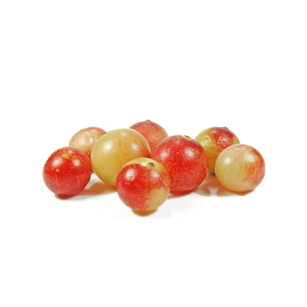 PotentPotentCamu-camuMyrciaria dubiaCarries the highest vitamin C load of any known fruit alongside a dense ellagitannin and anthocyanin fraction, and its juice lowered oxidative DNA damage in smokers where the same dose of vitamin C tablets did not.Mechanismascorbate + ellagitannin/anthocyanin radical scavenging; Nrf2/NQO1 ↑; oxidative DNA-damage (8-OHdG) ↓Read the evidenceShow lessMonograph →
The pulp carries roughly 1.8–6 g of ascorbic acid per 100 g 199,
The human result separates the fruit from its vitamin C. In 20 male smokers given 70 mL of juice (about 1050 mg vitamin C) or 1050 mg of vitamin C tablets for seven days, urinary 8-hydroxy-deoxyguanosine and total ROS fell significantly in the juice group and not in the tablet group 203Reference 203Tropical fruit camu-camu (Myrciaria dubia) has anti-oxidative and anti-inflammatory propertiesView study →. Non-ascorbate constituents, or altered ascorbate kinetics, are doing work that pure ascorbate does not.
Preclinically the signal is broad and replicated: Nrf2/NQO1 activation and ROS suppression in high-glucose human keratinocytes 204Reference 204In vitroCamu-camu fruit extract inhibits oxidative stress and inflammatory responses by regulating NFAT and Nrf2 signalling in high-glucose-induced human keratinocytes — in vitroView study →, and protection against hydrogen-peroxide and ethanol-induced DNA damage in mice with no genotoxicity of its own 205,
The human evidence is a single seven-day trial of 20 people under high oxidative load, and a randomised crossover of a blended açai, camu-camu and blackberry juice in 12 healthy non-smokers found no change in oxidative-defence markers 207Reference 207Bolus consumption of a specifically designed fruit juice rich in anthocyanins and ascorbic acid did not influence markers of antioxidative defense in healthy humansView study →. The effect may need an elevated baseline or camu-camu as the sole ingredient.
Ascorbate degrades fast, with up to a quarter lost in a month whether frozen or not 199Reference 199The Healing Power of Rainforest Herbs: A Guide to Understanding and Using Herbal Medicinals, so a stale spray-dried powder is a weaker product than the fresh pulp or juice the trials used.
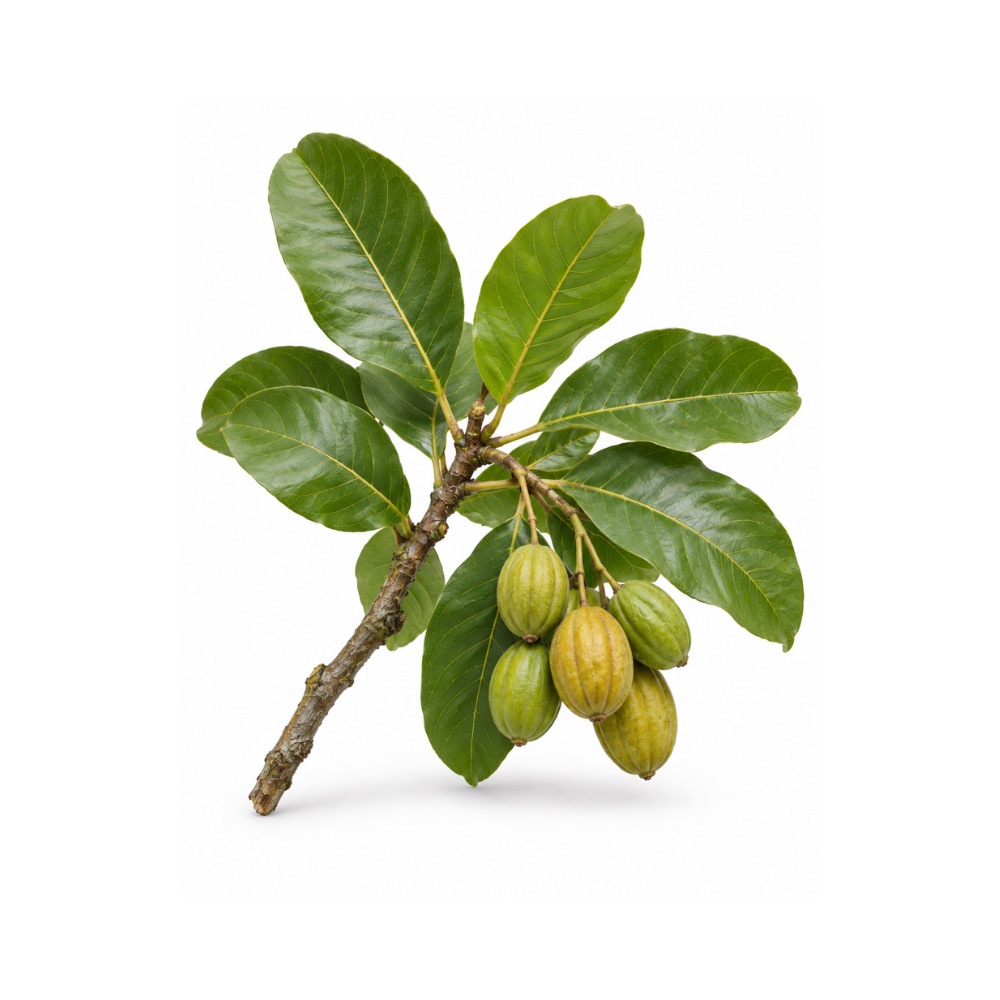 PotentPotentHaritakiTerminalia chebulaThe strongest antioxidant of the three Triphala fruits, with one of the richest hydrolysable-tannin loads in the materia medica, and reduced oxidative-stress markers in a diabetic trial.Mechanismradical scavenging (chebulagic/chebulinic acid, corilagin, gallic/ellagic acid); hepatic antioxidant-enzyme inductionRead the evidenceShow lessMonograph →
In the head-to-head comparison of the three Triphala fruits, T. chebula ranked highest, with very low antioxidant IC50 values and dose-dependent increases in hepatic antioxidant enzymes in rats 380Reference 380RCTComparative evaluation of Terminalia chebula extract mouthwash and chlorhexidine mouthwash on plaque and gingival inflammation — 4-week randomised controlled trialView study →, ahead of amla, the fruit usually credited as the antioxidant of the trio.
The route is chemical. The fruit is one of the richest natural sources of hydrolysable tannins, dense enough that the dried fruit is a commercial tanning material, and that gallate and ellagitannin fraction is what scavenges.
Human support is indirect. The type-2-diabetes RCT recorded reduced oxidative-stress markers on 250–500 mg twice daily 381Reference 381In vitroComparative study of the antioxidant and reactive oxygen species scavenging properties in the extracts of the fruits of Terminalia chebula, Terminalia belerica and Emblica officinalis — in vitro and animalView study →.
Antioxidant capacity is a laboratory property measured in a dish rather than a clinical outcome, and the enzyme-induction data are rodent.
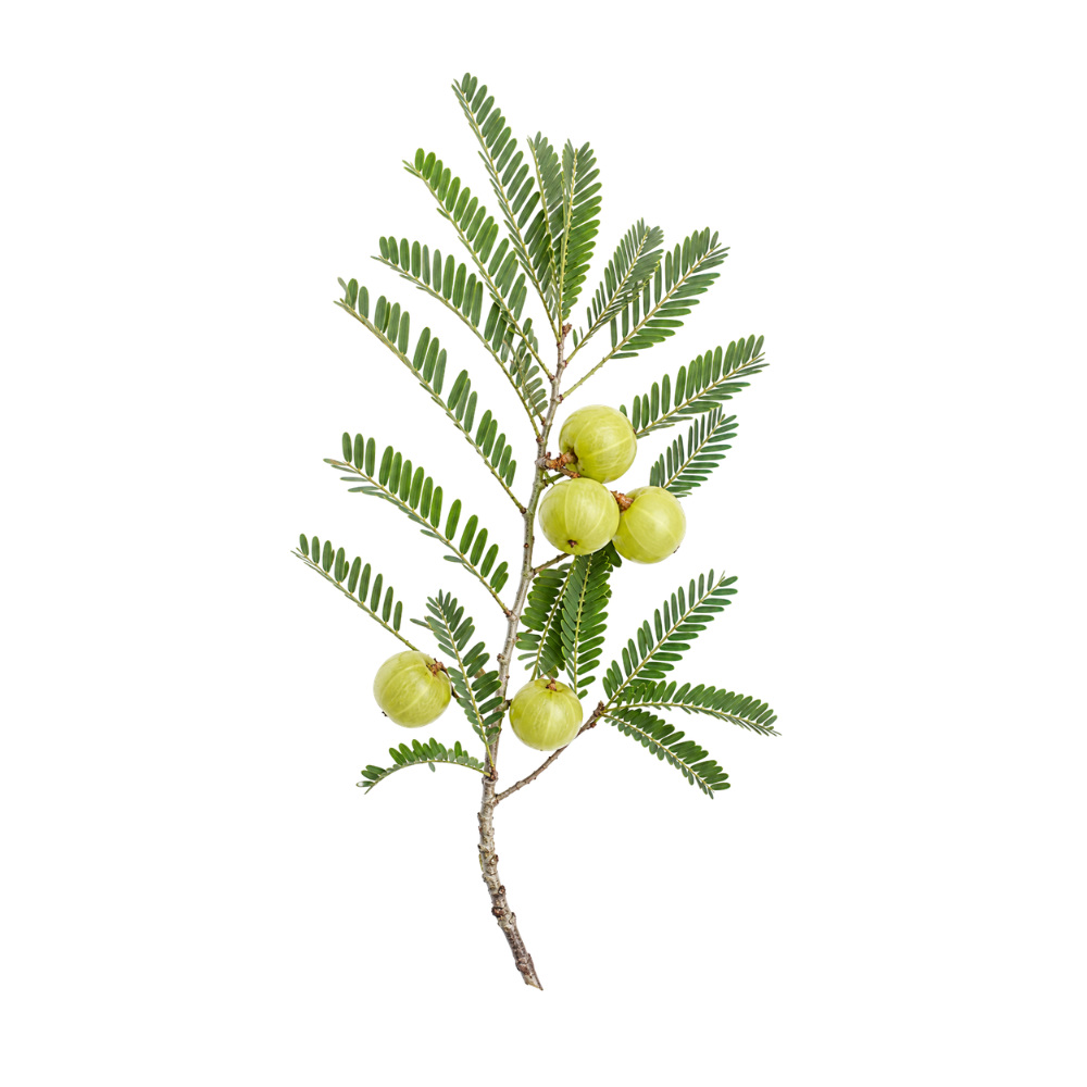 StrongStrongAmlaPhyllanthus emblicaLowers malondialdehyde and raises glutathione and total antioxidant status in people, through hydrolysable tannins that also stabilise its vitamin C.Mechanismhydrolysable-tannin (emblicanin A/B, pedunculagin, punigluconin) radical scavenging; ascorbate stabilized by the same tannins; MDA and 8-iso-PGF₂α ↓, glutathione/SOD/catalase ↑Read the evidenceShow lessMonograph →
The cardiometabolic RCTs recorded falls in malondialdehyde and rises in glutathione and total antioxidant status 149,
The chemistry is the tannins: emblicanin A and B, pedunculagin and punigluconin scavenge radicals and stabilise the fruit’s ascorbate. Amla’s vitamin C content is far lower than the popular figures suggest, and the tannins do most of the work.
The human signals are secondary markers inside lipid and renal trials, never an antioxidant endpoint in its own right, and none has been linked to a clinical outcome. The strongest antioxidant-adjacent results, the Amalaki-rasayana DNA-repair RCT in aged individuals 152Reference 152RCTEffect of Amalaki rasayana on DNA damage and repair in randomized aged human individualsView study → and the rodent geroprotection study 153Reference 153AnimalGeroprotection through modulation of heart rate variability, oxidative enzymes, tissue integrity and gene expression by Amalaki Rasayana — rat in vivo modelView study →, are multi-herb formulas rather than the fruit alone.
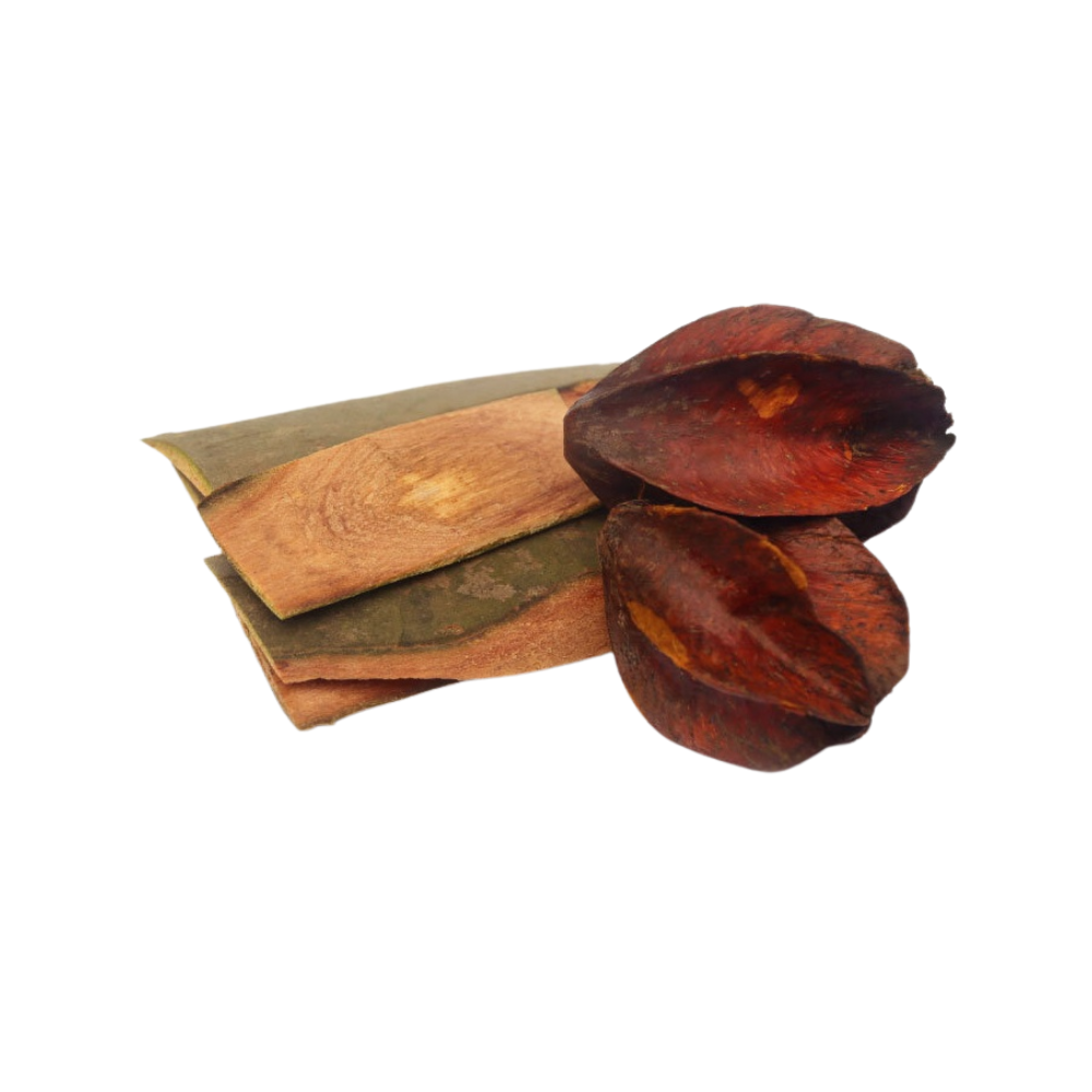 StrongStrongArjunaTerminalia arjunaRaised or preserved red-cell superoxide dismutase, catalase and glutathione against placebo in a 100-patient heart-failure trial, through a bark that is about 16% tannin.Mechanismtannin/flavonoid radical scavenging; endogenous antioxidant-enzyme preservation (RBC SOD, catalase, glutathione ↑ in humans); myocardial lipid peroxidation ↓Read the evidenceShow lessMonograph →
In the 100-patient chronic heart-failure RCT, add-on extract at 750 mg twice daily significantly raised or preserved red-blood-cell superoxide dismutase, catalase and glutathione versus placebo 155Reference 155RCTClinical efficacy of water extract of stem bark of Terminalia arjuna in patients of chronic heart failure — a double-blind, randomised controlled trialView study →. Rat isoproterenol cardiotoxicity models show the same thing in the target tissue, with restored myocardial antioxidant defences and limited lipid peroxidation 156Reference 156AnimalTerminalia arjuna augments cardioprotection via antioxidant and antiapoptotic cascade in isoproterenol-induced cardiotoxicity in rats — animal modelView study →.
The bark is dominated by phenolics, tannins at roughly 16% of an aqueous extract plus a large flavonoid complement including luteolin, baicalein, quercetin and proanthocyanidins, which scavenge reactive oxygen species and preserve the body’s own antioxidant enzymes 154Reference 154ReviewMedicinal properties of Terminalia arjuna (Roxb.) Wight & Arn. — a reviewView study →. The antioxidant effect is the presumed engine under the rest of what the bark does.
The human evidence is surrogate blood biomarkers from a single trial, and that trial was null on its own primary cardiac endpoint.
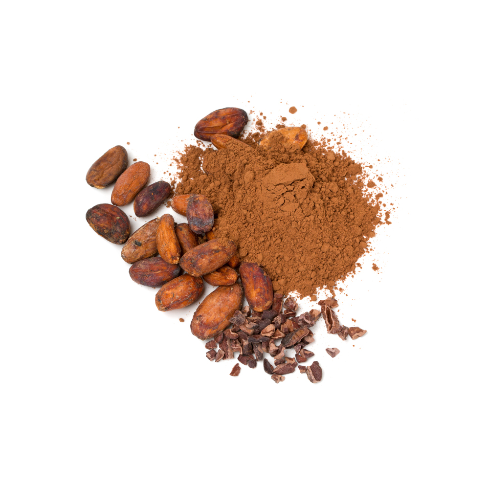 StrongStrongCacaoTheobroma cacaoFlavan-3-ols and procyanidins at 12–18% of the dry bean scavenge radicals in vitro, and pooled RCTs show cocoa flavanols shifting oxidative-status markers in people.Mechanismfree-radical scavenging; LDL oxidation ↓ (procyanidins, polyphenols)Read the evidenceShow lessMonograph →
A 2016 meta-analysis of RCTs found cocoa flavanol intake favourably shifted a panel of cardiometabolic biomarkers including oxidative-status, insulin-resistance and inflammation markers 197Reference 197Meta-analysisCocoa flavanol intake and biomarkers for cardiometabolic health: a systematic review and meta-analysis of randomised controlled trialsView study →.
The bean carries one of the densest polyphenol loads of any commonly eaten plant material, about 12–18% of the dry bean, of which procyanidins are about 58%, and those flavan-3-ols and procyanidins are potent radical scavengers in vitro 195,
Processing decides the dose. Radical-scavenging capacity concentrates in the least-processed plant parts, the leaf, cherelle and unfermented bean 195,
The human evidence stops at biomarkers rather than disease outcomes, and test-tube scavenging capacity is not a clinical antioxidant effect.
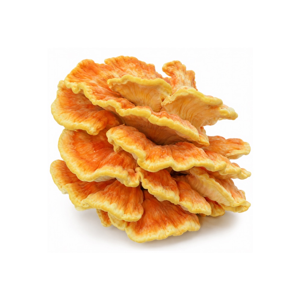 StrongStrongChicken Of The WoodsLaetiporus sulphureusScavenges radicals and chelates metals in vitro through an identified and unusually plant-like phenolic profile of quercetin, kaempferol, catechin and phenolic acids.Mechanismradical scavenging, reducing power, metal chelationRead the evidenceShow less
Strong DPPH, reducing-power and metal-chelating activity has been recorded across many independent extracts 295,
Metal chelation is a distinct antioxidant mechanism from radical scavenging, and fewer fungi demonstrate both.
Every measurement is an assay, and there are no human data.
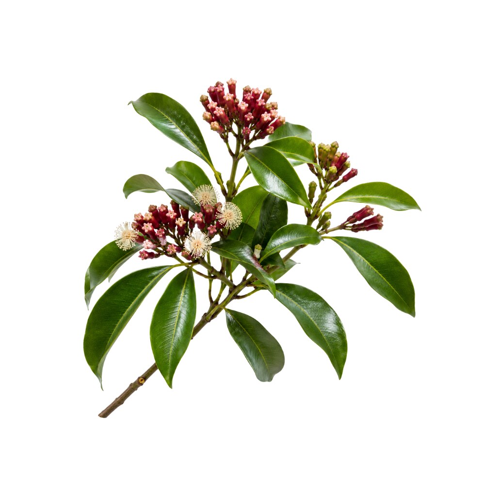 StrongStrongCloveSyzygium aromaticumAmong the most phenolic-rich spices there is; eugenol, gallic acid and the bud tannins scavenge radicals strongly in standard assays.Mechanismradical scavenging; metal chelationRead the evidenceShow lessMonograph →
Both the essential oil and the bud’s non-volatile fraction of gallic acid, hydrolysable tannins, caffeic acid and kaempferol show strong radical scavenging in standard assays 223,
Eugenol is itself a potent antioxidant, and its redox activity is proposed as the upstream driver of much of clove’s anti-inflammatory and tissue-protective behaviour 225Reference 225ReviewAn Overview on the Anti-inflammatory Potential and Antioxidant Profile of Eugenol — [review]View study →.
The activity is highly replicated but in vitro, with no human data.
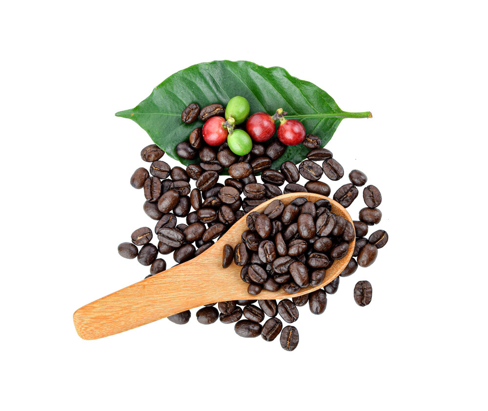 StrongStrongCoffeeCoffea arabicaFor many populations the single largest dietary source of antioxidants, through chlorogenic acid at 6–10% of the green bean and the melanoidins that roasting generates.Mechanismchlorogenic-acid radical scavenging; roasting melanoidinsRead the evidenceShow lessMonograph →
Green beans are 6–10% chlorogenic acid by dry weight, the largest bioactive fraction 226,
Roasting degrades native chlorogenic acid and generates radical-scavenging melanoidins in its place, so the brewed cup keeps much of its capacity.
Dietary dominance plus demonstrated bioavailability is a stronger footing than the in-vitro-only claims most herbs rest on.
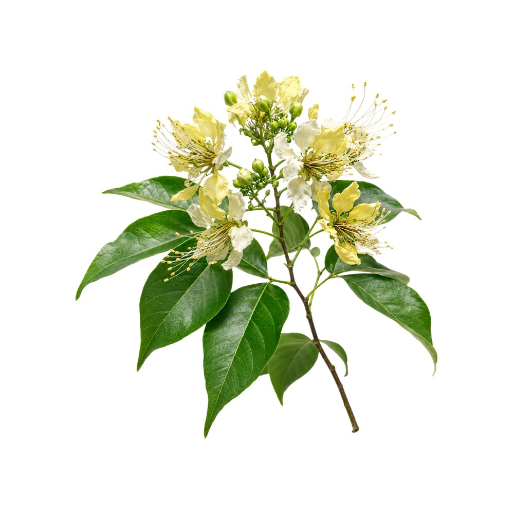 StrongStrongCrataevaCrataeva nurvalaThe stem bark had the highest phenolic content and the strongest superoxide-dismutase-mimetic activity of five Indian medicinal barks, and restores antioxidant enzymes in kidney and heart in vivo.Mechanismfree-radical scavenging; SOD/catalase/GPx restoration; lipid peroxidation ↓Read the evidenceShow lessMonograph →
Screened against four other Indian medicinal barks across multiple in-vitro assays, C. nurvala bark had the highest total phenolic and tannin content and the strongest superoxide-dismutase-mimetic activity of the cohort 233Reference 233In vitroScreening of antioxidant potential of selected barks of Indian medicinal plants — in vitroView study →.
In rodents the effect holds in tissue. Bark extract blunted testosterone/NMU-induced oxidative damage in the prostate 234Reference 234AnimalModulatory effects of Crataeva nurvala bark against testosterone- and NMU-induced oxidative damage in prostate — animal modelView study →, and in two-kidney-one-clip hypertensive rats it normalised malondialdehyde and nitric oxide while restoring antioxidant enzymes in both kidney and heart 235Reference 235AnimalCrataeva nurvala bark extract modulates oxidative-stress gene expression and restores antioxidant enzymes in kidney and heart of 2K1C rats — animal modelView study →.
The nephro-, cardio- and hepatoprotective effects are one redox mechanism pointed at different tissues, and the evidence uses the medicinal part.
The evidence is entirely preclinical, with no human biomarker or clinical antioxidant datum.
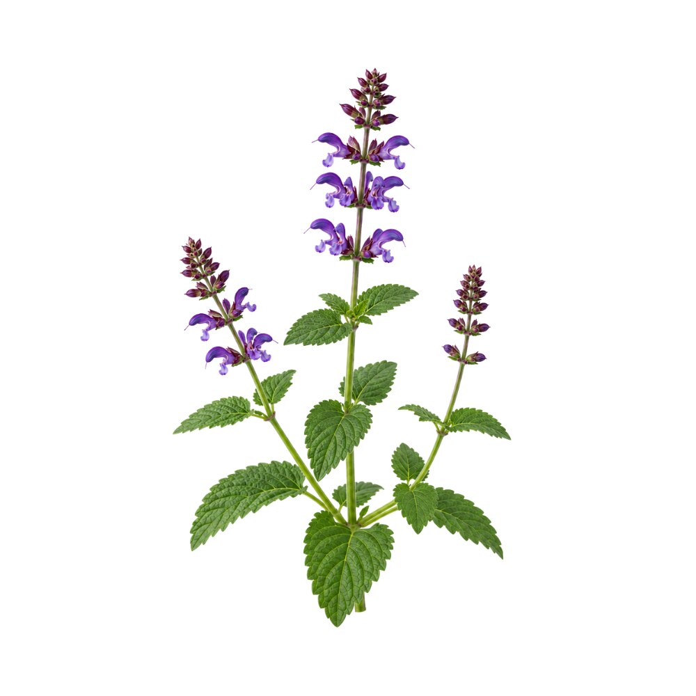 StrongStrongDan ShenSalvia miltiorrhizaTwo unrelated fractions act by two routes: the salvianolic acids scavenge radicals directly, and tanshinone IIA induces Nrf2 so the cell makes its own antioxidant enzymes.Mechanismdirect radical scavenging (salvianolic acids); Nrf2 pathway upregulation (tanshinone IIA)Read the evidenceShow lessMonograph →
The water-soluble salvianolic acids, especially salvianolic acid B, are potent direct free-radical scavengers, while the lipophilic tanshinone IIA works indirectly, upregulating the Nrf2 pathway so the cell makes more of its own antioxidant enzymes 101Reference 101ReviewPharmacological activity and mechanism of tanshinone IIA in related diseases — reviewView study →. That direct and indirect pairing in one root is unusual and is the reason the effect shows up in tissues as different as myocardium and liver.
The cardioprotective, hepatoprotective and neuroprotective signals all resolve back to this redox activity, and it is measured through downstream endpoints such as the SOD rise and malondialdehyde fall in the ischaemia-reperfusion models 102Reference 102Meta-analysisEfficacy of tanshinone IIA in rat models with myocardial ischemia-reperfusion injury — systematic mini-review and meta-analysis of 15 studiesView study →.
No human trial measures a clinical outcome attributable to the antioxidant activity. The Nrf2 result belongs to tanshinone IIA specifically and is routinely extended to tanshinone I, for which no equivalent finding has been published.
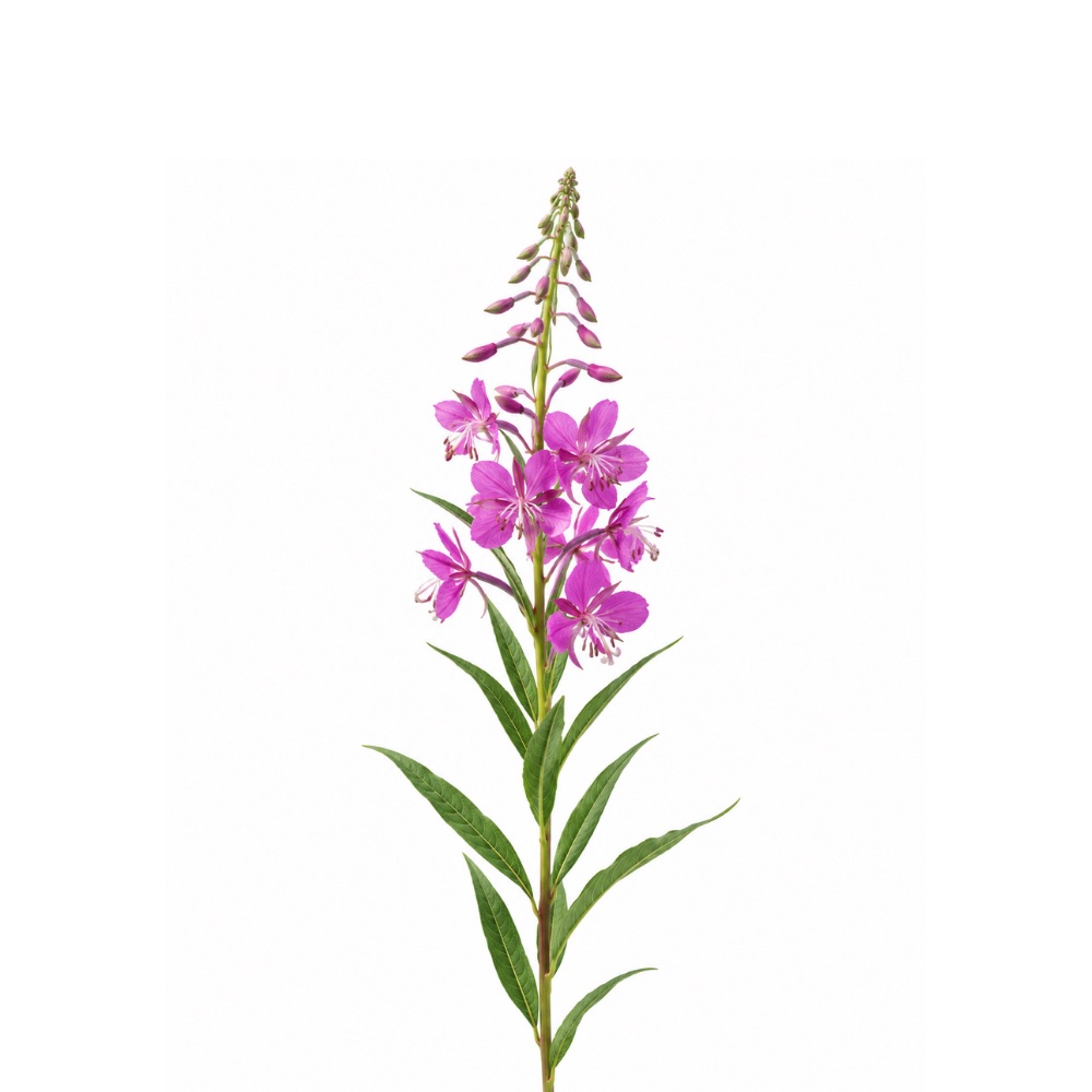 StrongStrongFireweedChamerion angustifoliumNear the top of the field on phenolic density and assay capacity, and one of the few herbs whose antioxidant claim is backed by enzyme rescue in a live animal.Mechanismradical scavenging; SOD/CAT/GPx enzyme rescue; metal chelationRead the evidenceShow less
Total phenolics run to about 85 mg GAE/g, with DPPH and ABTS scavenging at 310.7 and 466.8 mg Trolox-equivalent/g, plus high CUPRAC and FRAP reducing power and metal chelation 240Reference 240In vitroExploring the phytochemical profile and biological insights of Epilobium angustifolium L. herb — LC-Orbitrap profiling, antioxidant and enzyme-inhibition assays (in vitro)View study →. Activity tracks gallic acid, ellagic acid, flavonoids and oenothein B.
In DSS-colitis mice the extract raised colonic glutathione 64% and boosted catalase, SOD and glutathione peroxidase activity 36–70% 241Reference 241AnimalUlceroprotective effects of Epilobium angustifolium extract in DSS-induced colitis in mice (in vivo)View study →, which is what lifts it above the many herbs with a good DPPH number.
There are no human data.
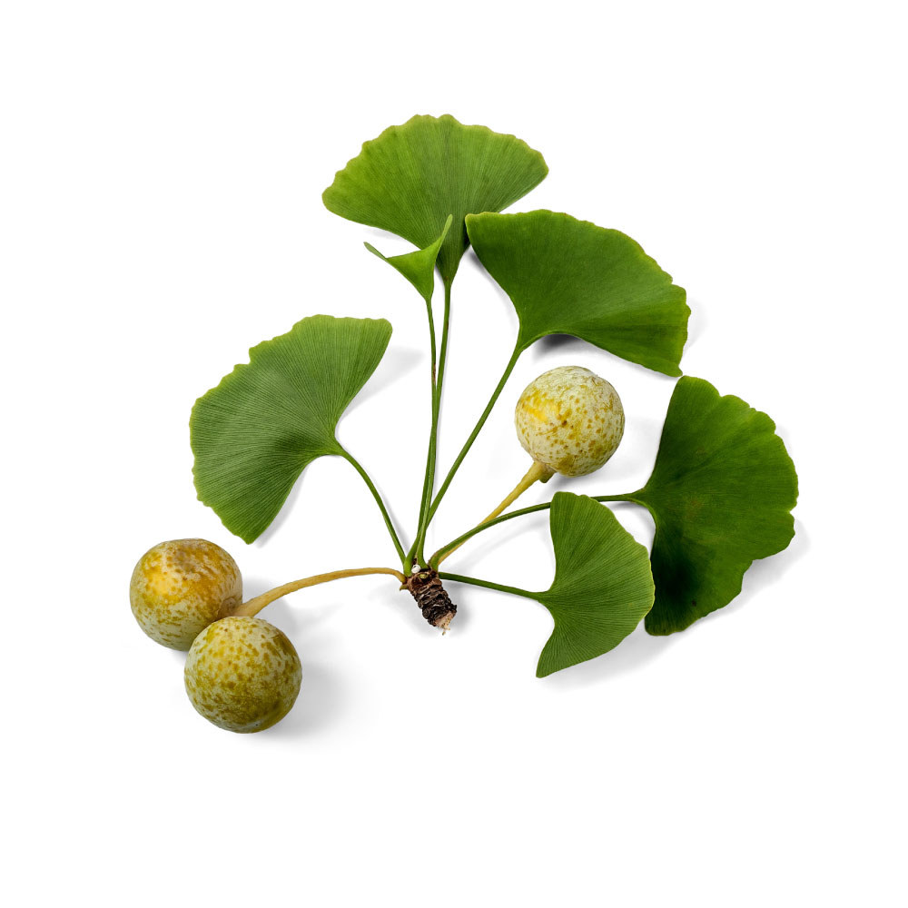 StrongStrongGinkgoGinkgo bilobaThe EGb 761 flavonol glycosides of quercetin, kaempferol and isorhamnetin scavenge peroxyl and hydroxyl radicals directly, and the whole extract cuts oxidative damage in neurons and retina.Mechanismradical scavenging; metal chelationRead the evidenceShow lessMonograph →
The flavonol glycosides of quercetin, kaempferol and isorhamnetin are direct peroxyl- and hydroxyl-radical scavengers in vitro 253Reference 253In vitroPeroxyl radical scavenging activity of Ginkgo biloba extract EGb 761 — in vitro studyView study →, and the whole extract cuts oxidative damage in neurons and retina 254,
Antioxidant activity is ginkgo’s mechanistic core and its best-replicated laboratory finding, and it underlies the neuroprotective and circulatory work.
The evidence is entirely in vitro and animal, and no human trial links ginkgo’s antioxidant action to a clinical outcome.
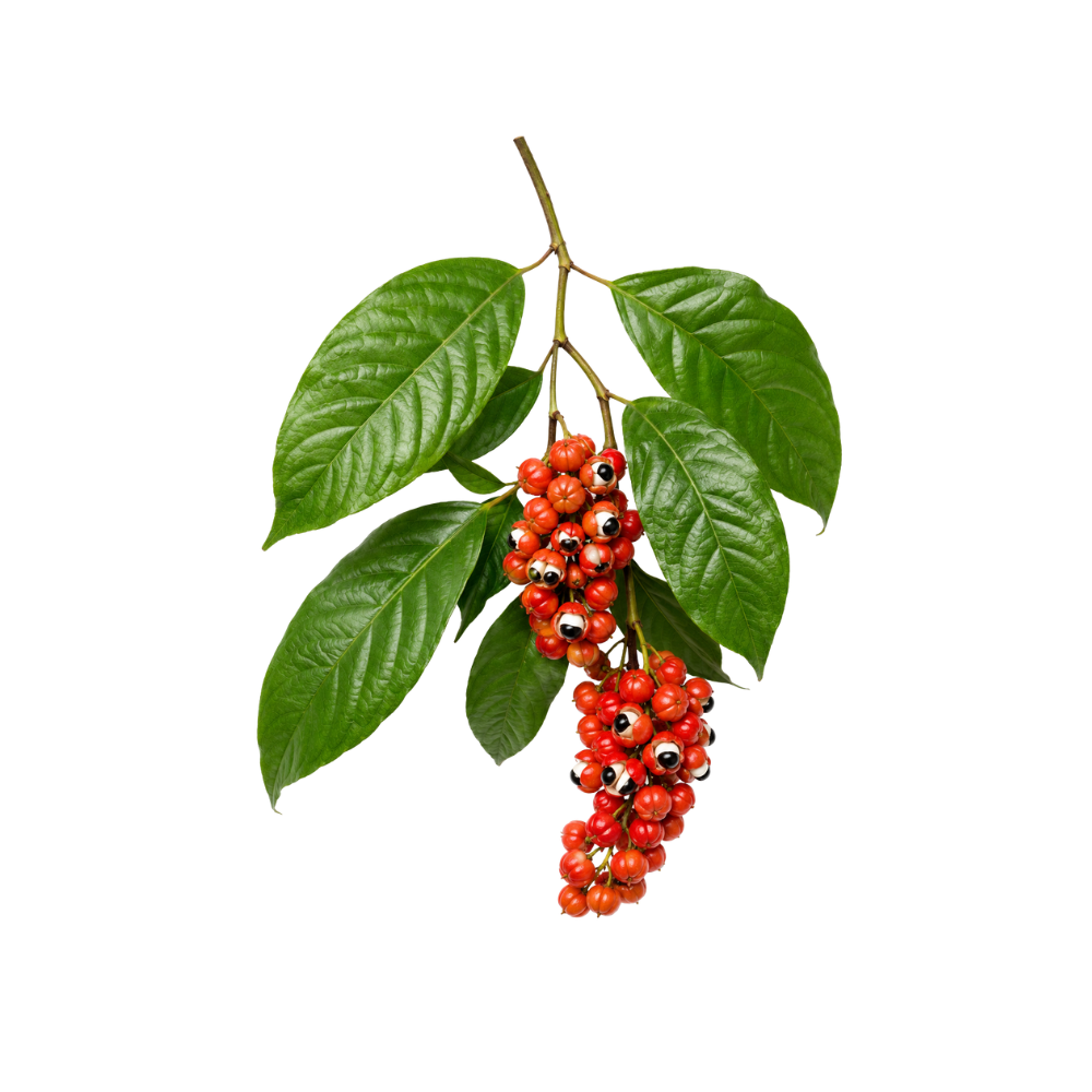 StrongStrongGuaranaPaullinia cupanaCondensed tannins at 8.5–9.5 g per 100 g of seed scavenge radicals strongly, a caffeine-free fraction keeps the full activity, and habitual consumers showed 27% lower LDL oxidation.Mechanismradical scavenging (condensed tannins, catechin/epicatechin); SOD & catalase modulation; glutathione preservationRead the evidenceShow lessMonograph →
The ethanol seed extract scavenges radicals strongly in vitro, attributed to the polyphenol fraction of catechol, catechin and epicatechin 265Reference 265In vitroAntibacterial and antioxidant activities of ethanol extract from Paullinia cupana — in vitroView study →, and isolated condensed tannins scavenge DPPH potently 266Reference 266In vitroAntioxidant capacity and in vitro prevention of dental plaque by extracts and condensed tannins of Paullinia cupana — in vitroView study →. A tannin-rich, methylxanthine-free fraction retains full antioxidant and anti-TNF-α activity, which shows the effect is polyphenol-driven rather than caffeine-driven 267Reference 267In vitroTNF-α inhibition, antioxidant effects and chemical analysis of guaraná seed powder — in vitroView study →.
The seed carries 8.5–9.5 g of condensed tannin per 100 g dried 268Reference 268ReviewGuarana: revisiting a highly caffeinated plant from the Amazon — reviewView study →.
The human signal is observational: elderly habitual guarana consumers showed 27% lower LDL oxidation than non-consumers, and the extract suppressed conjugated-diene and TBARS formation ex vivo 269Reference 269In vitroGuaraná effects on LDL oxidation in elderly people — in vitro and in vivo studyView study →. Redox-enzyme modulation shows up in the rodent hepatoprotection work, with preserved glutathione and modulated SOD and catalase against a paracetamol challenge 270Reference 270AnimalPreventive guarana powder mitigates acute paracetamol-induced hepatotoxicity in rats — animal modelView study →.
Reported potency varies several-fold with origin, roasting and fermentation 268Reference 268ReviewGuarana: revisiting a highly caffeinated plant from the Amazon — reviewView study →. Commercial energy-drink processing strips the tannins to avoid the brown colour, and sugary guarana soft drinks were pro-oxidant in dyslipidaemic mice 271Reference 271AnimalWorsening of oxidative stress, DNA damage and atherosclerotic lesions with guarana soft drinks in LDLr-knockout mice — animal modelView study →.
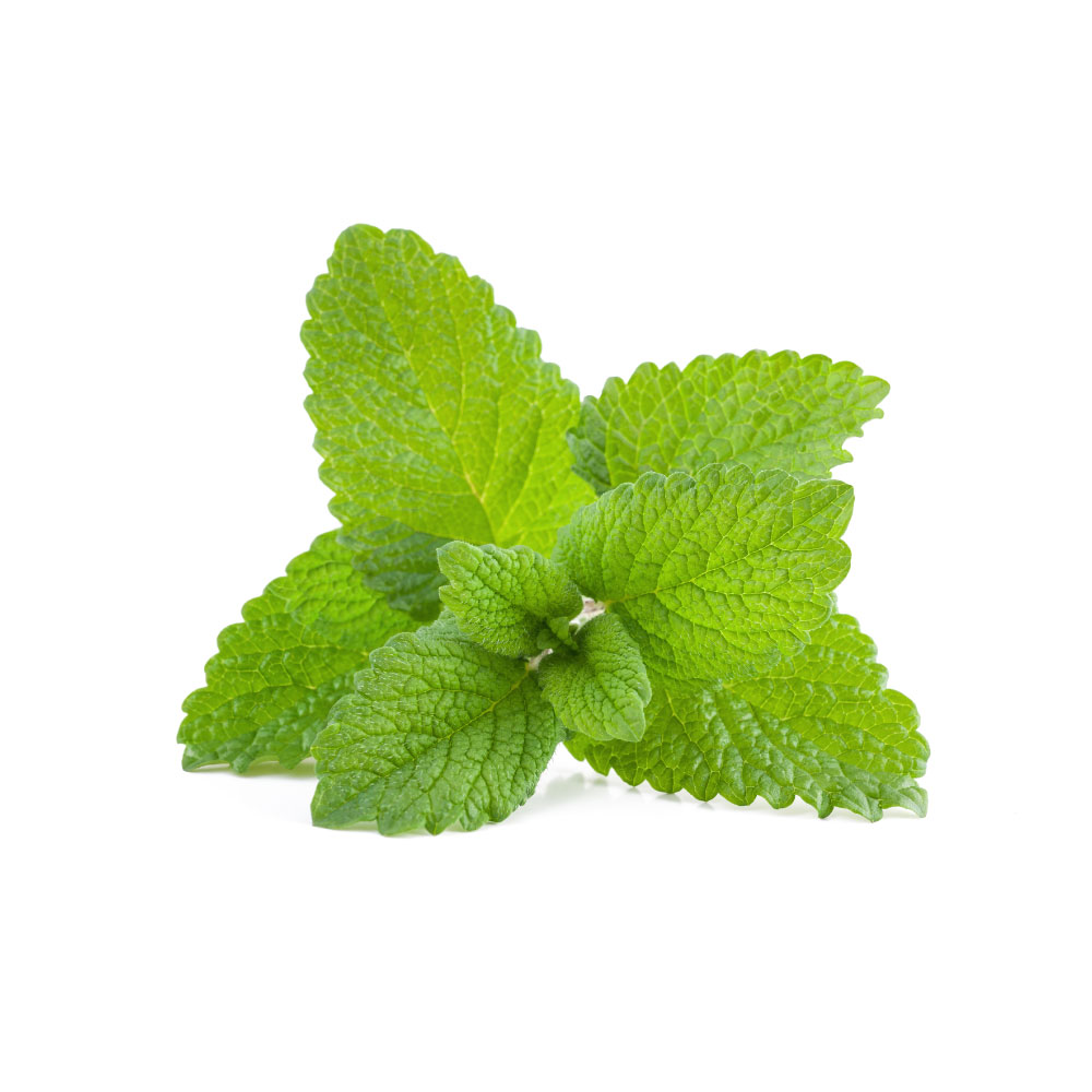 StrongStrongMelissaMelissa officinalisLowered serum malondialdehyde and raised paraoxonase-1, the HDL enzyme that protects LDL from oxidation, in an eight-week trial of powdered leaf in angina patients.Mechanismrosmarinic/caffeic/chlorogenic acid and luteolin-flavonoid radical scavenging; MDA ↓ and paraoxonase-1 ↑ (human)Read the evidenceShow lessMonograph →
Three grams a day of powdered leaf for eight weeks in chronic stable angina lowered serum malondialdehyde and raised paraoxonase-1 activity against placebo 320Reference 320RCTThe effects of Melissa officinalis in chronic stable angina on serum biomarkers of oxidative stress, inflammation and lipid profile — randomised placebo-controlled trialView study →. PON1 is an HDL-bound enzyme that protects LDL from oxidation, so this is enzyme induction rather than direct scavenging, and it is the mechanism most plausibly linking melissa’s antioxidant activity to its lipid and blood-pressure signals.
The activity belongs to the phenolic fraction, chiefly rosmarinic acid with caffeic and chlorogenic acids and luteolin-based flavonoids 319Reference 319ReviewMelissa officinalis L: a review study with an antioxidant prospective — reviewView study →, and it is lemon balm’s most heavily documented mechanism, more so than the nervine action it is famous for.
The single human study is small, and no antioxidant marker shift has been tied to a clinical outcome.
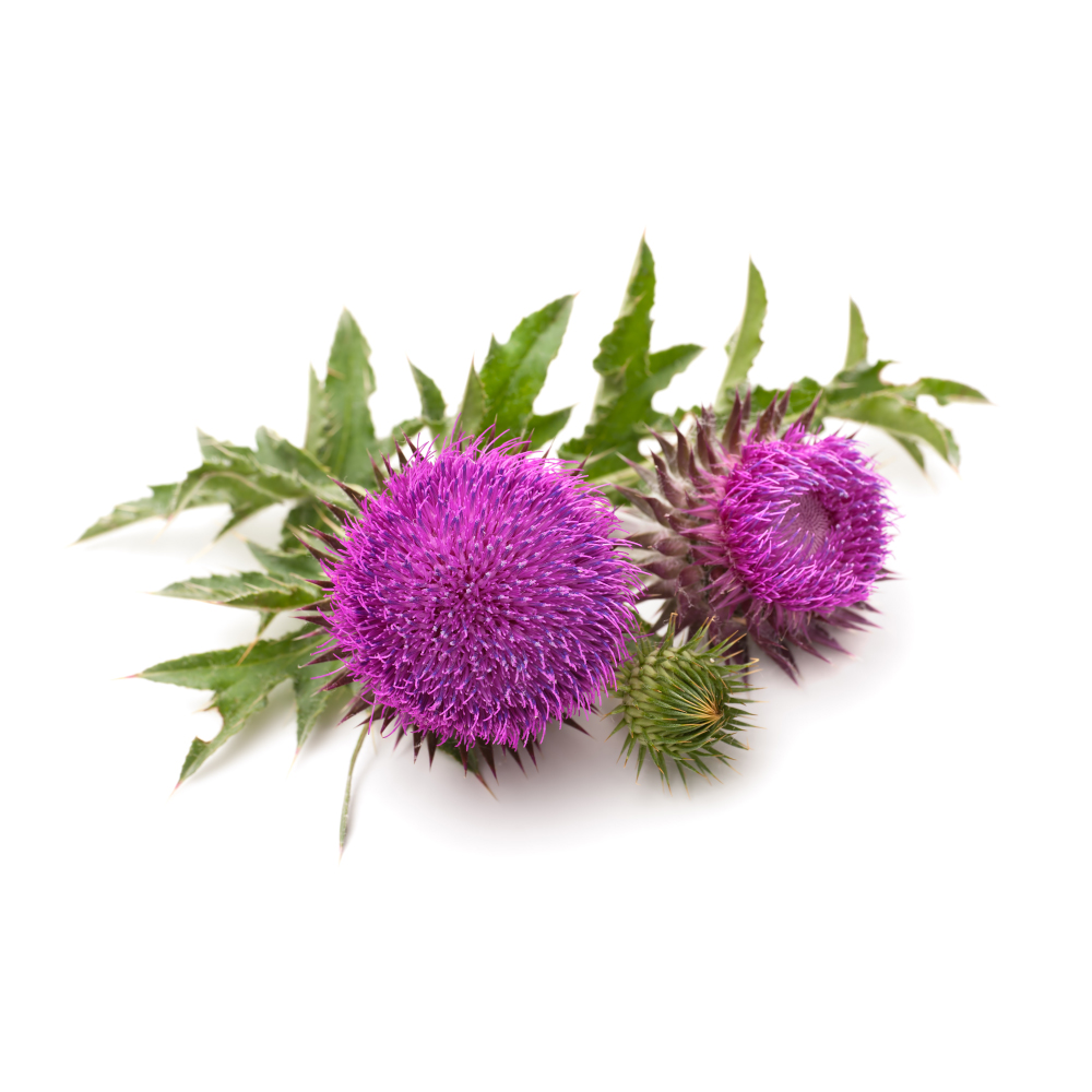 StrongStrongMilk ThistleSilybum marianumSilymarin scavenges free radicals, raises intracellular glutathione and stabilises hepatocyte membranes, which is the mechanism under its liver protection.Mechanismfree-radical scavenging; glutathione ↑; membrane stabilizationRead the evidenceShow lessMonograph →
Preclinically silymarin scavenges free radicals, raises intracellular glutathione, reduces lipid peroxidation and increases catalase, protecting hepatocytes from oxidative insult 321Reference 321ReviewRole of silymarin as antioxidant in clinical management of chronic liver diseases: a narrative review — reviewView study →.
Classic work established that the protection does not depend on inhibiting CYP2E1, pointing instead to direct radical scavenging and membrane stabilisation 322Reference 322In vitroHepatoprotective mechanism of silymarin: no evidence for involvement of cytochrome P450 2E1 — in vitro / animal mechanistic studyView study →, so it is not simply blocking toxin bioactivation.
In humans the antioxidant effect is inferred through enzyme and oxidative-stress markers rather than measured as an endpoint.
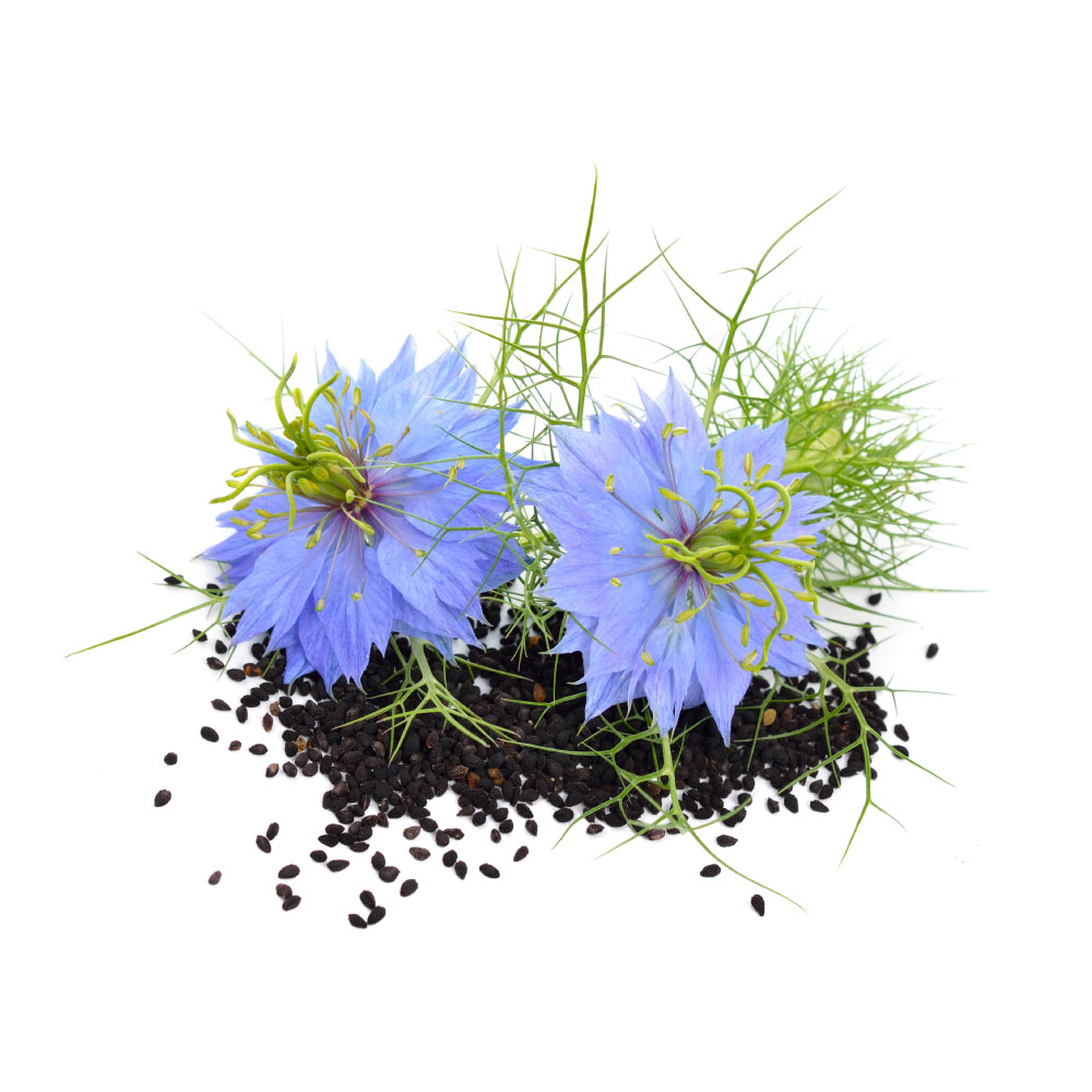 StrongStrongNigellaNigella sativaThymoquinone induces Nrf2 and the endogenous antioxidant enzymes, and pooled RCTs show falling malondialdehyde and rising total antioxidant capacity in people.MechanismNrf2/ARE activation (thymoquinone, thymohydroquinone); antioxidant-enzyme induction; lipid-peroxidation ↓Read the evidenceShow lessMonograph →
Pooled RCTs of the seed show falling malondialdehyde and rising total antioxidant capacity 324Reference 324Meta-analysisThe effect of Nigella sativa on biomarkers of inflammation and oxidative stress — systematic review and meta-analysis of randomized controlled trialsView study →.
Thymoquinone is a redox-cycling quinone in equilibrium with thymohydroquinone, and Nrf2/ARE induction of endogenous antioxidant enzymes is its best-characterised route 323Reference 323ReviewBiological and therapeutic activities of thymoquinone — focus on the Nrf2 signaling pathway — reviewView study →. The same pathway is invoked for the rodent hepatoprotection against chemical and drug-induced injury 325Reference 325AnimalProtective effect of Nigella sativa against 5-fluorouracil-induced liver damage in rats — animal studyView study →, and it is the same thymoquinone mechanism that carries nigella’s anti-inflammatory activity, read out as redox markers rather than cytokines.
The mechanistic work is on isolated thymoquinone rather than on the seed.
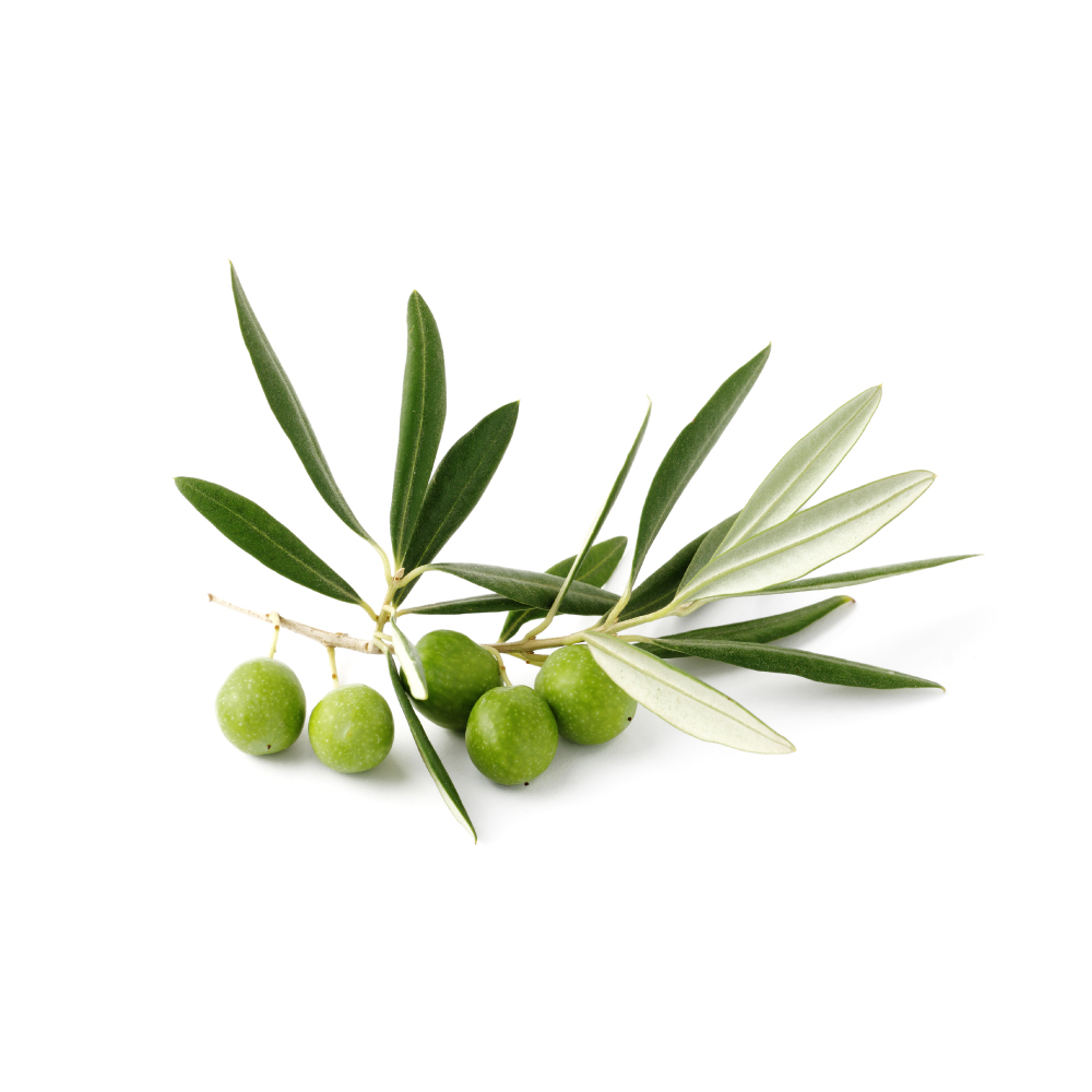 StrongStrongOlive LeafOlea europaeaOleuropein and its breakdown product hydroxytyrosol scavenge radicals and reduce iron in vitro, and 250 mg a day of extract lowered serum malondialdehyde in a 2025 RCT.Mechanismfree-radical scavenging; ferric reduction; lipid peroxidation (MDA) ↓Read the evidenceShow lessMonograph →
Olive leaf extract shows strong radical-scavenging and ferric-reducing activity in vitro that tracks its oleuropein content, with hydroxytyrosol, oleuropein’s hydrolysis product, which accumulates as an extract ages, carrying much of the capacity 332,
A 2025 RCT in women with obesity found 250 mg/day of extract added to a hypocaloric diet lowered serum malondialdehyde more than diet alone 334Reference 334RCTPhenolic-rich extract of olive leaf with a hypocaloric diet alleviates oxidative stress in obese females: a randomized double-blind placebo-controlled trialView study →, and a 2026 combination trial recorded about 23% lower oxidised LDL 335Reference 335RCTEffects of a combination of olive leaf extract and potassium on blood pressure in participants with mild to moderate hypertension: a double-blind, randomized, placebo-controlled trialView study →.
Total antioxidant capacity and liver enzymes were unchanged in the 2025 trial, so what moved was a single lipid-peroxidation marker rather than global redox status.
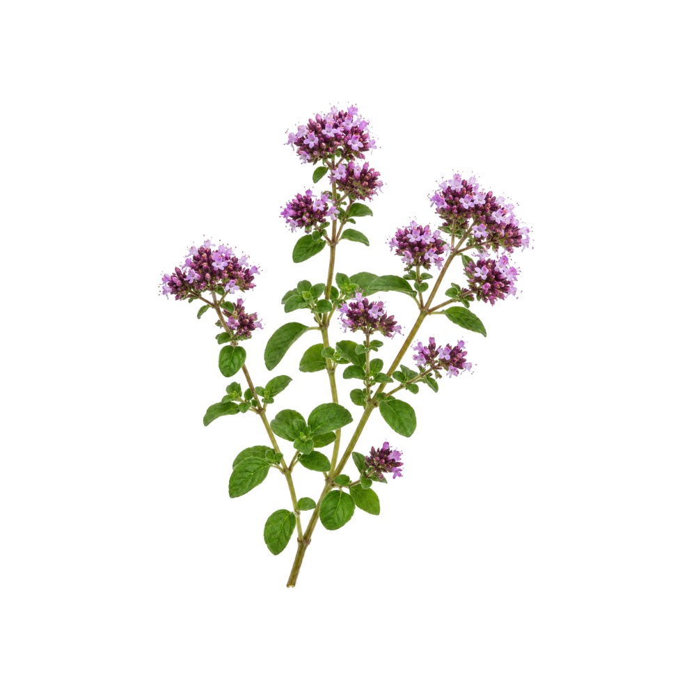 StrongStrongOreganoOriganum vulgareRosmarinic acid and the leaf flavones give the water-soluble fraction field-leading scavenging capacity among culinary mints; a single 500 mg capsule blunted post-exercise malondialdehyde in soldiers.Mechanismfree-radical scavenging and ferric reduction by rosmarinic acid and leaf flavones; ↑ endogenous antioxidant enzymes (SOD, GPX)Read the evidenceShow lessMonograph →
The water-soluble leaf fraction is dominated by rosmarinic acid alongside luteolin, apigenin and quercetin, which show strong radical-scavenging and ferric-reducing activity in DPPH and FRAP assays 336Reference 336In vitroPhenolic compounds from Origanum vulgare and their antioxidant and antiviral activities — in vitroView study →. This is the leaf action rather than the essential-oil one, and it runs on a different chemistry.
The human data are split. A single 500 mg O. vulgare capsule in 24 soldiers blunted post-exercise malondialdehyde and raised total antioxidant capacity 338Reference 338RCTOregano (Origanum vulgare) supplementation and antioxidant/muscle-damage markers after exercise — randomised controlled trialView study →, while four weeks of oregano-extract juice in 45 healthy men markedly increased urinary phenolic excretion yet produced no change in serum lipids or lipid-peroxidation biomarkers 337Reference 337RCTConsumption of juice fortified with oregano extract markedly increases excretion of phenolic acids but lacks short- and long-term effects on lipid peroxidation in healthy nonsmoking men — randomised controlled trialView study →.
Leaf rosmarinic acid ranges roughly 0.6–37 mg/g dry weight across accessions grown under identical conditions 339Reference 339Natural diversity in phenolic components and antioxidant properties of oregano (Origanum vulgare L.) accessions, grown under the same conditionsView study →, a 60-fold spread, so oregano is not one antioxidant material.
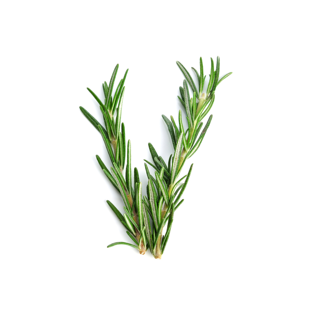 StrongStrongRosemaryRosmarinus officinalisCarnosic acid and carnosol scavenge radicals, chelate metals and induce the Nrf2 response, and standardised rosemary extract is a licensed food antioxidant because the activity holds up in real fats over shelf-life.MechanismNrf2 / phase-II antioxidant response induction; peroxyl and hydroxyl scavenging plus metal chelation (carnosic acid, carnosol)Read the evidenceShow lessMonograph →
Standardised rosemary extract is a licensed food and cosmetic antioxidant because its activity holds up in real matrices over shelf-life rather than only in an assay 354,
The actives are the non-volatile diterpene phenols carnosic acid and carnosol, with rosmarinic acid and ursolic acid. The essential oil that carries rosemary’s cognitive and rubefacient effects is not the fraction doing this.
Early work showed carnosol and carnosic acid scavenge peroxyl and hydroxyl radicals and chelate metals 6Reference 6In vitroAntioxidant and pro-oxidant properties of active rosemary constituents: carnosol and carnosic acid — in vitroView study →, and the activity is now largely understood to run through induction of the Nrf2/phase-II response as well, so the herb makes the cell build its own defences.
Carnosic acid can act as a pro-oxidant under some conditions 6Reference 6In vitroAntioxidant and pro-oxidant properties of active rosemary constituents: carnosol and carnosic acid — in vitroView study →, and almost all the data are in vitro or in food matrices, with little human pharmacodynamic confirmation at realistic oral doses.
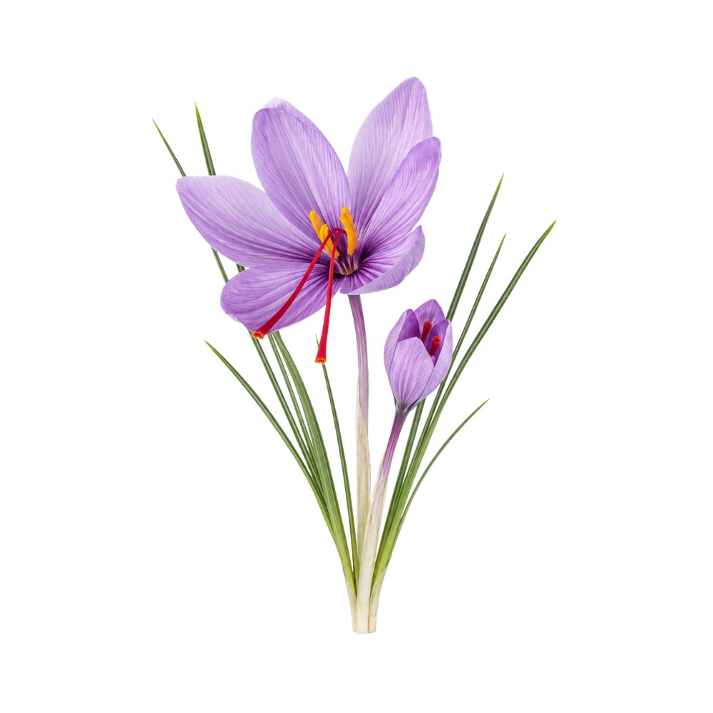 StrongStrongSaffronCrocus sativusCrocin and crocetin, its water-soluble carotenoids, scavenge radicals and activate Nrf2; 15 mg a day raised serum radical-scavenging activity in a prediabetes trial.Mechanismfree-radical scavenging (crocin/crocetin); Nrf2 pathway activationRead the evidenceShow lessMonograph →
The water-soluble carotenoid crocin and its aglycone crocetin scavenge free radicals and upregulate the Nrf2 antioxidant pathway 356Reference 356ReviewAnti-inflammatory, antioxidant, and immunomodulatory effects of Crocus sativus L. and its main constituents — reviewView study →. Apocarotenoids are capable radical scavengers, and much of the rest of saffron’s pharmacology, in the retina, in metabolism and in neuroprotection, runs downstream of this activity.
In humans it shows up as biomarker movement. A prediabetes RCT found 15 mg/day raised DPPH radical-scavenging activity alongside improved glycaemic markers 357Reference 357RCTEffect of saffron (Crocus sativus L.) on lipid profile, glycemic indices and antioxidant status among overweight/obese prediabetic individuals: a double-blinded, randomised controlled trialView study →.
No clinical antioxidant endpoint has been measured, only surrogate blood markers, and at 15–30 mg/day the absolute dose of scavenger delivered is tiny beside the gram-scale polyphenol herbs.
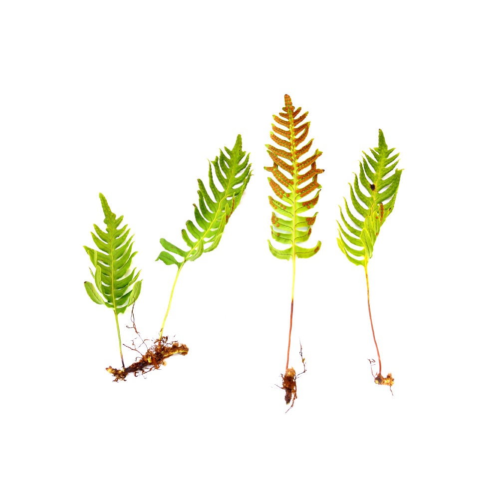 StrongStrongSamambaiaPolypodium leucotomosIts phenolic acids scavenge reactive oxygen species and the extract activates NRF2 in keratinocytes, which is the mechanism behind its reduction of UV-induced DNA damage in people.MechanismROS scavenging; NRF2 pathway activation; lipid-peroxidation inhibitionRead the evidenceShow lessMonograph →
HPLC fractionation of the aqueous extract identifies caffeic acid, ferulic acid, p-coumaric acid, vanillic acid and chlorogenic acid, which directly scavenge reactive oxygen species and inhibit lipid peroxidation 358,
Beyond direct scavenging, the extract upregulates the NRF2 pathway in keratinocytes and blunts both immediate and delayed cyclobutane-pyrimidine-dimer formation after UVA 360,
No standalone trial of systemic oxidative-stress biomarkers exists, so the human relevance is confined to the skin.
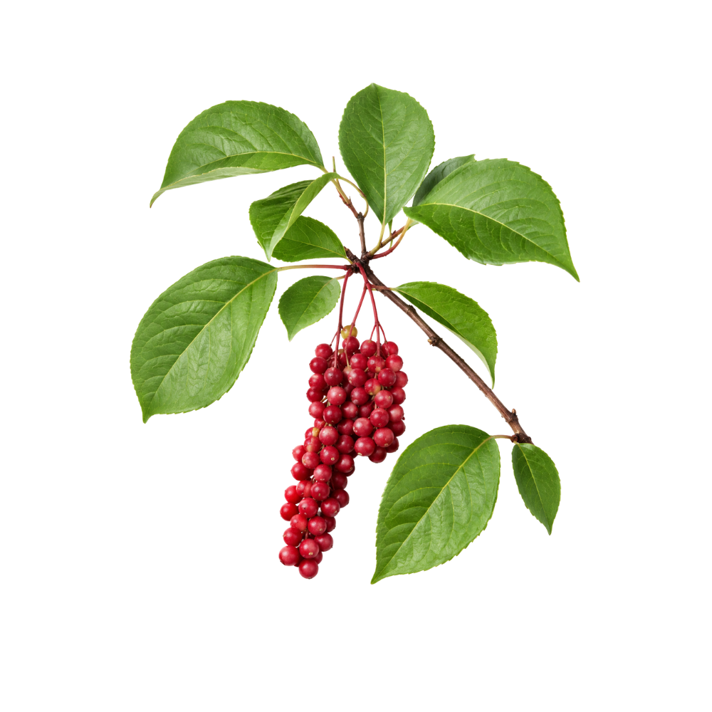 StrongStrongSchisandraSchisandra chinensisIts lignans activate the Nrf2/ARE programme, raising glutathione and SOD while lowering lipid peroxidation in liver, skin and neuronal models.MechanismNrf2/ARE; glutathione & SOD ↑; MDA ↓Read the evidenceShow lessMonograph →
Across liver 366Reference 366AnimalSchisandrol B protects against acetaminophen-induced acute hepatotoxicity in mice via activation of the NRF2/ARE signaling pathway — animal (mouse) studyView study →, skin 367Reference 367In vitroSchisandrin A alleviates UVB-induced photoaging via AMPK/Nrf2-mediated antioxidant and autophagy pathways — in vitro and mouse studyView study → and neuronal models, the lignans switch on the Nrf2/ARE transcriptional programme, raising glutathione, SOD and downstream antioxidant enzymes while lowering lipid-peroxidation markers, with schisandrin A working through AMPK/Nrf2 in UVB-stressed keratinocytes 367Reference 367In vitroSchisandrin A alleviates UVB-induced photoaging via AMPK/Nrf2-mediated antioxidant and autophagy pathways — in vitro and mouse studyView study →.
This is the mechanism beneath most of schisandra’s other actions, and it is consistently reproduced across tissue types.
The human translation is thin, as it is for the liver action.
 StrongStrongTeaCamellia sinensisIts catechins, led by EGCG, scavenge radicals in vitro and in vivo in proportion to polyphenol content, and a controlled supplementation trial cut oxidative DNA damage in people.Mechanismfree-radical scavenging; DNA protectionRead the evidenceShow lessMonograph →
Green, oolong and black extracts scavenge radicals in vitro and in vivo with activity tracking total polyphenol content, highest in green at about 23% polyphenol and lowest in black at about 14%, with EGCG the single most active contributor 377Reference 377In vitroAntioxidant and antibacterial properties of green, black, and herbal teas of Camellia sinensis — in vitroView study →.
A controlled supplementation trial cut oxidative DNA damage in human subjects 378Reference 378Clinical trialGenoprotective effects of green tea (Camellia sinensis) in human subjects: results of a controlled supplementation trialView study →, and the flowers are antioxidant too 379Reference 379AnimalAnti-inflammatory effects of a polyphenols-rich extract from tea (Camellia sinensis) flowers in acute and chronic mice models — animalView study →. Green, unoxidised leaf is the higher-potency chemotype.
Low and variable plasma catechin levels limit how much reaches tissue, and tube antioxidant capacity does not translate into disease prevention.
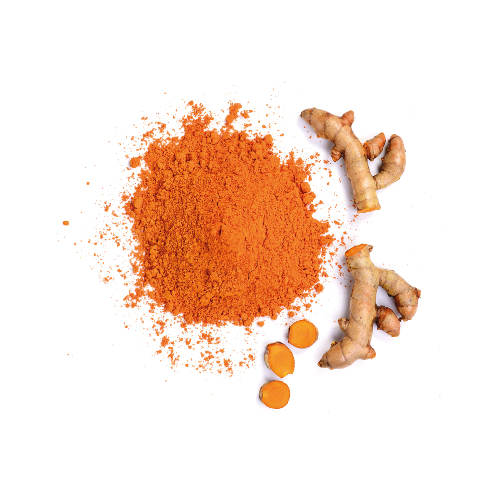 StrongStrongTurmericCurcuma longaCurcumin activates Nrf2 and scavenges radicals, and a 66-trial meta-analysis found it raised total antioxidant capacity, lowered malondialdehyde and increased superoxide dismutase in people.MechanismNrf2/ARE activation; lipid-peroxidation ↓ (MDA); SOD ↑Read the evidenceShow lessMonograph →
The 66-RCT GRADE meta-analysis found curcumin and turmeric significantly raised total antioxidant capacity, lowered malondialdehyde and increased superoxide dismutase activity 389Reference 389Meta-analysisAntioxidant and anti-inflammatory effects of curcumin/turmeric supplementation in adults: a GRADE-assessed systematic review and dose-response meta-analysis of randomized controlled trialsView study →, and a separate 15-RCT analysis echoed the reduced malondialdehyde 390Reference 390Meta-analysisThe effects of curcumin-containing supplements on biomarkers of inflammation and oxidative stress: a systematic review and meta-analysis of randomized controlled trialsView study →.
Mechanistically the curcuminoids scavenge radicals and activate the Nrf2/ARE cytoprotective pathway 389Reference 389Meta-analysisAntioxidant and anti-inflammatory effects of curcumin/turmeric supplementation in adults: a GRADE-assessed systematic review and dose-response meta-analysis of randomized controlled trialsView study →, and this activity is the core beneath most of turmeric’s other effects.
The endpoints are surrogate oxidative-stress markers with high heterogeneity, and no durable clinical outcome has been isolated to the antioxidant action specifically.
 StrongStrongYerba MatéIlex paraguariensisRoughly 10% of the dry leaf is phenolics, dominated by chlorogenic acid and caffeoylquinic acids, and four weeks of extract shifted antioxidant gene networks in men at cardiovascular risk.Mechanismfree-radical scavenging (caffeoylquinic acids, flavonols); antioxidant gene-network upregulation; lipid-peroxidation ↓Read the evidenceShow lessMonograph →
Phenolics make up roughly 10% of the dry leaf, dominated by chlorogenic acid and caffeoylquinic acids with rutin, quercetin and caffeic acid 397,
Four weeks of a standardised polyphenol-rich maté extract in middle-aged men at cardiovascular risk shifted expression of thousands of genes in circulating immune cells, including antioxidant and inflammatory regulatory networks 399Reference 399RCTMultigenomic modifications in human circulating immune cells in response to consumption of polyphenol-rich extract of yerba mate — randomized controlled trialView study →. In C. elegans an aqueous extract extended lifespan and protected against paraquat toxicity 400Reference 400AnimalIlex paraguariensis extract increases lifespan and protects against paraquat toxicity in Caenorhabditis elegans — in vivo modelView study →, and maté lowered oxidative-stress markers in rat bone 401Reference 401AnimalIlex paraguariensis decreases oxidative stress in bone and mitigates damage in rats — in vivo studyView study →.
The substantial human study measured gene expression rather than a clinical antioxidant endpoint. Habitual very-hot maté drinking is an established risk factor for oesophageal squamous cell carcinoma 402,
 StrongStrongPomegranatePunica granatumPunicalagin and the fruit anthocyanins give the juice one of the highest antioxidant capacities of any common beverage, and three years of daily juice reduced LDL oxidation in patients with carotid stenosis.Mechanismellagitannin (punicalagin) and anthocyanin radical scavenging; serum and LDL lipid peroxidation ↓ (human)Read the evidenceShow less
In a three-year controlled trial in patients with carotid artery stenosis, daily juice substantially reduced serum and LDL lipid peroxidation and raised serum antioxidant status, alongside a fall in intima-media thickness and blood pressure 412Reference 412Clinical trialPomegranate juice consumption for 3 years by patients with carotid artery stenosis reduces common carotid intima-media thickness, blood pressure and LDL oxidation — clinical trialView study →.
The chemistry is the ellagitannin punicalagin, its hydrolysis product ellagic acid and the fruit anthocyanins, which give the juice one of the highest measured antioxidant capacities of any commonly consumed beverage.
The evidence belongs to juice and standardised fruit extracts. Polyphenol delivery from tea and whole-fruit intake is lower and more variable, and the larger endpoint trials of carotid progression were null, so the antioxidant markers have not been carried through to a disease outcome.
 ModerateModerateAbutaCissampelos pareiraThe alkaloid root fraction scavenges DPPH and superoxide radicals and inhibits iron-induced lipid peroxidation in rat-liver homogenate, the tissue its liver protection acts on.Mechanismradical scavenging; lipid-peroxidation ↓ (alkaloid fraction)Read the evidenceShow lessMonograph →
The alkaloidal root fraction, containing about 0.18% berberine, strongly scavenged DPPH and superoxide radicals and inhibited iron-induced lipid peroxidation in rat-liver homogenate 132Reference 132In vitroAntioxidant and immunomodulatory activity of the alkaloidal fraction of Cissampelos pareira Linn — in vitro and in vivo (mice)View study →, which matches the tissue of the hepatoprotective finding.
Stem and leaf extracts show high total phenolic content of about 96 mg GAE/g and flavonoid content with corresponding in-vitro capacity 133Reference 133In vitroPhytochemical screening and in vitro antioxidant and anticancer evaluation of stem and leaf extracts of Cissampelos pareira L. — in vitroView study →.
The antioxidant activity is the mechanism beneath the liver action, and it has not been measured in people.
 ModerateModerateAcaciaAcacia spp.Gum arabic at 30 g a day raised total antioxidant capacity and lowered malondialdehyde in haemodialysis patients, while the radical-scavenging chemistry belongs to the tannin-rich bark of a different species.Mechanismtotal antioxidant capacity ↑ / MDA ↓ (gum, human); condensed-tannin and flavan-3-ol radical scavenging (bark, in vitro)Read the evidenceShow lessMonograph →
Thirty grams a day of gum arabic for 12 weeks raised total antioxidant capacity and lowered malondialdehyde in haemodialysis patients 134Reference 134RCTGum Arabic Augmented Total Antioxidant Capacity and Reduced C-Reactive Protein among Haemodialysis Patients in a Phase II Trial — randomised trialView study →, and gum arabic cut oxidative and nitrosative stress markers in the gut of CKD mice 135Reference 135AnimalGum arabic reduces inflammation, oxidative and nitrosative stress in the gastrointestinal tract of mice with chronic kidney disease — mouse, in vivoView study →.
The scavenging chemistry belongs to the bark. The condensed tannins and flavan-3-ols of A. mearnsii are potent direct radical scavengers 136Reference 136In vitroTannins from Acacia mearnsii De WildView study →, as are gallic acid, ellagic acid, quercetin and kaempferol.
The gum contains essentially none of them, so the human effect most likely runs indirectly, through fermentation and a reduced inflammatory load.
The two preparations do not share their evidence, and the human endpoint has no established mechanism.
 ModerateModerateAmaranthAmaranthus blitumThe betalain pigments of the red and purple leaf forms, with rutin, ascorbate and beta-carotene, scavenge most of the DPPH and superoxide radicals in a tube and preserve catalase in rat kidney.Mechanismfree-radical scavenging (DPPH/superoxide) by betalain pigments; flavonoid/phenolic-acid radical scavenging; catalase preservationRead the evidenceShow lessMonograph →
An A. lividus (= A. blitum) leaf extract scavenged about 76% of DPPH and 85% of superoxide radicals, with gallic acid, caffeic acid, quercetin, kaempferol and β-sitosterol identified in the same extract 142Reference 142In vitroAmaranthus lividus leaf extract radical scavenging and phytochemical profile — in vitroView study →, and a 16-genotype characterisation found pigment content tracking antioxidant activity 143Reference 143Nutrients, minerals, pigments, phytochemicals and radical-scavenging activity in Amaranthus blitum — analytical characterisation of 16 genotypesView study →. The red and purple forms owe their colour to the betalains betacyanin and betaxanthin, which with β-carotene, ascorbate and rutin carry the capacity, so the pigmented cultivars are the chemotype that matters and green forms are weaker.
A water extract of A. lividus reduced CCl₄-induced kidney lipid peroxidation and preserved catalase in rats 144Reference 144AnimalWater extract of A. lividus protects rat kidney against CCl₄ injury — animal studyView study →, and methanol extract protected SH-SY5Y neuroblastoma cells against hydrogen peroxide 145Reference 145In vitroMethanol extracts of A. lividus and A. tricolor protect SH-SY5Y neuroblastoma cells against H₂O₂ — in vitroView study →.
No human study has shown a measurable antioxidant effect from eating the leaf, CCl₄ is a toxicology assay rather than a disease model, and the better-characterised hepatoprotective work is on a different species, A. spinosus 146,
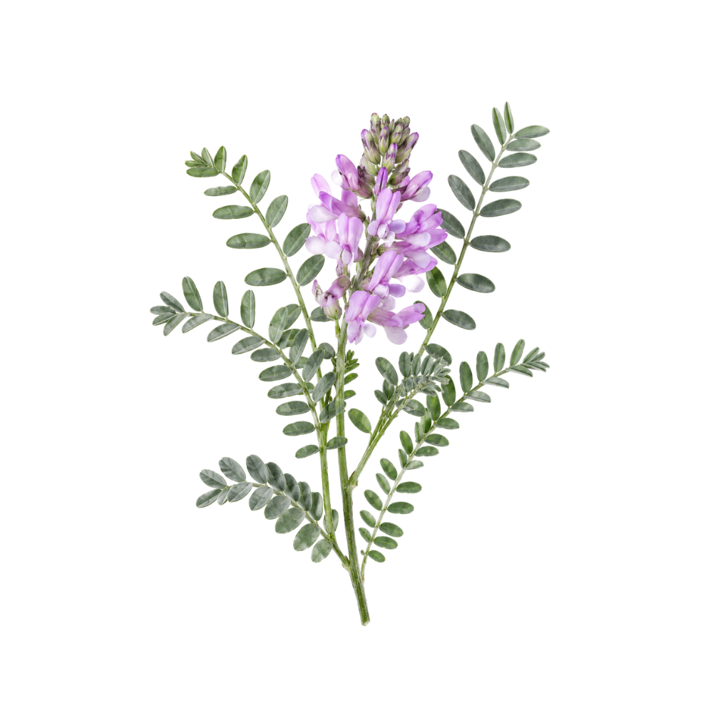 ModerateModerateAstragalusAstragalus membranaceusAstragaloside IV and the polysaccharides upregulate Nrf2/HO-1 and lower reactive oxygen species and lipid peroxidation in cells and animals, the shared mechanism under its kidney, liver and heart protection.MechanismNrf2/HO-1 ↑; ROS ↓; lipid peroxidation ↓Read the evidenceShow lessMonograph →
Astragaloside IV and astragalus polysaccharide reduce reactive oxygen species and lipid peroxidation and upregulate the Nrf2/HO-1 cytoprotective pathway in endothelial and other cell models 159,
This is the engine shared by the renal, hepatic and cardiac signals rather than a headline action of the root.
The evidence is entirely in vitro and animal, attributed to isolated constituents, and no human study measures an antioxidant endpoint after oral astragalus 159,
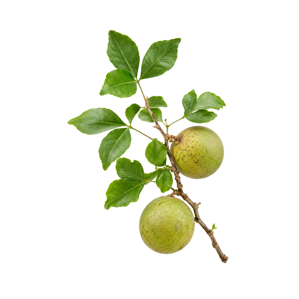 ModerateModerateBaelAegle marmelosRestores SOD and catalase alongside its glucose-lowering in diabetic rats, and the unripe fruit's ulcer protection is attributed to the same redox activity.Mechanismendogenous antioxidant-enzyme restoration (SOD/CAT); lipid peroxidation ↓Read the evidenceShow lessMonograph →
Leaf extract restored antioxidant enzymes alongside its glucose-lowering in diabetic rats 166Reference 166In vitroIsolation and partial characterisation of a novel lectin from Aegle marmelos fruit and its effect on adherence and invasion of Shigellae to HT29 cells — [in vitro]View study →, and the unripe fruit’s ulcer protection is attributed to antioxidant mechanisms 167Reference 167Antidiabetic activity of Aegle marmelos and its relationship with its antioxidant properties — [animal]View study →.
The chemistry supports it: coumarins, the flavonoids quercetin and rutin, and a tannin load.
Antioxidant activity has not been measured as an endpoint in its own right, and no human oxidative-stress markers exist.
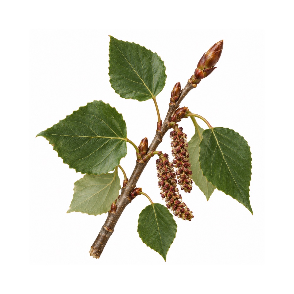 ModerateModerateBalsam PoplarPopulus balsamiferaThe propolis-type flavonoids of the buds, pinobanksin, pinocembrin, chrysin and galangin, scavenge radicals in laboratory assays.Mechanismpropolis flavonoid radical scavengingRead the evidenceShow less
Bud extracts show antioxidant activity in laboratory assays 170Reference 170In vitroComparison of ethanolic and aqueous Populus balsamifera L. bud extracts by different extraction methods: chemical composition, antioxidant and antibacterial activities — [in vitro]View study →, carried by the propolis flavonoids pinobanksin, pinocembrin, chrysin and galangin.
The evidence is assay-level, with no animal or human data.
ModerateBarbatimãoStryphnodendron adstringensA bark rich enough in flavan-3-ols to score high in any antioxidant assay, through the same proanthocyanidin chemistry that makes it astringent.Mechanismflavan-3-ol and phenolic radical scavengingRead the evidenceShow less
The bark’s flavan-3-ol and proanthocyanidin fraction gives it substantial radical-scavenging capacity in vitro, the same polyphenol chemistry behind its astringency.
The activity is generic to tannin-rich barks, and it has not been measured beyond the assay.
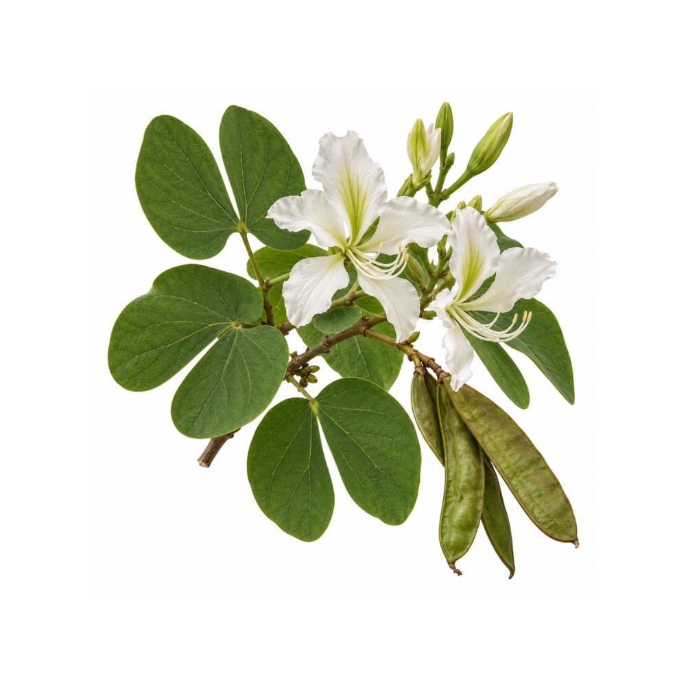 ModerateModerateBauhinia ForficataBauhinia forficataScavenges radicals across the standard assays and protects against lipid peroxidation and DNA damage in vivo, a stronger endpoint than assay scavenging.Mechanismradical scavenging (DPPH/ABTS/FRAP); lipid-peroxidation and genoprotective effects in vivoRead the evidenceShow less
Alongside the usual in-vitro panel there are in-vivo lipid-peroxidation and genoprotective results 174,
Genoprotection, protection against DNA damage, is a different endpoint from radical scavenging in a cuvette, and relatively few herbs have it.
There are no human data.
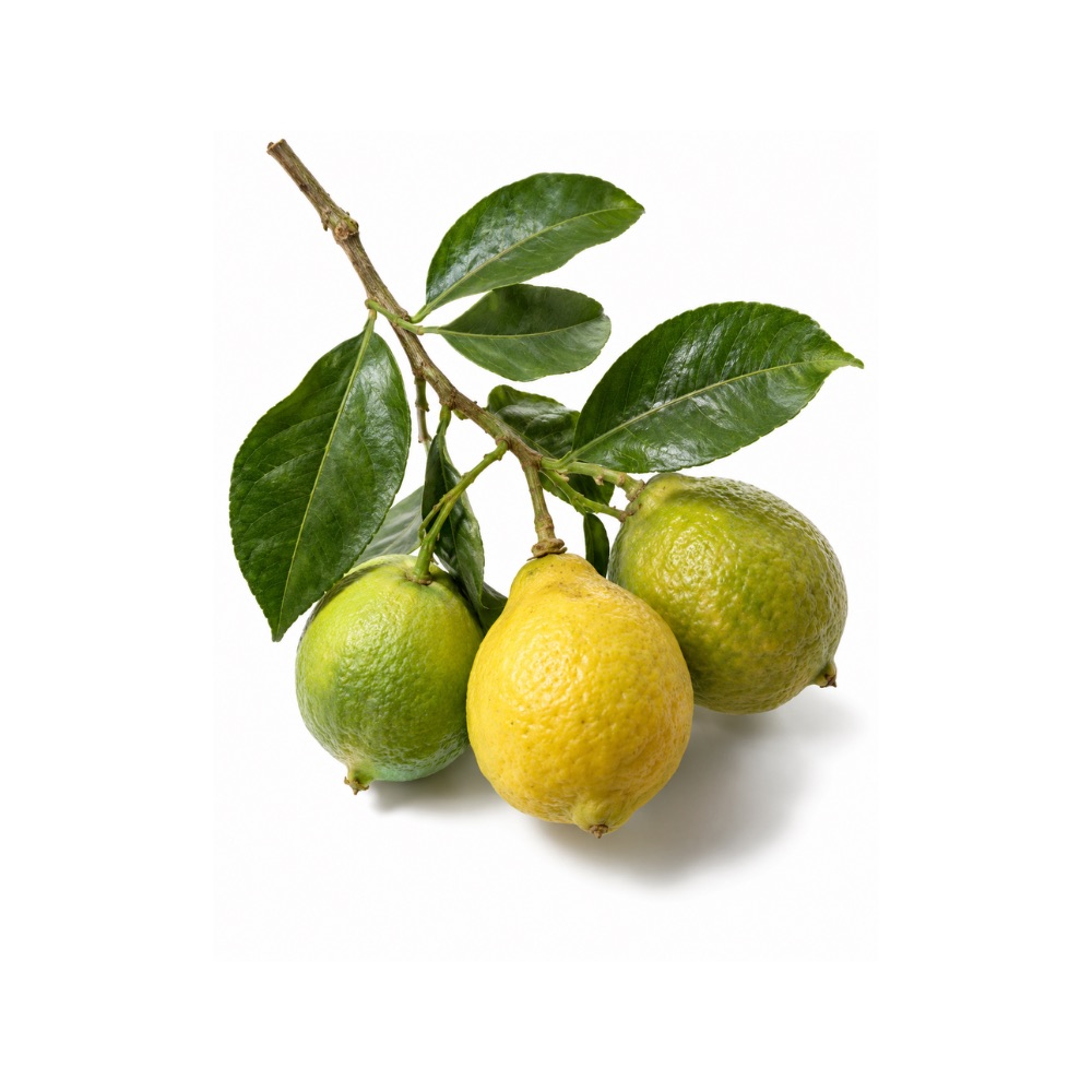 ModerateModerateBergamotCitrus bergamiaThe juice flavanones protect the LDL particle: added to rosuvastatin, bergamot polyphenol fraction lowered malondialdehyde and the oxidised-LDL receptor LOX-1 in people.MechanismLDL-particle antioxidation, oxLDL receptor LOX-1 ↓, MDA ↓, endothelial NO ↑ (flavanone glycosides neoeriocitrin, neohesperidin, naringin)Read the evidenceShow lessMonograph →
In the statin-adjunct RCT, adding bergamot polyphenol fraction to rosuvastatin reduced malondialdehyde, the oxidised-LDL receptor LOX-1 and phospho-PKB in peripheral cells alongside the lipid change 178Reference 178Bergamot polyphenolic fraction enhances rosuvastatin-induced effect on LDL-cholesterol, LOX-1 expression and protein kinase B phosphorylationView study →. A small randomised pilot found a BPF-Gold and L-citrulline combination raised nitric oxide, reactive hyperaemia index and VO₂max 179Reference 179The antioxidant power of bergamot polyphenolic fraction Gold potentiates the effects of L-citrulline in athlete performance and vasodilationView study →, and a combination-product RCT improved endothelial reactivity and hs-CRP against baseline 180Reference 180Effect of dietary supplementation with Eufortyn Colesterolo Plus on serum lipids, endothelial reactivity and NAFLD indices (ANEMONE study)View study →.
The target is specific: oxidative modification of the LDL particle, a sub-mechanism of the cardiometabolic action rather than general-purpose scavenging. The actives are the juice and albedo flavanone glycosides neoeriocitrin, neohesperidin and naringin, not the peel-oil constituents, and every lipid reference is Citrus bergamia rather than Monarda.
Every endpoint is a surrogate biomarker in a small arm, two of the three trials are confounded by a co-administered statin or citrulline, and nothing links the antioxidant activity to a vascular clinical outcome.
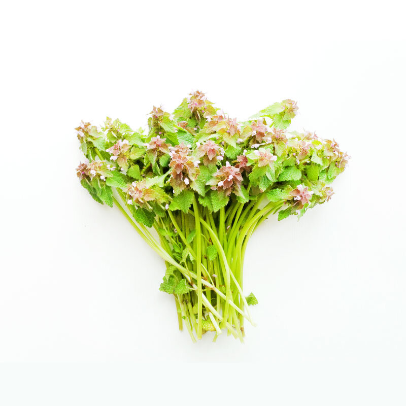 ModerateModerateBlack HorehoundBallota nigraThe phenylpropanoid glycosides of the flowering tops, verbascoside above all, block copper-driven LDL oxidation in vitro.MechanismCu2+-mediated LDL peroxidation inhibition; phenylpropanoid-glycoside radical scavengingRead the evidenceShow lessMonograph →
The characteristic phenylpropanoid glycosides verbascoside, forsythoside B, arenarioside and ballotetroside inhibit copper-mediated oxidation of LDL in vitro 181Reference 181A phenylpropanoid glycoside from Ballota nigra — phytochemicalView study →, the initiating event in atherosclerosis 182Reference 182In vitroPhenylpropanoid glycosides from Ballota nigra — in vitro / phytochemicalView study →. Antioxidant activity for the same fraction is also reported alongside the neurosedative work 183Reference 183In vitroPhenylpropanoids from Ballota nigra L. inhibit in vitro LDL peroxidation — in vitroView study →.
Verbascoside runs about 7–8.5% and forsythoside B about 3.8–6.0% of dried leaf, peaking around flowering in June, so a flowering-tops harvest is doing the work and out-of-season material is substantially weaker.
Every measurement is a cell-free or in-vitro assay, with no animal or human oxidative-stress endpoint.
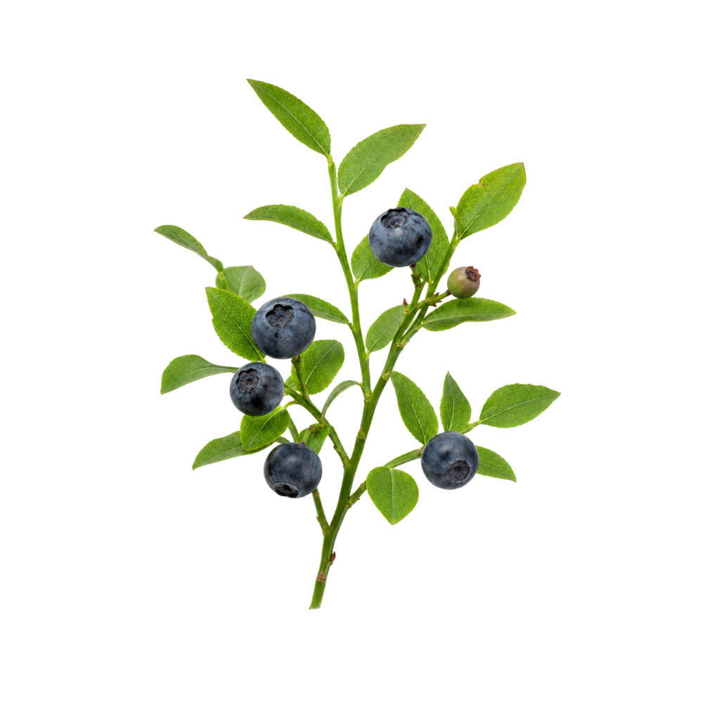 ModerateModerateBilberryVaccinium myrtillusConsistent free-radical and peroxynitrite scavenging by the anthocyanidins, with the whole-berry effect smaller than the isolated compounds.Mechanismfree-radical and peroxynitrite scavenging (anthocyanidins)Read the evidenceShow lessMonograph →
Bilberry’s dark red-purple flesh is the point: it is unusually rich in anthocyanins, and nearly all of its claimed activity is attributed to them. Cultivated blueberries are pale inside and carry far less.
The scavenging is consistent across free radicals and peroxynitrite, and it is the best-characterised of the herb’s mechanisms.
Two limits follow the chemistry. Human data are biomarker measurements rather than outcomes, and the whole-berry effect is smaller than what the isolated compounds do in a cuvette.
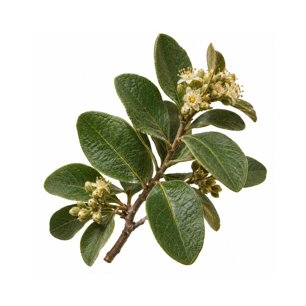 ModerateModerateBoldoPeumus boldusBoldine scavenges radicals and supports the endogenous antioxidant enzymes, replicated across independent animal and cell models.Mechanismboldine radical scavenging; endogenous antioxidant-enzyme supportRead the evidenceShow less
The antioxidant activity of boldo, carried by the aporphine alkaloid boldine, is replicated across independent animal and cell models 186,
It is the mechanistic root of the hepatoprotective and neuroprotective findings.
There are no human data.
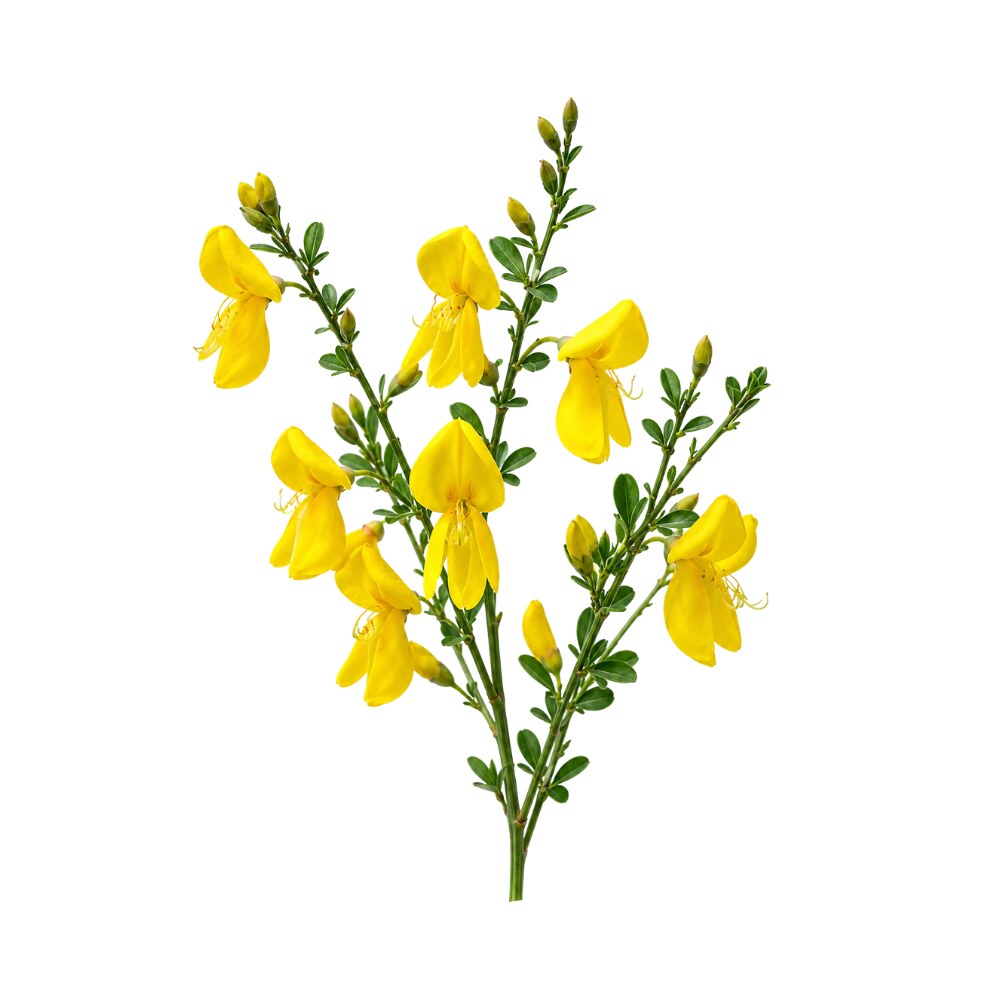 ModerateModerateBroomCytisus scopariusIts flavonoids, not its alkaloids, scavenge radicals consistently across four models and restore SOD, catalase and glutathione peroxidase in stressed rats.Mechanismflavonoid/polyphenol radical scavenging (DPPH IC₅₀ ≈ 1.5 µg/mL); endogenous antioxidant-enzyme restoration (SOD, catalase, GPx); lipid peroxidation ↓Read the evidenceShow lessMonograph →
Aerial-part extract scavenges dose-dependently in vitro against DPPH (IC₅₀ ≈ 1.5 µg/mL), nitric oxide and superoxide, with reducing power and protection against lipid peroxidation in rat liver microsomes 188Reference 188In vitroCytisus scoparius link — a natural antioxidant — in vitroView study →. The activity belongs to the flavonoid and polyphenol fraction rather than the alkaloids.
In vivo the picture repeats across unrelated models. In carbon-tetrachloride liver injury, 250 and 500 mg/kg lowered TBARS and restored superoxide dismutase, catalase and glutathione peroxidase 189Reference 189AnimalAntioxidant effect of Cytisus scoparius against carbon tetrachloride treated liver injury — animal model, in vivoView study →.
Under chronic unpredictable mild stress a 60% methanolic extract raised brain and organ antioxidant-enzyme activity while lowering TBARS 190Reference 190AnimalEvaluation of behavioural and antioxidant activity of Cytisus scoparius in rats exposed to chronic unpredictable mild stress — animal model, in vivoView study →, and flavonoid- and carotenoid-rich extracts scavenged reactive oxygen and nitrogen species topically without skin irritation 191Reference 191In vitroPotential use of Cytisus scoparius extracts in topical applications for skin protection against oxidative damage — in vitroView study →.
There are no human data and no standardised extract. The tested preparations differ in solvent and plant part and none reports alkaloid content, so the antioxidant fraction describes a polyphenol extract rather than any product a person could obtain.
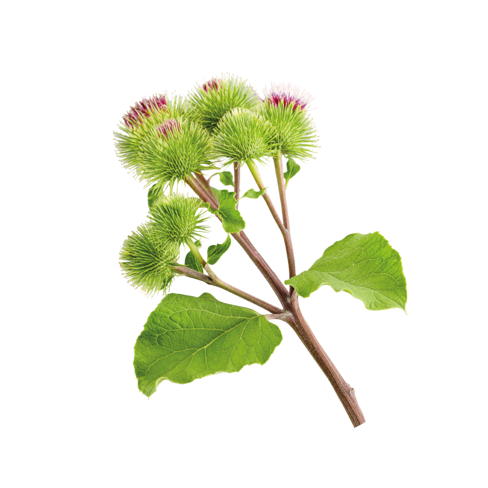 ModerateModerateBurdockArctium lappaRaises the body's own antioxidant enzymes rather than scavenging directly: root tea increased total antioxidant capacity and superoxide dismutase in osteoarthritis patients.Mechanismfructan and phenolic-acid radical scavenging; total antioxidant capacity and SOD ↑ (human); lipid peroxidation ↓Read the evidenceShow lessMonograph →
Root tea significantly raised total antioxidant capacity and superoxide dismutase activity in osteoarthritis patients, measured directly in the anti-inflammatory trial 194Reference 194RCTEffects of Arctium lappa L. (Burdock) root tea on inflammatory status and oxidative stress in patients with knee osteoarthritis — randomised controlled trialView study →.
The activity comes from an unusual source. Most of it is attributed to the storage fructan inulin and the phenolic-acid fraction rather than to a classic polyphenol scavenger: a purified root fructan raised antioxidant enzyme activity and lowered lipid peroxidation in animals 192Reference 192In vitroIn vitro and in vivo antioxidant activity of a fructan from the roots of Arctium lappa L. — in vitro and animalView study →, and crude root polysaccharide fractions scavenge dose-dependently in vitro 193Reference 193AnimalExtraction and antioxidant activities of polysaccharides from roots of Arctium lappa L. — animal modelView study →.
An enzyme-induction effect distributed across systems is the most likely modern mechanism behind the traditional alterative use.
The human data are biomarkers from one trial, and most of the rest is in vitro or animal work on purified polysaccharide fractions rather than the crude root as drunk.
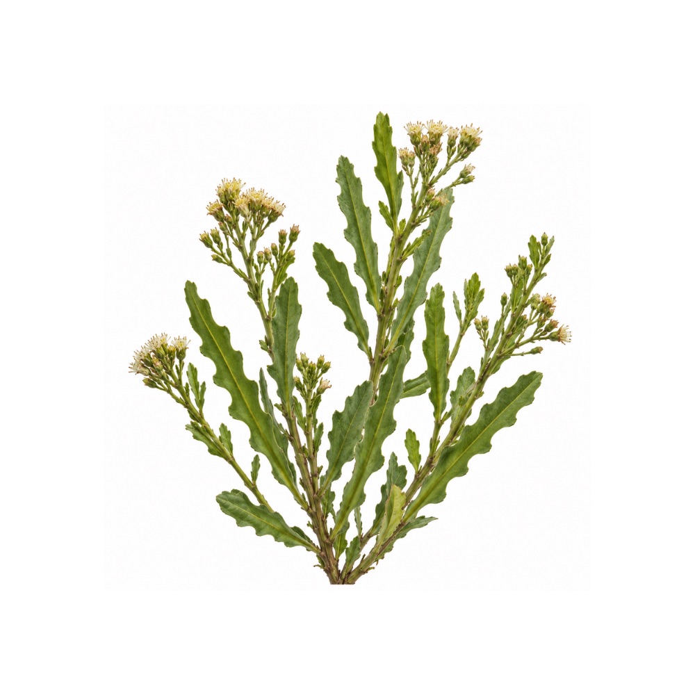 ModerateModerateCarquejaBaccharis trimeraScavenges radicals in vitro and raises SOD, catalase and glutathione in vivo, the mechanism under its liver protection.Mechanismradical scavenging; endogenous antioxidant-enzyme induction; Nrf2Read the evidenceShow less
Radical scavenging in vitro is joined by raised SOD, catalase and glutathione in vivo 162,
In-vivo enzyme induction is a better endpoint than assay scavenging alone, and it is the mechanistic root of the hepatoprotective action.
There are no human data.
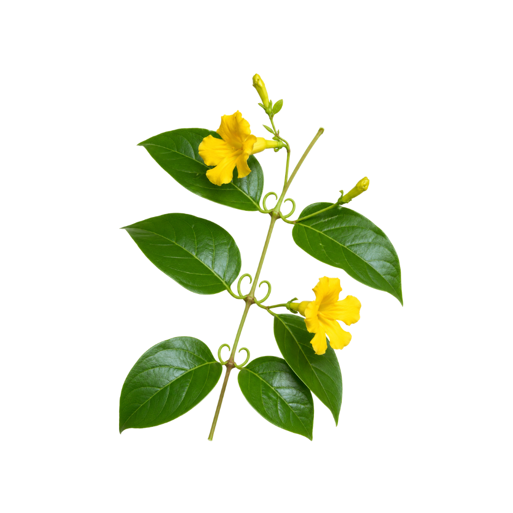 ModerateModerateCat’s ClawUncaria tomentosaBark tannins, catechins and procyanidins scavenge radicals dose-dependently, and whole extracts outperform alkaloid-enriched or alkaloid-free fractions.Mechanismfree-radical scavenging (tannins, catechins, procyanidins); oxidative-stress modulationRead the evidenceShow lessMonograph →
Ethanolic and aqueous bark extracts scavenge radicals dose-dependently, attributed to tannins, catechins and procyanidins 211Reference 211In vitroAntioxidant activity of ethanolic and aqueous extracts of Uncaria tomentosa — in vitroView study →, with comparable DPPH activity between U. tomentosa and U. guianensis 212Reference 212RCTEfficacy and safety of freeze-dried cat’s claw in osteoarthritis of the knee — randomised controlled trialView study →.
Whole hydroalcoholic extracts repeatedly outperform fractions enriched in either alkaloid or non-alkaloid components 213,
Every datum is in vitro or animal, and no human study has measured an oxidative-stress endpoint.
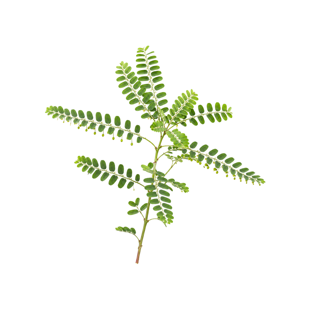 ModerateModerateChanca PiedraPhyllanthus niruriEllagitannins and flavonoids scavenge radicals near the potency of ascorbic acid in vitro, preserve SOD, catalase and glutathione peroxidase in diabetic rat kidney, and the tea raised plasma gallic acid in volunteers.Mechanismfree-radical scavenging (ellagitannins/flavonoids); SOD, catalase, glutathione-peroxidase preservation; lipid peroxidation ↓Read the evidenceShow lessMonograph →
In a randomised crossover study, healthy volunteers drinking the tea (5 g/750 mL) showed significantly raised plasma gallic acid and ascorbic acid, while erythrocyte catalase and superoxide dismutase were unchanged, in five people 219Reference 219RCTAntioxidant effects of Phyllanthus niruri tea on healthy subjects — randomized crossover trialView study →.
In diabetic rats an aqueous leaf extract prevented the rise in kidney lipid peroxidation and the fall in SOD, catalase and glutathione peroxidase, with in-vitro radical-scavenging potency near ascorbic acid 220Reference 220In vitroAqueous extract of Phyllanthus niruri leaves displays in vitro antioxidant activity and prevents oxidative stress in diabetic rat kidney — in vitro and animal modelView study →. Activity is attributed to the ellagitannins geraniin and corilagin and the flavonoids.
The only human study moved circulating markers and not enzyme-level defences, and no clinical antioxidant outcome exists.
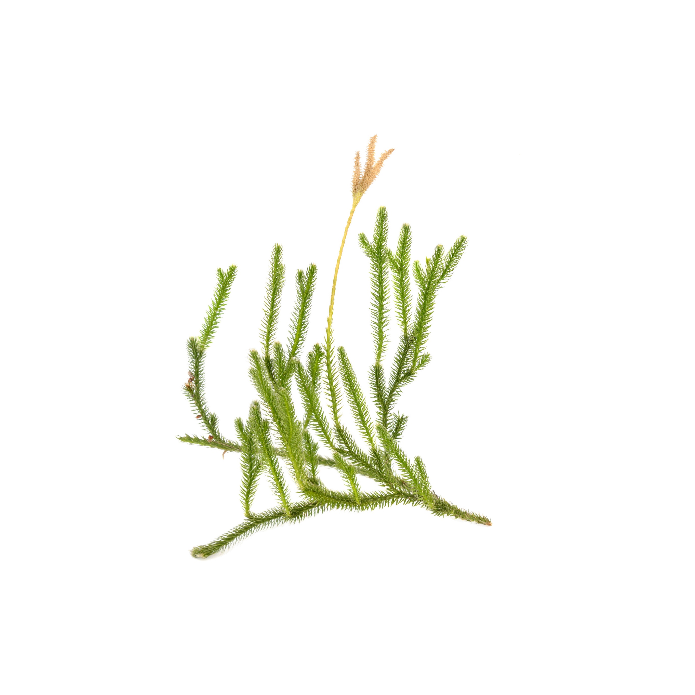 ModerateModerateChinese ClubmossHuperzia serrataHuperzine A lowers malondialdehyde and raises glutathione peroxidase by activating the Nrf2-ARE programme in injured mouse brain, an inductive rather than scavenging action.MechanismNrf2-ARE activation; lipid-peroxidation reductionRead the evidenceShow lessMonograph →
In a mouse traumatic-brain-injury model huperzine A lowered malondialdehyde and raised glutathione peroxidase by activating the Nrf2-ARE antioxidant programme 242Reference 242AnimalHuperzine A protects against traumatic brain injury through anti-oxidative effects via the Nrf2-ARE pathway — mouse in vivoView study →, and it reduces amyloid-beta-induced oxidative injury in cell studies 243Reference 243ReviewHuperzine A: is it an effective disease-modifying drug for Alzheimer’s disease? — reviewView study →.
The total alkaloid fraction of the herb, not just the isolated molecule, shows antioxidant and anti-amnesic activity in scopolamine-treated mice 244Reference 244AnimalNeuroprotective effects of total alkaloids fraction of Huperzia serrata on scopolamine-induced neurodegenerative animals — mouse in vivoView study →, the one thread that touches the crude material.
The action is modest and entirely preclinical, with no human data linking it to any outcome.
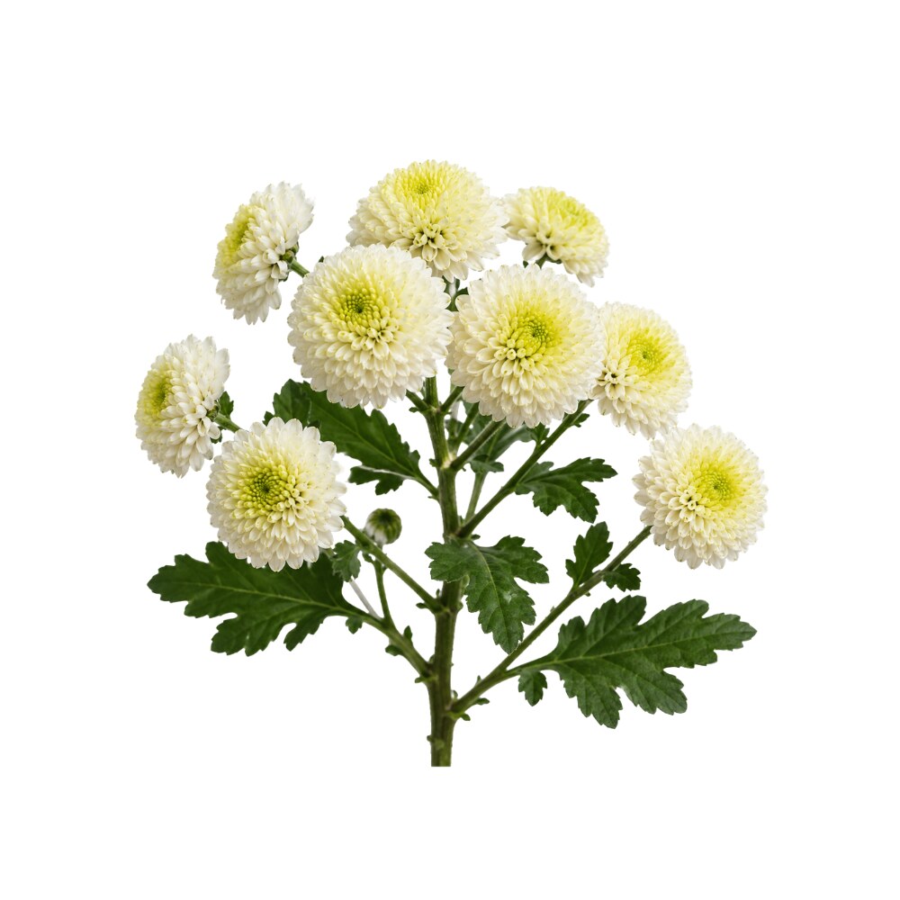 ModerateModerateChrysanthemumChrysanthemum morifoliumThe flower's luteolin and apigenin glycosides scavenge radicals in the standard in-vitro assays, the substrate under its hepatoprotective and anti-inflammatory use.Mechanismflavonoid radical scavengingRead the evidenceShow lessMonograph →
Chrysanthemum flower is flavonoid-rich, with luteolin and apigenin glycosides prominent, and shows the standard in-vitro scavenging profile.
The activity is the substrate under the hepatoprotective and anti-inflammatory actions rather than a distinct effect.
There is no human antioxidant endpoint.
ModerateDouglas FirPseudotsuga menziesiiNeedle and bark extracts scavenge free radicals about as well as ascorbic acid in the DPPH assay, through a taxifolin-dominant phenolic profile.Mechanismtaxifolin and condensed-tannin radical scavengingRead the evidenceShow less
In the DPPH assay, needle and bark extracts matched ascorbic acid 236Reference 236In vitroComparative antioxidant and antimicrobial activities of several conifer needles and bark extracts — in vitro (DPPH, FRAP, ABTS; MIC)View study →, driven by a taxifolin-dominant phenolic profile with catechin, epicatechin and condensed tannins 237,
A direct comparison against vitamin C is more informative than a bare IC50, and it is the strongest research finding on the tree.
It remains an assay result, with no animal or human data.
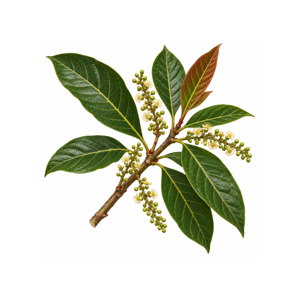 ModerateModerateDragon’s BloodCroton lechleriThe catechin, epicatechin and gallocatechin monomers of its proanthocyanidin fraction give the latex substantial flavan-3-ol radical-scavenging capacity.Mechanismflavan-3-ol radical scavengingRead the evidenceShow less
The capacity is carried by the catechin, epicatechin and gallocatechin monomers of the proanthocyanidin fraction, which is substantial in quantity.
The mechanism is generic flavan-3-ol chemistry, and it has not been measured beyond the assay.
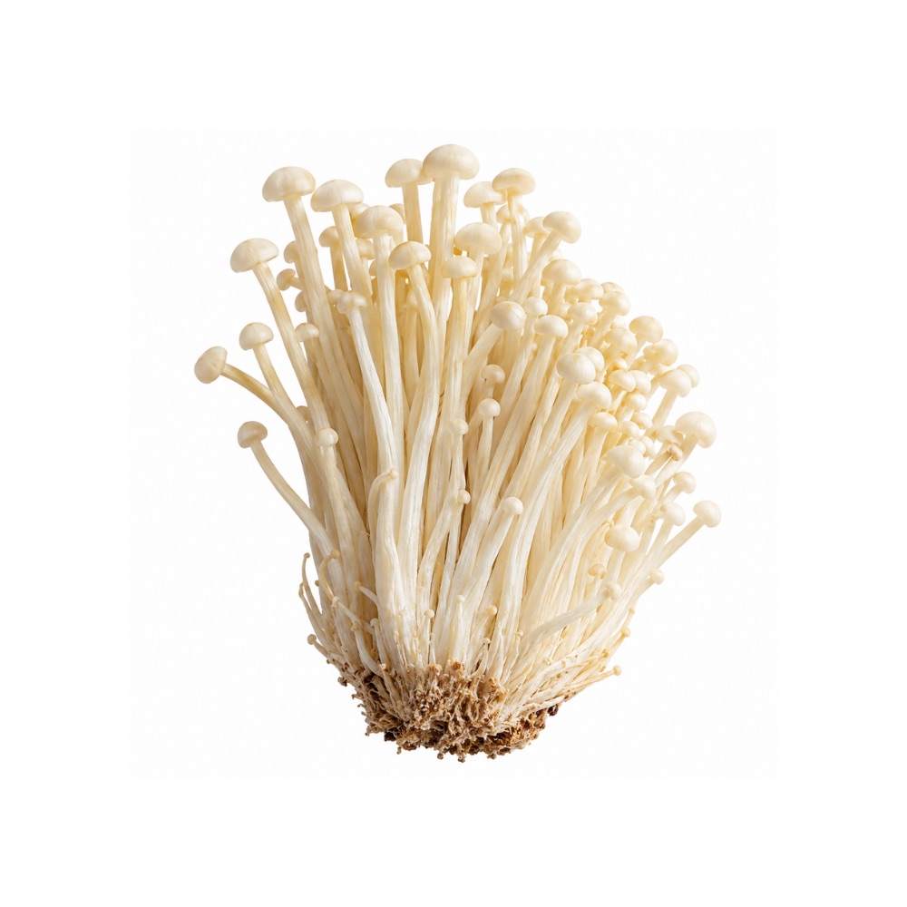 ModerateModerateEnokiFlammulina velutipesPolysaccharides, phenolic acids and hydrolysate peptides scavenge radicals in vitro, and in animals the mushroom raises SOD and catalase and lowers malondialdehyde through Keap1/Nrf2.Mechanismradical scavenging; SOD/CAT/GSH ↑, MDA ↓; Keap1/Nrf2 activationRead the evidenceShow less
Alongside in-vitro radical scavenging, animal studies show raised SOD and catalase and lowered malondialdehyde in tissue, with Keap1/Nrf2 activation as the mapped pathway.
The contributors are the polysaccharides, phenolic acids and hydrolysate peptides.
The work is constituent-level throughout, with no human data.
ModerateModerateForsythiaForsythia suspensaActivates Nrf2/HO-1, the inductive antioxidant response that is the common thread under its anti-inflammatory and hepatoprotective work.MechanismNrf2/HO-1 inductionRead the evidenceShow less
The Nrf2/HO-1 activation reported in both the anti-inflammatory and hepatoprotective work 245,
The action is inductive rather than scavenging, and it has been shown in cells and animals rather than in people.
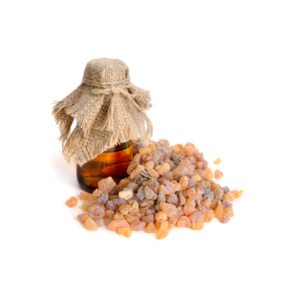 ModerateModerateFrankincenseBoswellia serrataAKBA activates the Nrf2/HO-1 axis, raising the body's own antioxidant enzymes and lowering oxidative-stress markers in rodent nerve-injury and Alzheimer's models.MechanismNrf2/HO-1 activationRead the evidenceShow lessMonograph →
The Nrf2/HO-1 axis is the cell’s master antioxidant response, and AKBA switches it on, upregulating endogenous antioxidant enzymes and reducing oxidative-stress markers in rodent nerve-injury and Alzheimer’s models 247,
The action is inductive rather than direct scavenging.
It has only been shown for the isolated compound in animals and cells, never for the resin as taken.
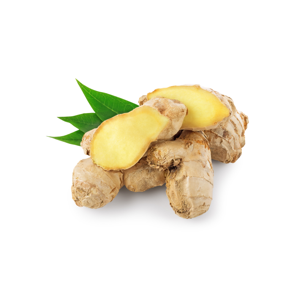 ModerateModerateGingerZingiber officinaleThe meta-analyses that found ginger lowering inflammatory cytokines also report improved total antioxidant capacity and lowered malondialdehyde in people.Mechanism↑ glutathione peroxidase; ↑ total antioxidant capacity; ↓ malondialdehydeRead the evidenceShow lessMonograph →
The same meta-analyses that found the cytokine reductions also report improved total antioxidant capacity and lowered malondialdehyde 251,
It is a secondary action that travels with the anti-inflammatory one.
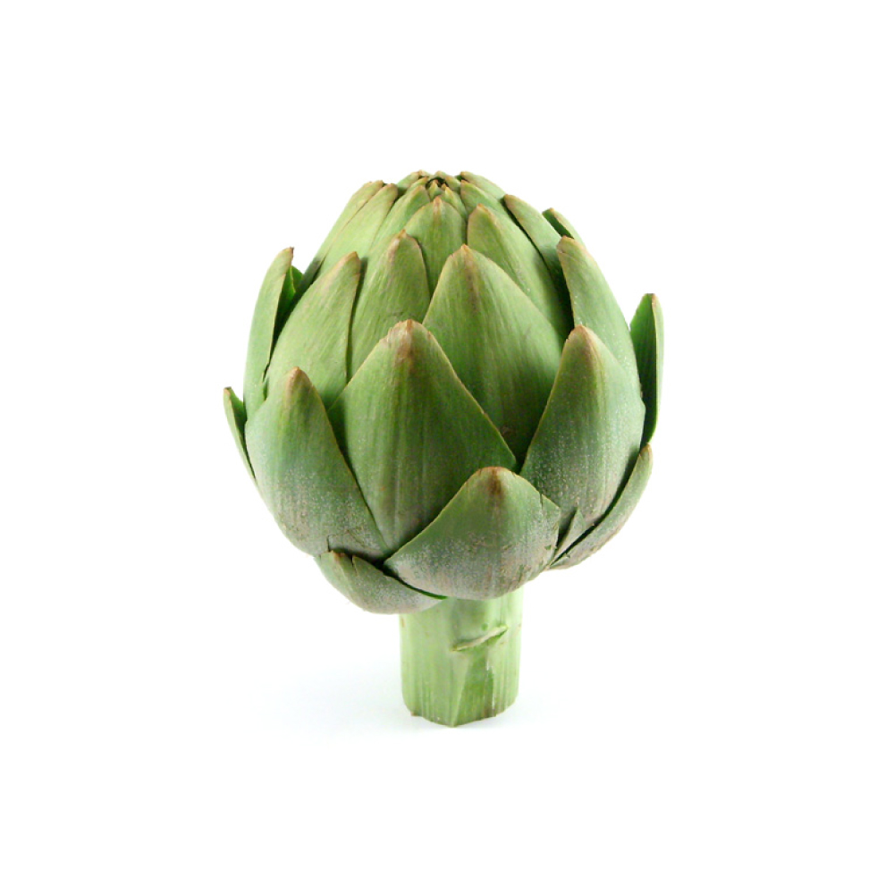 ModerateModerateGlobe ArtichokeCynara scolymusRaises SOD, catalase, glutathione and glutathione peroxidase and lowers malondialdehyde in pooled animal studies, while human trials in the same review showed little or no change.MechanismROS scavenging; SOD/GSH ↑; LDL-oxidation ↓Read the evidenceShow lessMonograph →
A meta-analysis of animal studies found supplementation raised superoxide dismutase, catalase, glutathione and glutathione peroxidase and lowered malondialdehyde in liver and plasma 158Reference 158Meta-analysisThe antioxidant activity of artichoke (Cynara scolymus) — systematic review and meta-analysis of animal studiesView study →.
In vitro, chlorogenic acid and caffeic acid scavenge ROS and inhibit LDL oxidation in endothelial cells.
The same review noted that human trials showed no change or only slight improvement in antioxidant status 158Reference 158Meta-analysisThe antioxidant activity of artichoke (Cynara scolymus) — systematic review and meta-analysis of animal studiesView study →, and the gap between the two is unusually well documented.
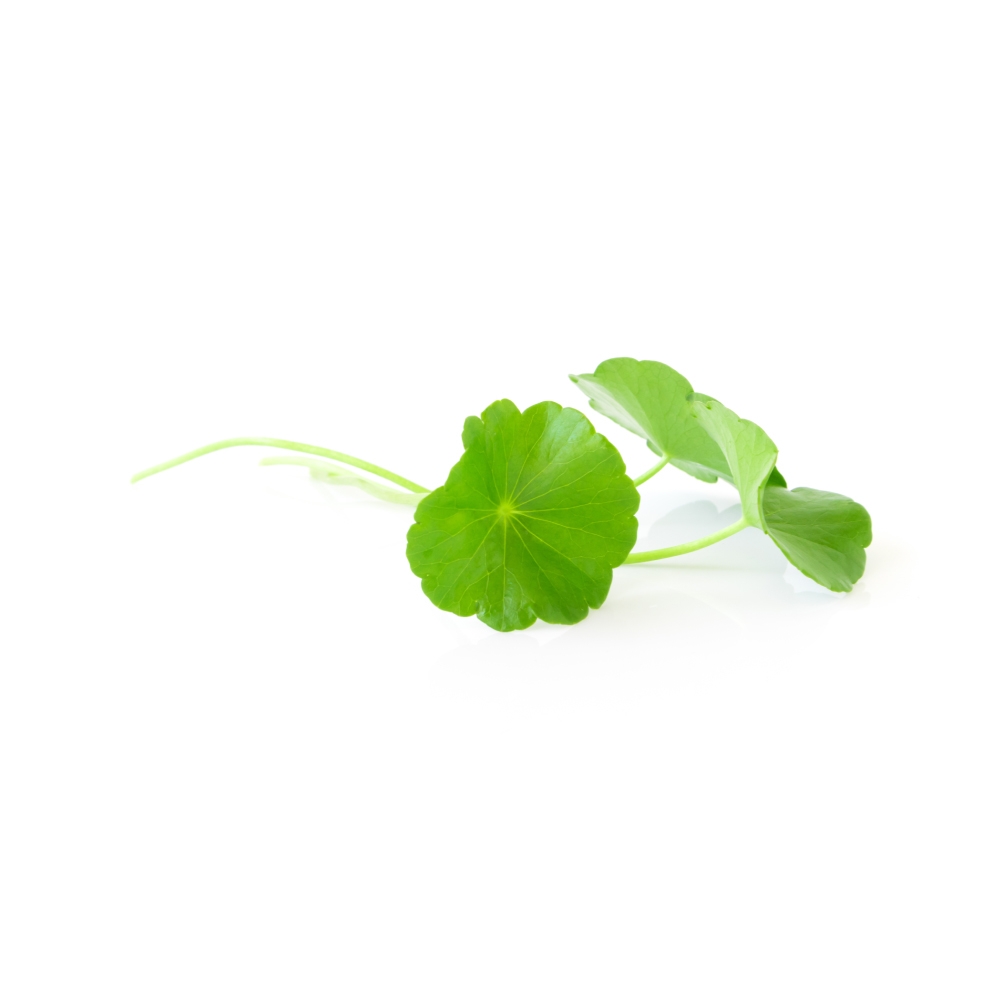 ModerateModerateGotu KolaCentella asiaticaAsiaticoside raises SOD, catalase, glutathione peroxidase, vitamin E and ascorbate in wound tissue, and the water extract activates NRF2 in Alzheimer's-model mice.MechanismNRF2 activation; SOD/catalase/GPx ↑; lipid peroxidation ↓Read the evidenceShow lessMonograph →
In wound tissue, asiaticoside raised SOD, catalase, glutathione peroxidase, vitamin E and ascorbate while cutting lipid peroxidation 256Reference 256AnimalAsiaticoside-induced elevation of antioxidant levels in healing wounds — animal (rat) modelView study →, and in Alzheimer’s-model mice water extract activated the NRF2 antioxidant-response pathway and improved hippocampal mitochondrial antioxidant markers 257,
Antioxidant activity is one of gotu kola’s most reproducible preclinical properties and underlies several of its other effects.
Direct human antioxidant-endpoint data are limited to one RCT raising macular pigment optical density 260Reference 260RCTLong-term supplementation with Centella asiatica extract (CA-HE50) on macular pigment optical density — randomised, double-blind, placebo-controlled trialView study →.
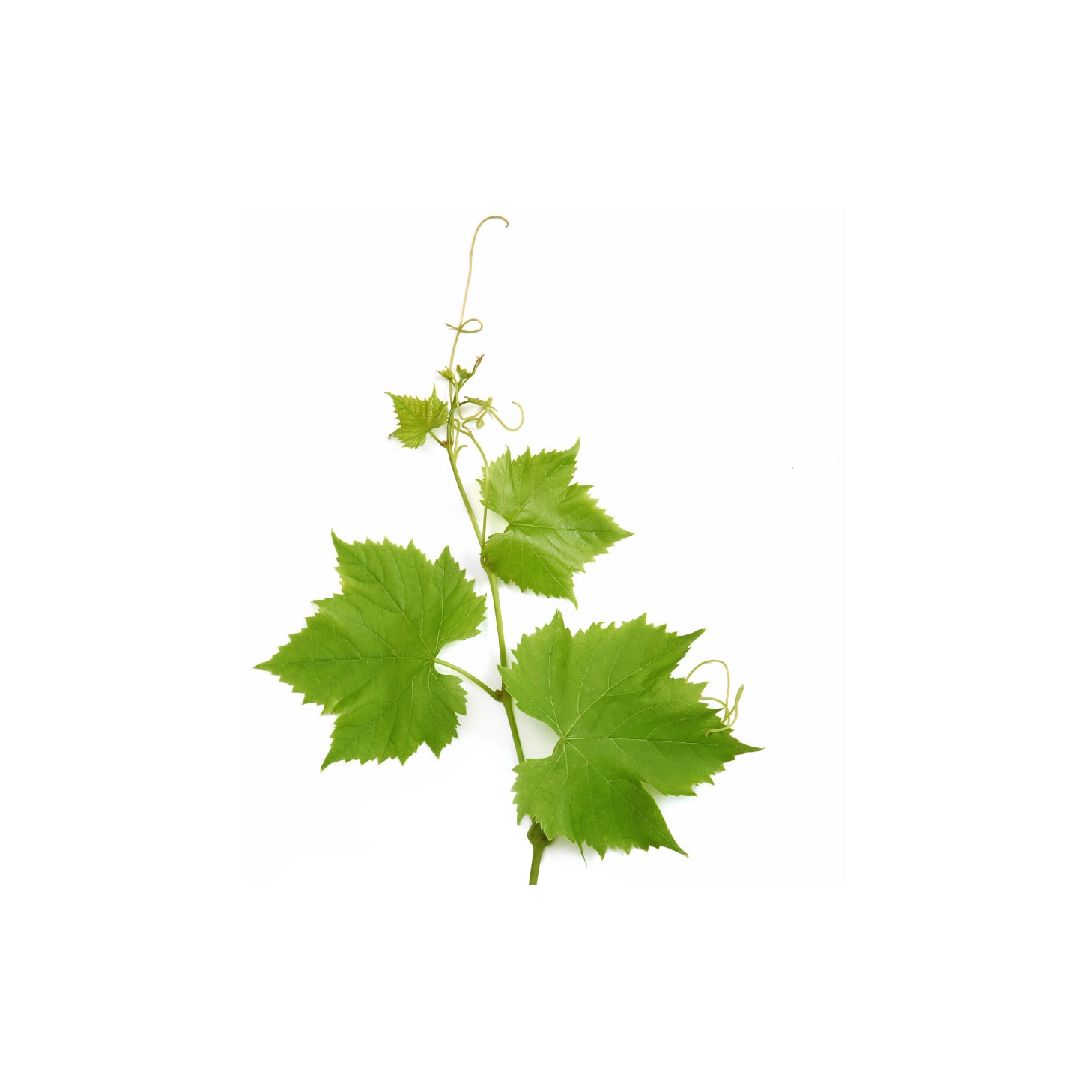 ModerateModerateGrape Seed ExtractVitis viniferaA concentrated flavanol preparation with oligomeric proanthocyanidins at about 75%, standardised on that figure because the proanthocyanidins are the radical-scavenging fraction.Mechanismflavanol radical scavenging; polyphenol redox chemistryRead the evidenceShow lessMonograph →
Oligomeric proanthocyanidins run to about 75% of the extract, built from catechin and epicatechin monomers at about 12% each, with procyanidin B2 as the dimeric marker and free gallic acid completing the phenolic profile. That chemistry is the reason the extract is standardised to a proanthocyanidin figure at all (100–300 mg/day).
The general reviews cover grape-seed polyphenol biochemistry and functionality at review level 261,
OPC bioavailability is poor. Only monomers and small dimers are absorbed intact, and most of the oligomeric fraction reaches the colon and acts through gut-microbial phenyl-γ-valerolactone catabolites rather than the parent molecules, so in-tube antioxidant capacity over-reads the systemic effect.
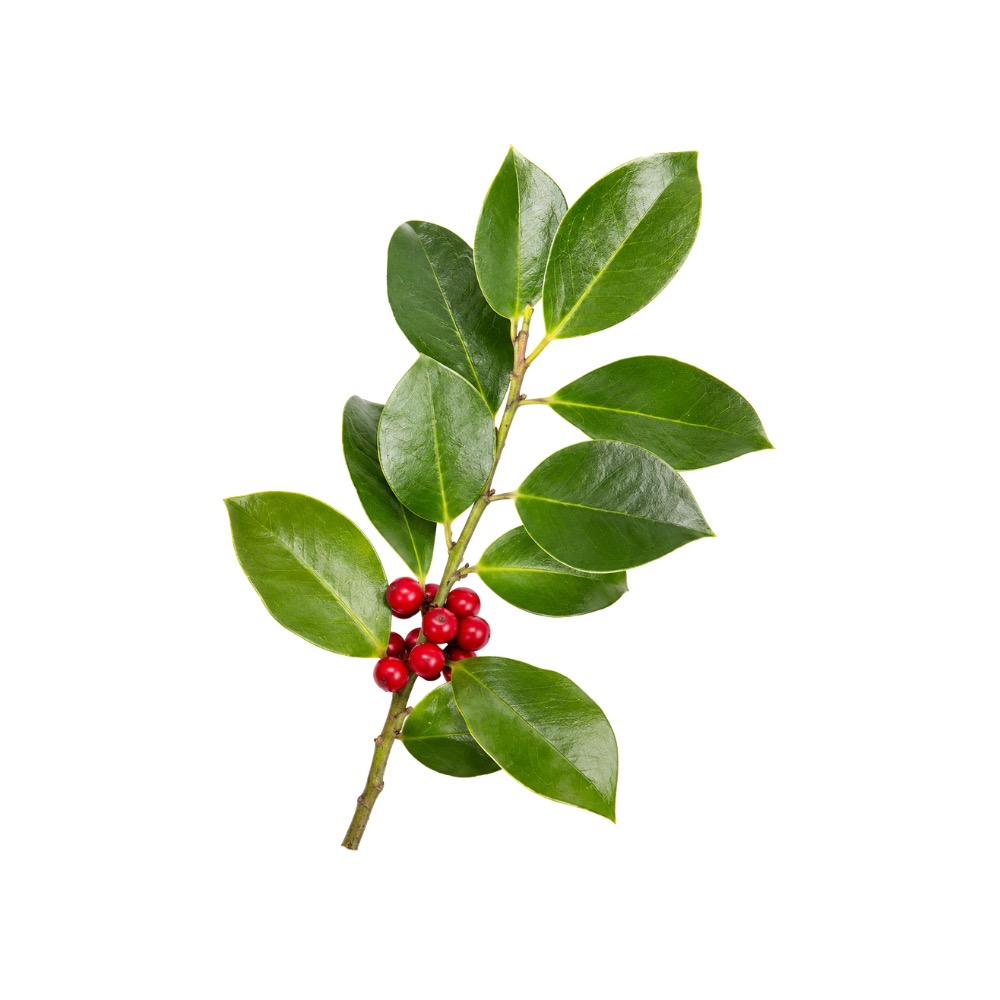 ModerateModerateGuayusaIlex guayusaLeaf infusions carry 54–67 mg of polyphenol per gram, led by chlorogenic acid, with peroxyl-radical scavenging about half that of green tea and cellular protection comparable to it.Mechanismchlorogenic-/caffeoylquinic-acid radical scavenging; quercetin-glycoside and lutein redox activityRead the evidenceShow lessMonograph →
Total polyphenol runs to 54–67 mg gallic-acid-equivalents per gram of leaf infusion, dominated by chlorogenic acid and its di-caffeoylquinic derivatives with quercetin glycosides and the carotenoid lutein 272,
Peroxyl-radical scavenging capacity is roughly half that of green tea, though Caco-2 cellular protection (60–80%) is comparable to Camellia sinensis 272Reference 272In vitroAntioxidant and anti-inflammatory properties of Ilex guayusa tea preparations: a comparison to Camellia sinensis teas — in vitro comparative studyView study →. Water-based extraction gives the highest phenolic yield 277Reference 277In vitroGreen-based methods to obtain bioactive compounds from guayusa (caffeine 2.27% of raw leaf) — in vitroView study →, and younger leaves and particular growing sites carry markedly more phenolic load 278,
No human study measures any antioxidant biomarker after guayusa intake, and cellular assays do not establish systemic benefit at drinking doses.
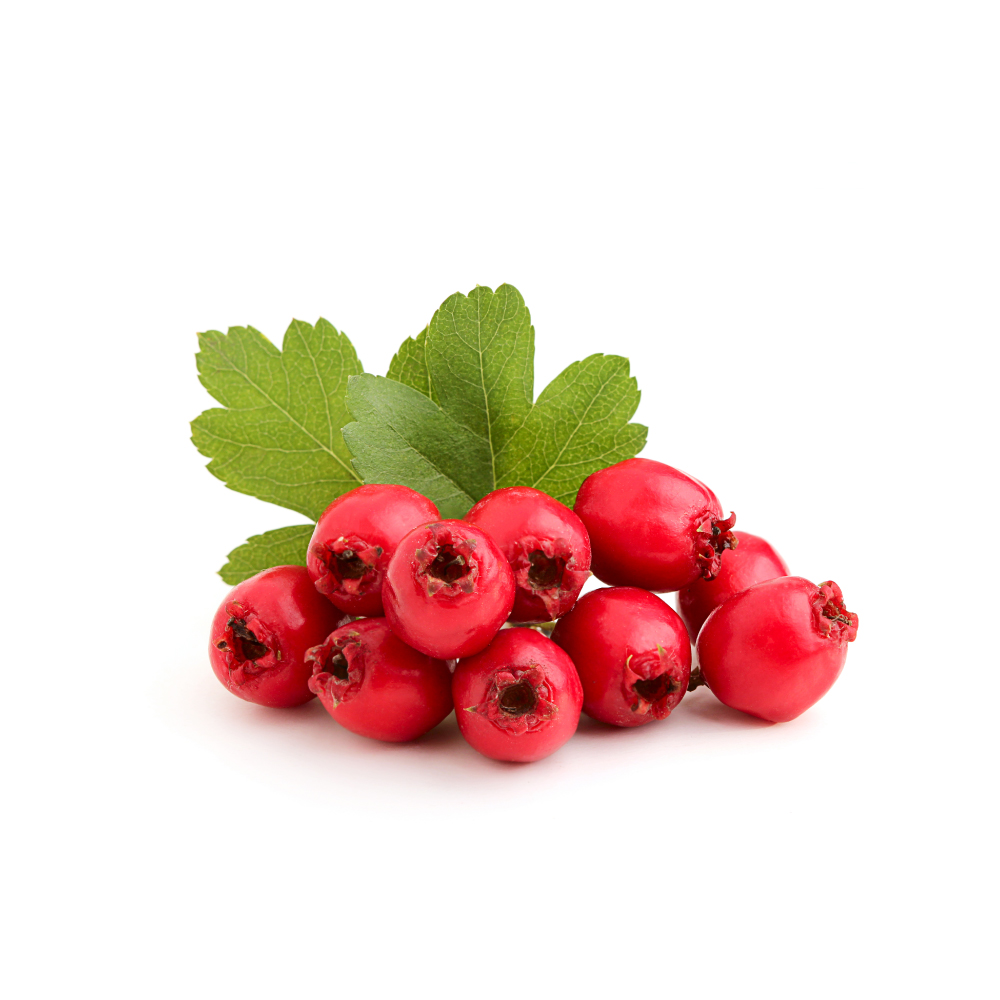 ModerateModerateHawthornCrataegus oxyacanthaIts procyanidins and flavonoids scavenge radicals, and the WS 1442 extract activates endothelial nitric-oxide synthase through a redox-sensitive route, a functional antioxidant action on the vessel wall.Mechanismredox-sensitive eNOS activation; vascular protectionRead the evidenceShow lessMonograph →
The standardised extract WS 1442 activates endothelial nitric-oxide synthase through phosphorylation at serine 1177 280Reference 280In vitroCrataegus special extract WS 1442 induces an endothelium-dependent, NO-mediated vasorelaxation via eNOS-phosphorylation at serine 1177 — in vitroView study →, and a follow-up showed this proceeds via redox-sensitive Src- and Akt-dependent eNOS activation 416Reference 416In vitroCrataegus special extract WS 1442 causes endothelium-dependent relaxation via a redox-sensitive Src- and Akt-dependent activation of endothelial NO synthase — in vitroView study →. This is a functional antioxidant action on the endothelium rather than a DPPH score.
The oligomeric procyanidins and the flavonoids hyperoside, rutin and vitexin carry the herb’s radical-scavenging capacity, which is unremarkable in raw terms next to the tannin-rich fruits.
The HERB CHF trial measured oxidative-stress and inflammation markers in heart-failure patients and found no difference from placebo 415Reference 415RCTHawthorn Extract Randomized Blinded Chronic Heart Failure (HERB CHF) trial — randomised controlled trialView study →, so the preclinical mechanism has been looked for in people and not found.
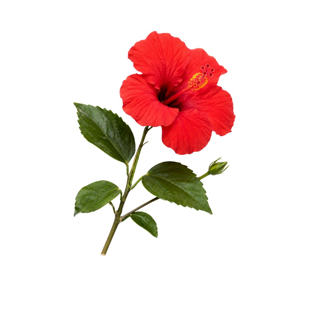 ModerateModerateHibiscusHibiscus rosa-sinensisThe cyanidin anthocyanins of the red, pink and orange flowers scavenge radicals and reduce iron in vitro, and a root extract restored antioxidant enzymes in diabetic rats.Mechanismradical scavenging (DPPH/ABTS); metal chelation; antioxidant-enzyme (SOD/GSH) supportRead the evidenceShow lessMonograph →
A 2023 UHPLC-Orbitrap-MS survey of 16 cultivars mapped the pigment chemistry: red, pink, orange and lilac flowers carry cyanidin-based anthocyanins, chiefly the sophoroside, white and yellow forms carry essentially none, and flavonol content (quercetin and kaempferol glycosides) runs inversely to anthocyanin content 281Reference 281Color, antioxidant capacity and flavonoid composition in Hibiscus rosa-sinensis cultivars — analytical (UHPLC-MS)View study →.
Flower extracts give concentration-dependent DPPH and ABTS scavenging and reducing power in vitro 282Reference 282In vitroAntioxidant and antibacterial activities of Hibiscus rosa-sinensis Linn. flower extracts — in vitroView study →, and a root extract restored antioxidant enzymes while lowering lipids in diabetic rats 283Reference 283AnimalAntidyslipidemic and antioxidant activity of Hibiscus rosa-sinensis root extract in alloxan-induced diabetic rats — in vivoView study →. The cultivar decides the dose: the activity belongs to pigmented flowers, not white or yellow ornamentals and not the leaf.
The effect is measured in chemical and rodent assays only, with no human bioavailability or antioxidant-status data, and anthocyanins are poorly absorbed.
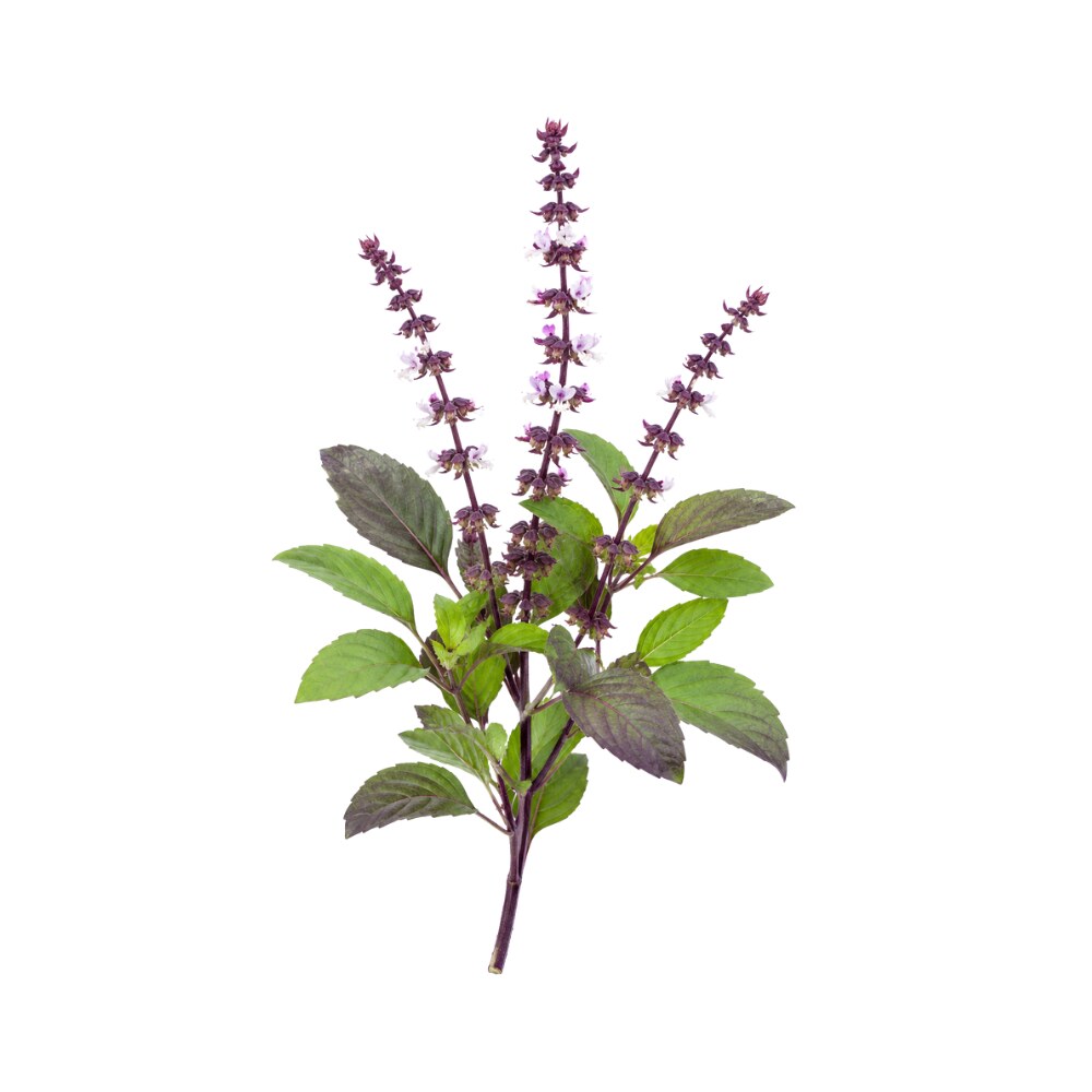 ModerateModerateHoly BasilOcimum tenuiflorumThe essential oil ranked among the more active of ten Thai aromatic oils in DPPH and TBARS tests, through eugenol, rosmarinic acid and the flavonoids.Mechanismfree-radical scavenging (DPPH); lipid-peroxidation ↓ (TBARS)Read the evidenceShow lessMonograph →
Among ten Thai aromatic-plant oils tested in DPPH radical-scavenging and TBARS lipid-peroxidation assays, holy basil oil ranked among the more active 284Reference 284In vitroPotential of native Thai aromatic plant extracts (including holy basil oil) in antiwrinkle body creams: antioxidant assays — in vitroView study →, with activity tracking eugenol, rosmarinic acid and the flavonoids.
The potent data were generated on the essential oil, whereas tulsi is overwhelmingly drunk as leaf tea or taken as an aqueous or ethanolic extract.
There is no evidence of a meaningful antioxidant effect at tissue level in people.
ModerateModerateHoneysuckleLonicera japonicaThe bud's very high load of chlorogenic, neochlorogenic and isochlorogenic acids gives it strong radical-scavenging capacity in vitro.Mechanismchlorogenic-acid radical scavenging; phenolic-acid poolRead the evidenceShow lessMonograph →
The bud carries a very high phenolic-acid load of chlorogenic, neochlorogenic and isochlorogenic acids, giving strong radical-scavenging capacity in vitro, consistent with the cooling traditional character 285,
There is no human biomarker study, and high in-vitro ORAC-style activity is close to universal among phenolic-rich botanicals, so it does not distinguish honeysuckle from other high-chlorogenic plants such as coffee, artichoke and yerba maté.
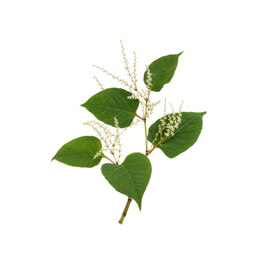 ModerateModerateJapanese KnotweedReynoutria japonicaPolydatin activates the Nrf2 pathway in vascular tissue, and Nrf2-mediated suppression of ferroptosis reduced plaque in ApoE-knockout mice.MechanismNrf2 activation; free-radical scavenging; ferroptosis ↓Read the evidenceShow lessMonograph →
Optimised resveratrol-rich extracts show concentration-dependent radical scavenging in vitro 290Reference 290In vitroOptimisation of extraction and antioxidant activities of resveratrol from Polygonum cuspidatum — in vitroView study →, and a resveratrol-enriched knotweed extract retained measurable antioxidant bioactivity in a functional assay 291Reference 291In vitroResveratrol-enriched Polygonum cuspidatum extract enhances functional bioactivity — in vitroView study →.
The sharpest mechanism is polydatin activating the Nrf2 antioxidant-response pathway in vascular tissue, where Nrf2-mediated suppression of ferroptosis reduced plaque in ApoE-knockout mice 292Reference 292AnimalNrf2-mediated inhibition of ferroptosis contributes to amelioration of atherosclerosis by polydatin — ApoE-knockout mouse, in vivoView study →.
The work is almost entirely in vitro and constituent-level, whole-root antioxidant benefit has never been shown in people, and free-resveratrol data overstate what a polydatin-dominant root delivers. A standardised extract built to a resveratrol specification and a 9–30 g decoction are not the same intervention.
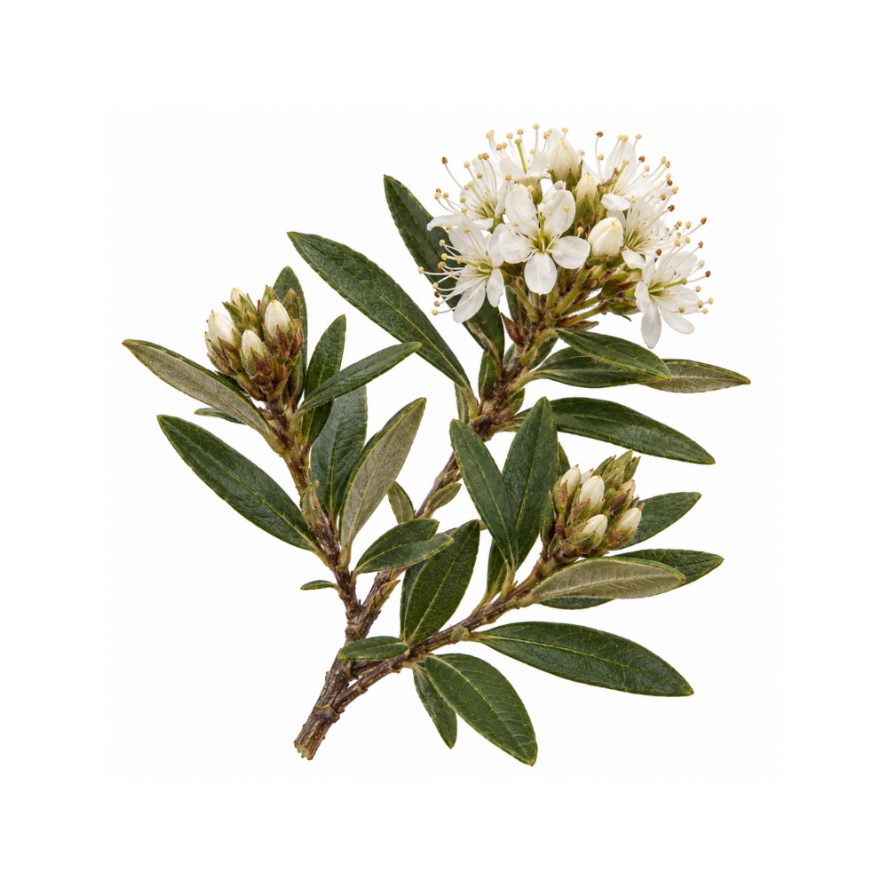 ModerateModerateLabrador TeaRhododendron groenlandicumPolyphenol radical scavenging in vitro, mostly measured in methanol and essential-oil preparations rather than the water infusion it is drunk as.Mechanismpolyphenol radical scavengingRead the evidenceShow less
In-vitro antioxidant activity has been recorded for the leaf 347,
The measurements are mostly on methanol or essential-oil preparations, the furthest of any of this herb’s data from the water infusion it is drunk as.
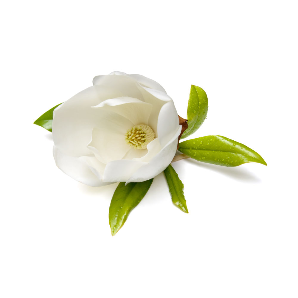 ModerateModerateMagnoliaMagnolia officinalisThe phenolic hydroxyls of magnolol and honokiol scavenge radicals directly, and reduced lipid peroxidation recurs across the bark's pharmacology.Mechanismphenolic-hydroxyl radical scavenging (magnolol/honokiol); ↓ lipid peroxidationRead the evidenceShow lessMonograph →
Direct radical-scavenging capacity comes from the phenolic hydroxyls of magnolol and honokiol, measured for instance in a controlled polymerisation-inhibition assay run alongside their COX-2 work 302Reference 302In vitroComparative inhibitory effects of magnolol, honokiol, eugenol and bis-eugenol on cyclooxygenase-2 expression and nuclear factor-kappa B activation in RAW264.7 macrophage-like cells — in vitroView study →, and reviews repeatedly cite reduced lipid peroxidation and boosted endogenous antioxidant defences as a recurring theme across the bark’s pharmacology 303,
This is the rationale layer underneath the neuroprotective and hepatoprotective claims.
No human antioxidant or oxidative-stress-outcome study exists for the herb, and the scavenging is measured on isolated neolignans rather than on a bark decoction where they sit at single-digit percent.
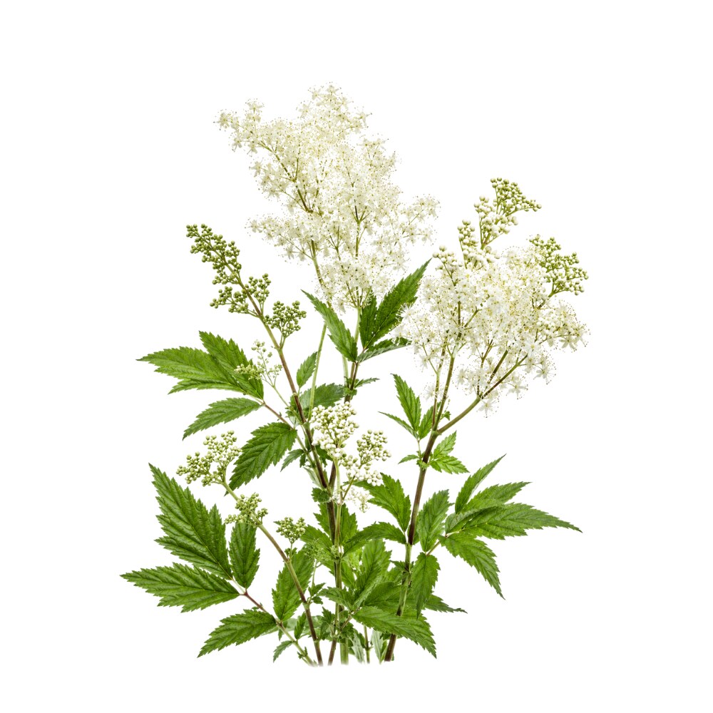 ModerateModerateMeadowsweetFilipendula ulmariaFlowers and leaves carry up to 65 mg/g of phenolics and over 100 mg/g of flavonoids, scavenge radicals across every standard assay, and oral extract cut DNA damage in rat liver and kidney after cisplatin.Mechanismphenolic/flavonoid radical scavenging (DPPH, hydroxyl, FRAP); lipid-peroxidation ↓; DNA protectionRead the evidenceShow lessMonograph →
Antioxidant activity turns up across DPPH, hydroxyl-radical, FRAP and lipid-peroxidation assays in many independent extracts of flowers, aerial parts and roots 313,
Two in-vivo results give it more weight than an assay panel alone. Oral extract cut oxidative-stress markers and DNA damage in rat liver and kidney after cisplatin 317Reference 317AnimalFilipendula ulmaria extracts attenuate cisplatin-induced liver and kidney oxidative stress in rats — in vivoView study →, and a Bulgarian dry tincture was DNA-protective in vitro 318Reference 318In vitroDNA-Protective, Antioxidant and Anti-Carcinogenic Potential of Meadowsweet (Filipendula ulmaria) Dry Tincture — in vitroView study →.
There are no human data of any kind, and radical-scavenging capacity in a cuvette says nothing about what survives digestion at a realistic tea dose.
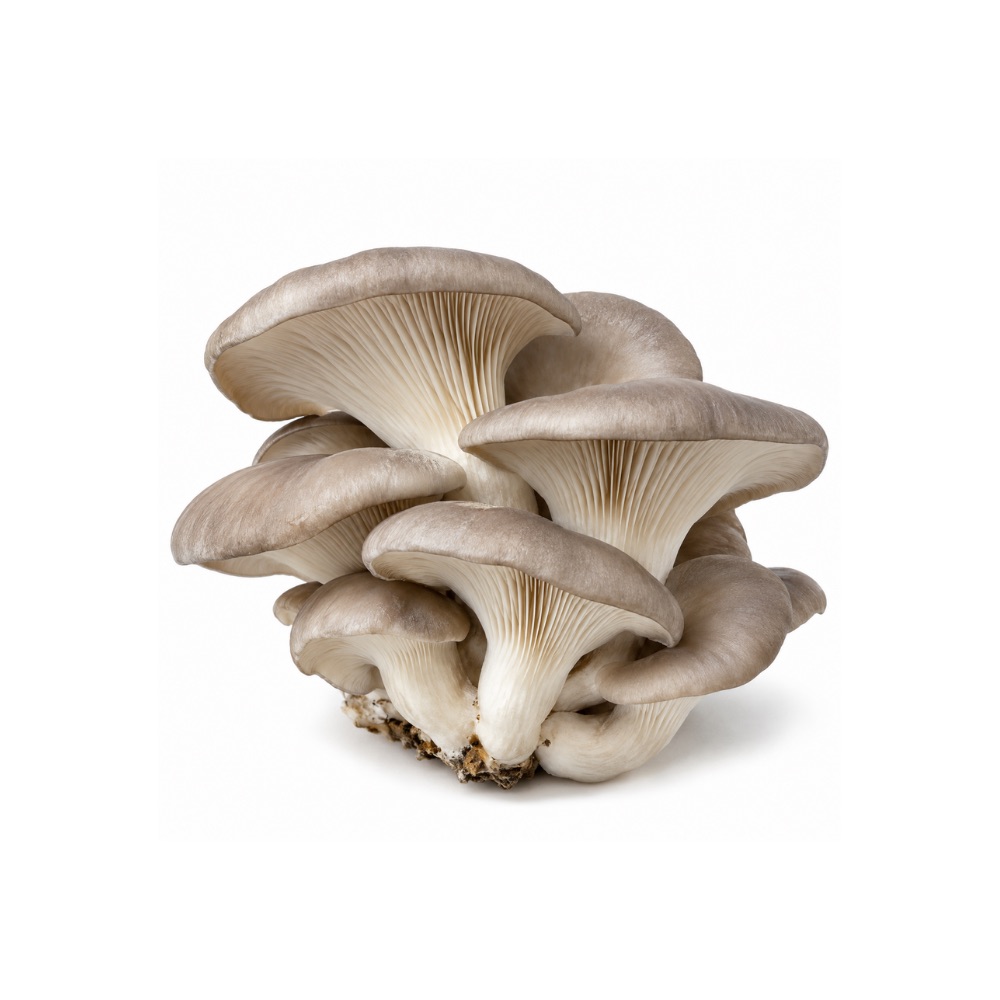 ModerateModerateOyster MushroomPleurotus ostreatusPolyphenols, ergothioneine and ergosterol scavenge radicals in vitro, and extracts restore SOD, catalase and glutathione in rodent liver-injury models.Mechanismradical scavenging; SOD/CAT/GSH restoration, MDA ↓Read the evidenceShow less
Methanol extract gave the strongest DPPH (87.7%) and hydrogen-peroxide scavenging, while the chloroform extract was highest in flavonoids 340Reference 340In vitroComparative antioxidant activity and phytochemical content of five extracts of Pleurotus ostreatus — in vitroView study →, so there is no single oyster-mushroom antioxidant value. The contributors are polyphenols at about 487 mg GAE/100 g dry matter, ergothioneine at about 1.9 mg/g and ergosterol.
Extracts restored SOD, catalase and glutathione while lowering malondialdehyde in rodent liver-injury models 341Reference 341AnimalAntioxidant activity of the oyster mushroom, Pleurotus ostreatus, on CCl4-induced liver injury — rat model in vivoView study →, which lifts it above assay-only herbs.
There are no human data.
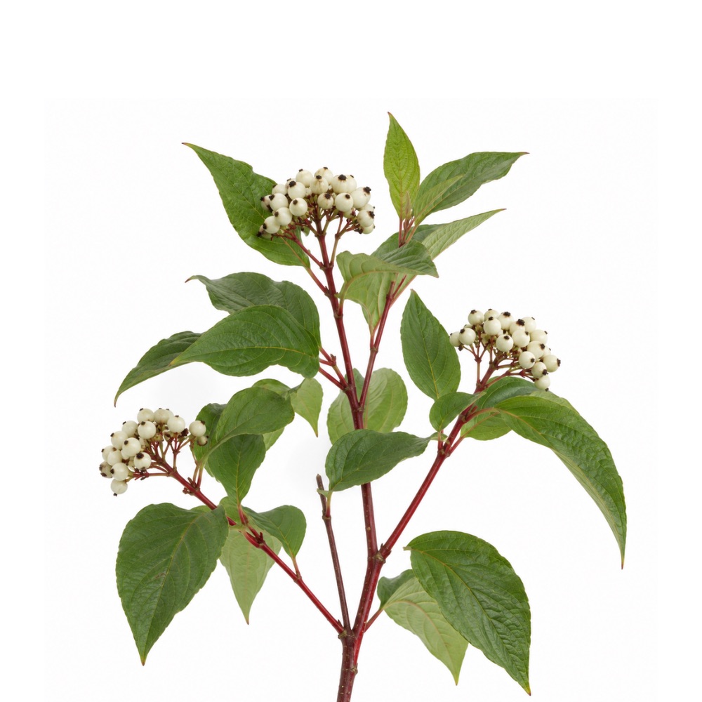 ModerateModerateRed-Osier DogwoodCornus sericeaPolyphenol radical scavenging replicated across chemical assays, human cell lines and animal blood antioxidant measures.Mechanismpolyphenol radical scavenging; in-vivo blood antioxidant capacityRead the evidenceShow less
The antioxidant activity is consistent across chemical assays, human cell lines and animal blood-antioxidant measures 228,
Three independent evidence types is unusual for a preclinical antioxidant claim, and the in-vivo blood measure is a real endpoint rather than a cuvette result.
There are no human data.
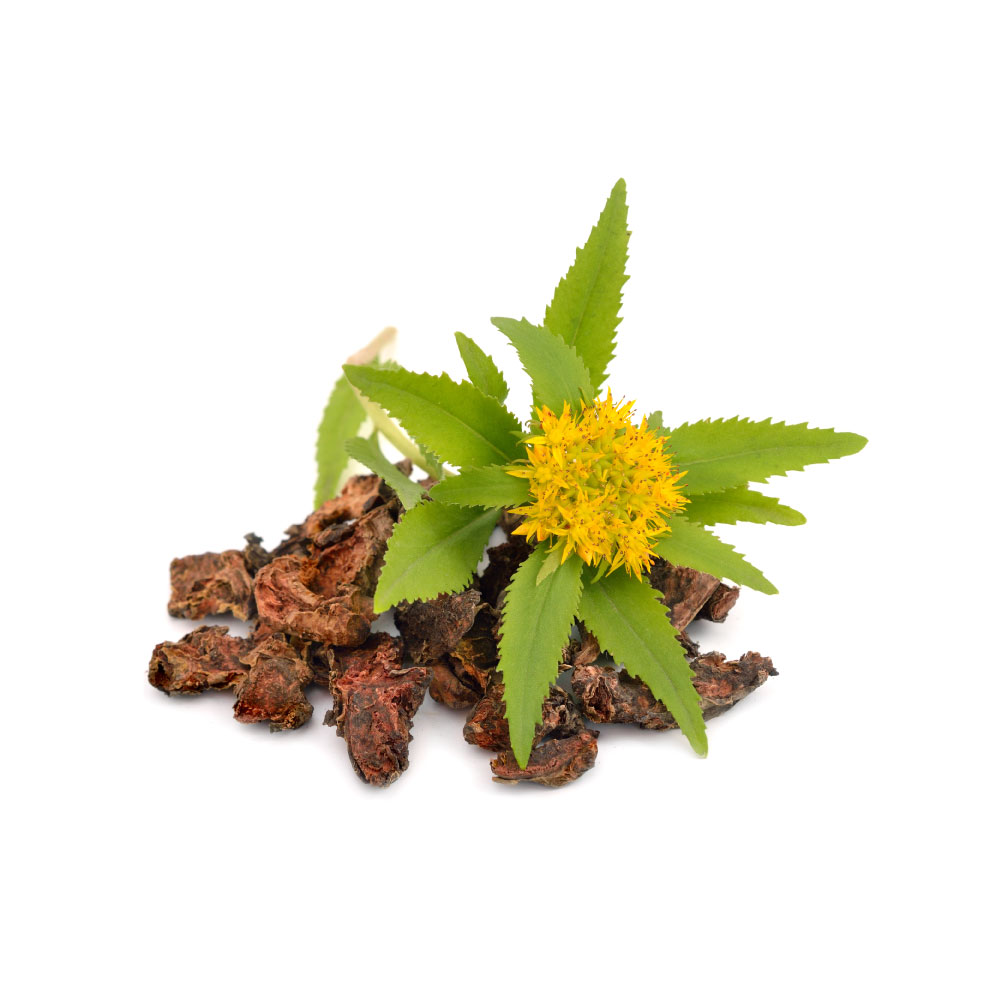 ModerateModerateRhodiolaRhodiola roseaPooled exercise trials show it raising total antioxidant capacity and superoxide dismutase and lowering malondialdehyde in athletes, with salidroside activating Nrf2 at constituent level.Mechanismtotal antioxidant capacity and SOD ↑, MDA ↓ (human, exercise); Nrf2 pathway activation by salidrosideRead the evidenceShow lessMonograph →
A 2025 endurance meta-analysis found supplementation raised total antioxidant capacity and superoxide dismutase while lowering malondialdehyde across exercise trials 344Reference 344Meta-analysisThe effect of Rhodiola rosea supplementation on endurance performance and related biomarkers — systematic review and meta-analysisView study →, and an exercise systematic review reported the same directionally 345Reference 345Systematic reviewEffects of Rhodiola rosea supplementation on exercise and sport: a systematic reviewView study →. The effect shows up in pooled human data rather than a single study.
At constituent level the mechanism is indirect: salidroside activates the Nrf2 pathway in cell and animal models 346Reference 346Meta-analysisNeuroprotective mechanisms of salidroside in Alzheimer’s disease — systematic review and meta-analysis of preclinical studiesView study →, enzyme induction rather than direct scavenging, the same route underlying the herb’s neuro- and cardioprotective claims.
Every human measurement is in the exercise setting, where oxidative stress is acutely provoked and easier to move than at baseline, and the broader claim leans on salidroside in vitro rather than the whole root in people.
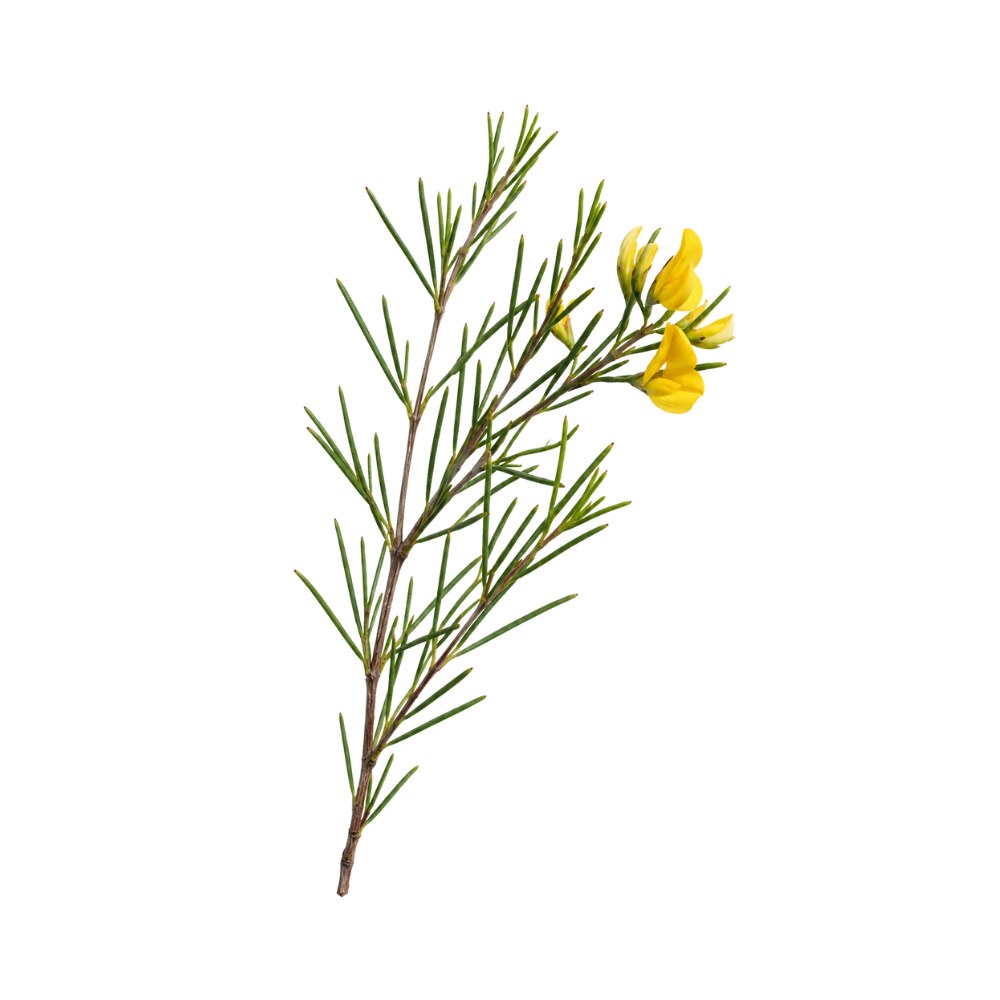 ModerateModerateRooibosAspalathus linearisSix cups a day of fermented rooibos for six weeks lowered conjugated dienes and TBARS and improved the glutathione redox ratio in adults at cardiovascular risk.Mechanismfree-radical scavenging; glutathione redox ratio ↑; lipid-peroxidation ↓Read the evidenceShow lessMonograph →
Forty adults at cardiovascular risk drinking six cups of fermented rooibos daily for six weeks raised plasma total polyphenols (about 79.8 to 89.8 mg/L), significantly lowered conjugated dienes and TBARS, and improved the reduced-to-oxidised glutathione ratio, while overall plasma antioxidant capacity did not change 349Reference 349Clinical trialEffects of rooibos (Aspalathus linearis) on oxidative stress and biochemical parameters in adults at risk for cardiovascular disease — controlled clinical trialView study →. A scoping review of the small human literature agrees on direction without adding weight 350Reference 350ReviewThe health benefits of rooibos tea in humans (Aspalathus linearis) — a scoping review. https://pubmed.ncbi.nlm.nih.gov/38204815/View study →.
The active chemistry is the dihydrochalcone aspalathin with nothofagin and the C-glycosides orientin and isoorientin. Preparation dominates: unfermented green leaf carries far higher antioxidant and antimutagenic potential, with aspalathin falling by over 90% during the oxidative fermentation that makes red bush tea 351,
Rooibos was consistently weaker than Camellia sinensis teas in comparative antimutagenicity work 353Reference 353In vitroA comparative study on the antimutagenic properties of aqueous extracts of Aspalathus linearis and Cyclopia spp. — in vitro (Salmonella). https://pubmed.ncbi.nlm.nih.gov/16949333/View study →, and the only human endpoint is a surrogate biomarker panel.
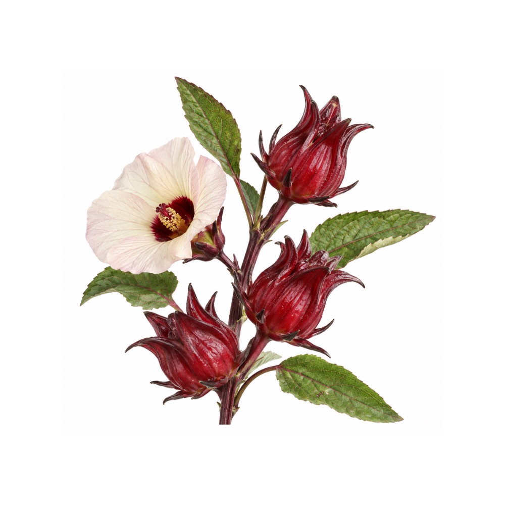 ModerateModerateRoselleHibiscus sabdariffaThe calyx anthocyanins and phenolic acids scavenge radicals in vitro and induce Nrf2 in rodents, and the extract shifted serum oxidative-stress markers in a double-blind trial in metabolic syndrome.Mechanismanthocyanin and phenolic-acid radical scavenging; Nrf2 induction, SOD/catalase/glutathione ↑ and MDA ↓ (rodent); serum oxidative-stress markers (human)Read the evidenceShow less
In a double-blind placebo-controlled trial in metabolic-syndrome patients, roselle extract shifted serum oxidative-stress markers alongside lipids, insulin and hs-CRP 408Reference 408RCTEvaluation of the effects of roselle (Hibiscus sabdariffa L.) on oxidative stress and serum levels of lipids, insulin and hs-CRP in adult patients with metabolic syndrome — double-blind placebo-controlled trialView study →.
The calyx is a concentrated source of anthocyanins and phenolic acids, chiefly delphinidin and cyanidin sambubiosides with protocatechuic and chlorogenic acids, and its radical-scavenging activity is the best-characterised of its properties in vitro and in animal tissue. In high-fat-diet rats the extract induced the Nrf2 pathway and raised superoxide dismutase, catalase and glutathione while lowering malondialdehyde 409Reference 409AnimalHibiscus sabdariffa extract improves hepatic steatosis, partially through IRS-1/Akt and Nrf2 signaling pathways in rats fed a high fat diet — animal modelView study →.
Human evidence stops at the biomarker level, and no clinical outcome has been tied to the antioxidant effect.
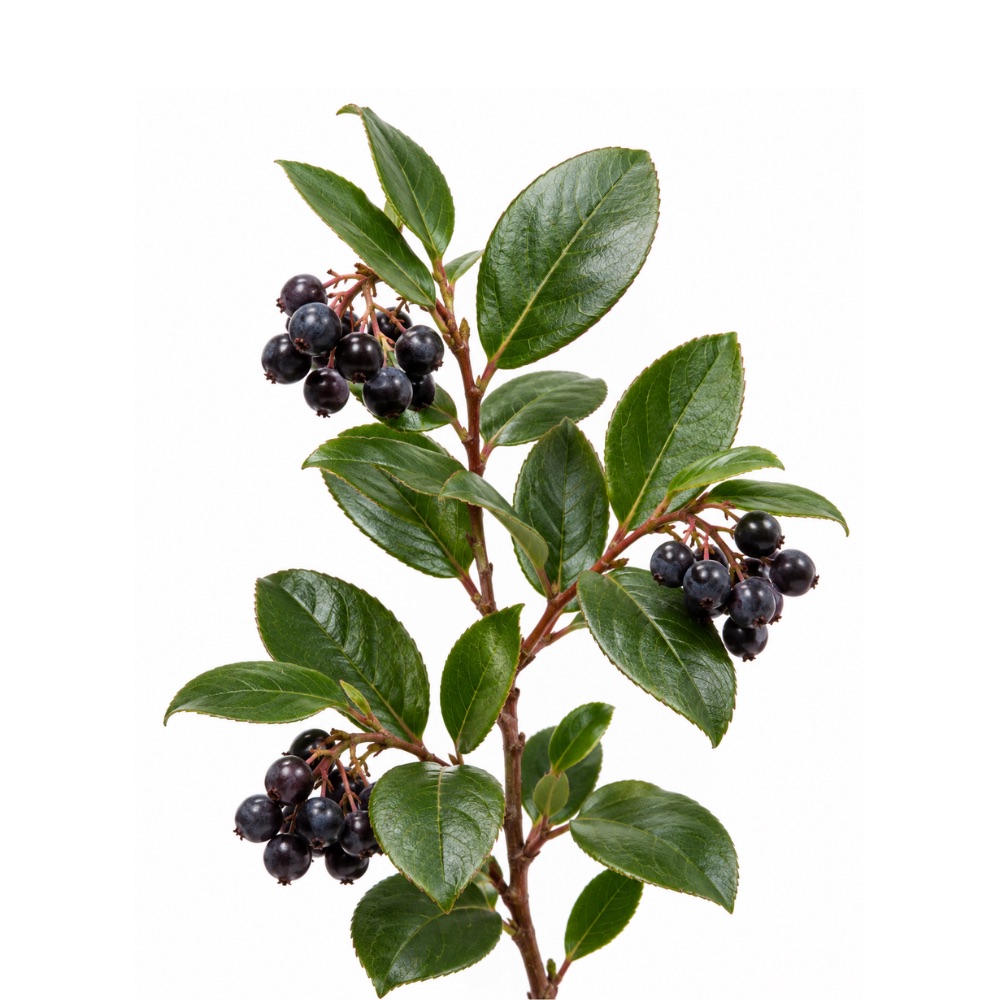 ModerateModerateSalalGaultheria shallonThe berries carry proanthocyanidins and anthocyanins at concentrations exceeding many blueberries and huckleberries, with the test-tube antioxidant capacity that follows.Mechanismproanthocyanidin and anthocyanin radical scavengingRead the evidenceShow less
Proanthocyanidin and anthocyanin concentrations in the berry exceed those of many Vaccinium fruits 249,
The finding is compositional and assay-level, and it describes the berry eaten as food rather than a medicinal preparation.
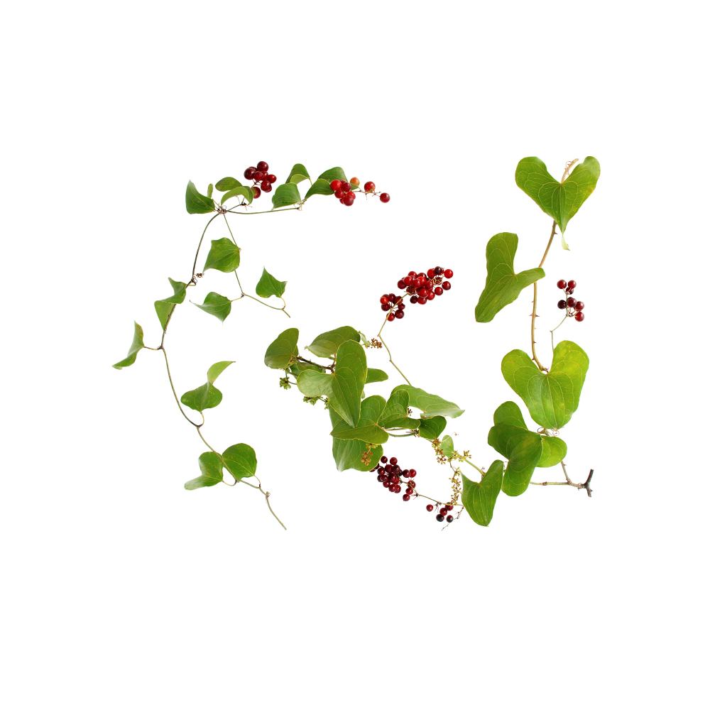 ModerateModerateSarsaparillaSmilax spp.Phenolic-enriched S. glabra extract matched or beat ascorbic acid on ABTS, with astilbin the principal contributor, and eased cisplatin kidney injury in mice through Nrf2/HO-1.MechanismDPPH/ABTS radical scavenging; ferric-reducing power; Nrf2/HO-1 ↑Read the evidenceShow lessMonograph →
On ABTS, phenolic-enriched S. glabra extract matched or exceeded ascorbic acid, and it scavenged DPPH and reduced ferric iron as well 363Reference 363In vitroAntioxidant and Anti-Inflammatory Activities of Phenolic-Enriched Extracts of Smilax glabra — in vitroView study →, with screening of six purified flavonoids confirming astilbin and its stereoisomers as the principal contributors 364Reference 364In vitroAntioxidant and Anti-Inflammatory Activities of Six Flavonoids from Smilax glabra Roxb. — in vitroView study →. Catechins and epicatechin plausibly add to net capacity 363Reference 363In vitroAntioxidant and Anti-Inflammatory Activities of Phenolic-Enriched Extracts of Smilax glabra — in vitroView study →.
In vivo, S. glabra eased cisplatin-induced acute kidney injury in mice via Nrf2/HO-1 activation 365Reference 365AnimalSmilax glabra Roxb. alleviates cisplatin-induced acute kidney injury in mice by activating the Nrf2/HO-1 signalling pathway — animal modelView study →, an induced endogenous antioxidant response rather than stoichiometric scavenging.
The bulk of the work is cuvette and cell assay, and no human oxidative-stress biomarker study exists.
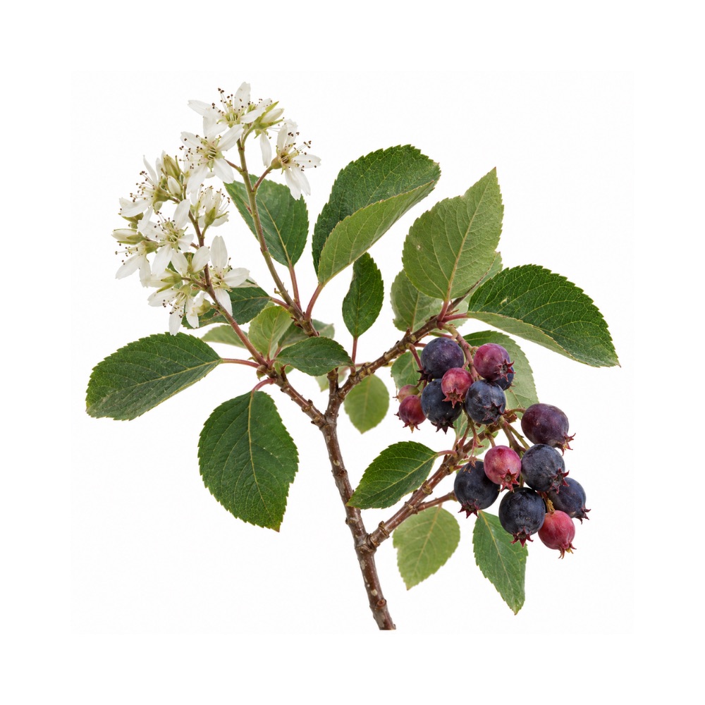 ModerateModerateSaskatoon BerryAmelanchier alnifoliaCarries the full anthocyanidin set of cyanidin, delphinidin, peonidin, petunidin, malvidin and pelargonidin, a broader spread than most berries, with the assay capacity that goes with it.Mechanismanthocyanin and phenolic radical scavengingRead the evidenceShow less
Dozens of analytical papers profile the fruit’s polyphenols and antioxidant capacity. Saskatoon carries the full anthocyanidin set of cyanidin, delphinidin, peonidin, petunidin, malvidin and pelargonidin, a broader spread than most berries.
The finding is compositional and assay-level: a statement about what is in the fruit rather than about what it does in a person.
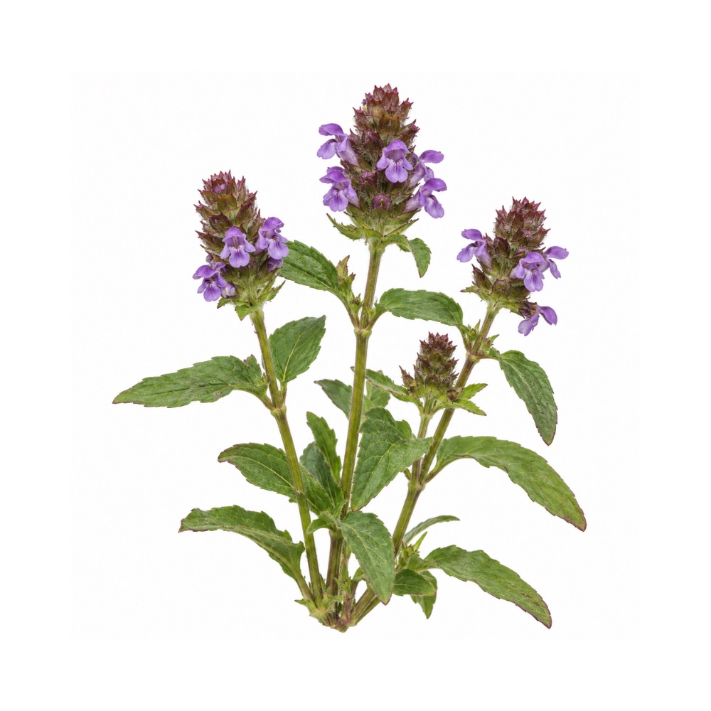 ModerateModerateSelf-HealPrunella vulgarisRosmarinic acid and the flavonoids give it the extensive in-vitro radical scavenging typical of the mint family.Mechanismrosmarinic acid and flavonoid radical scavengingRead the evidenceShow less
The in-vitro antioxidant activity of self-heal is extensively documented and rests on rosmarinic acid and the flavonoids.
The activity is typical of a rosmarinic-acid-rich mint, and it has not been measured beyond the assay.
ModerateShiitakeLentinula edodesOne of the richest dietary sources of ergothioneine, a thiol antioxidant with its own transporter that concentrates it in tissue.Mechanismergothioneine ROS scavenging via OCTN1 tissue uptake; mitochondrial supportRead the evidenceShow less
Ergothioneine is a thiol antioxidant with a dedicated transporter, OCTN1, that concentrates it in tissue, and shiitake is among its richest dietary sources, which puts it ahead of fungi whose antioxidant claim rests only on assay capacity.
Ergothioneine-rich shiitake extract restored mitochondrial function in senescent HT22 neurons 369Reference 369In vitroErgothioneine-rich Lentinula edodes mushroom extract restores mitochondrial functions in senescent HT22 cells — in vitro. https://pubmed.ncbi.nlm.nih.gov/39643233/View study →, and isolated ergothioneine showed a cognition-decline signal in a mild-cognitive-impairment pilot RCT 370Reference 370RCTInvestigating the efficacy of ergothioneine to delay cognitive decline in mild cognitively impaired subjects: a pilot study — randomised controlled trialView study →.
Content is cultivar- and light-dependent, about 1.2–2.8 mg/g dry and roughly doubled by blue-light cultivation.
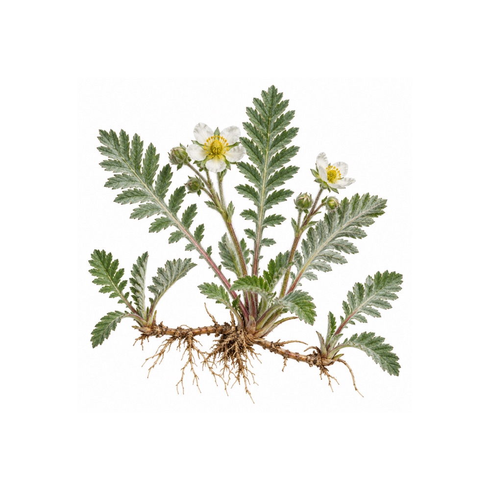 ModerateModerateSilverweedArgentina anserinaPolyphenol and triterpene antioxidant activity replicated across in-vitro assays, extract testing and in-vivo stress models by independent groups.Mechanismpolyphenol and triterpene radical scavenging; in-vivo stress-model protectionRead the evidenceShow less
Independent groups have reproduced the effect at three levels, in-vitro assays, extract testing and in-vivo stress models, and it is the most reproducible signal the plant has.
Three-level replication makes the claim stronger than most, though there are no human data.
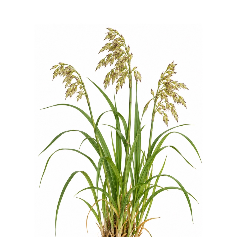 ModerateModerateSweetgrassHierochloe odorataIts 5,8-dihydroxycoumarin scavenges radicals in vitro and protected against ethanol-induced oxidative stress in three rat studies.Mechanism5,8-dihydroxycoumarin and its glucoside; radical scavengingRead the evidenceShow less
The antioxidant effect is reproduced across in-vitro radical scavenging and three rat studies of ethanol-induced oxidative stress 371,
In-vivo replication across three studies is a stronger position than most assay-only claims.
The same coumarin is weakly genotoxic and pro-oxidant-cytotoxic at concentrated-extract levels 376Reference 376In vitroEvaluation of the biological activity of naturally occurring 5,8-dihydroxycoumarin — in vitro and in vivo genotoxicityView study →, so the active and the hazard are one molecule at different concentrations.
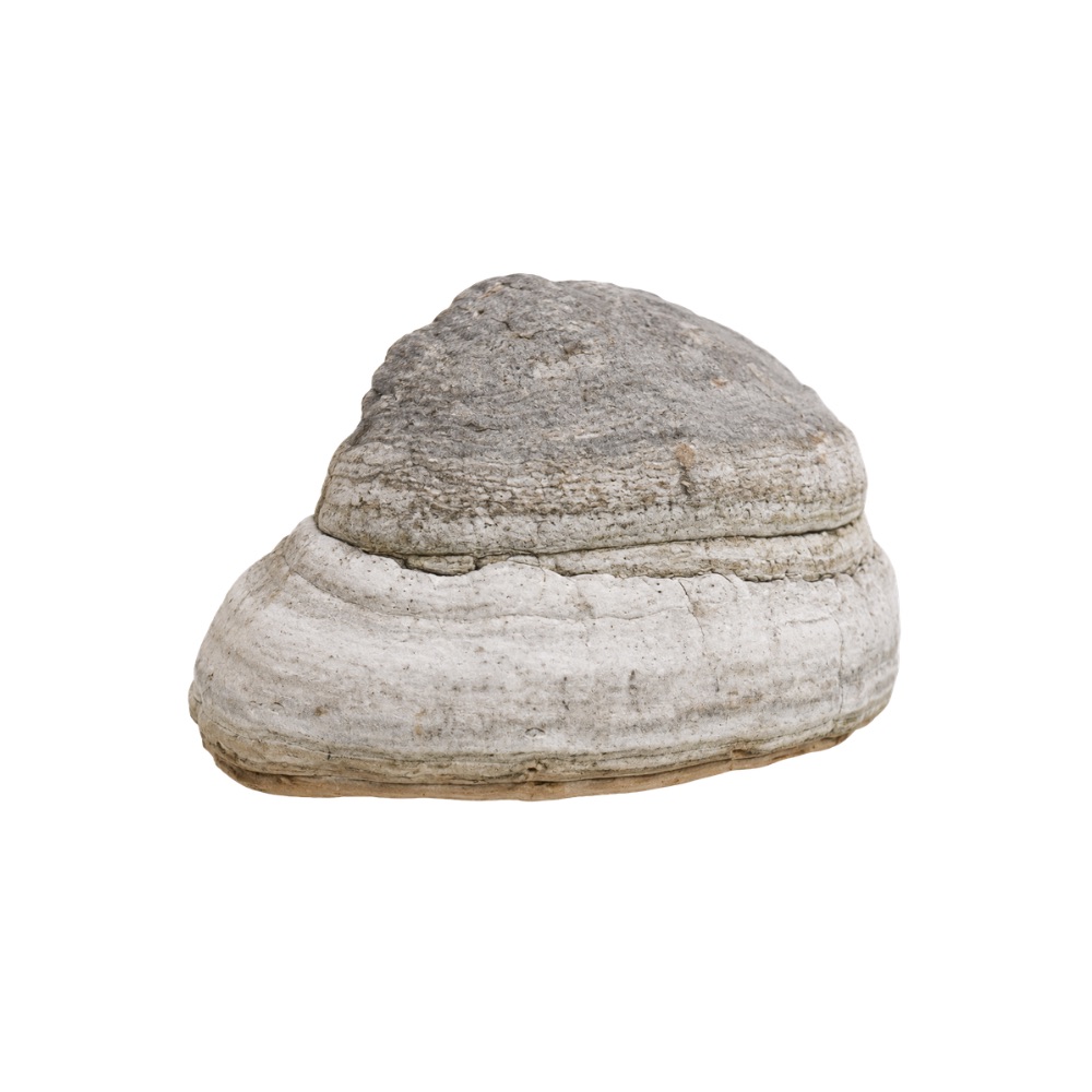 ModerateModerateTinder ConkFomes fomentariusHot-water and aqueous-alkali extracts, close to the traditional decoction, had the strongest radical scavenging of several wild wood fungi, with the melanin pigments also absorbing UVB.Mechanismdirect radical scavenging (DPPH, FRAP, ROS); melanin-pigment UVB absorptionRead the evidenceShow lessMonograph →
Crude extracts had the strongest radical-scavenging capacity among several wild lignicolous fungi (DPPH IC50 ≈ 10.7 µg/mL) alongside the highest total phenol and flavonoid content 382Reference 382In vitroWild-growing lignicolous mushroom species as sources of novel agents with antioxidative and antibacterial potentials — in vitroView study →. Purified polysaccharides carry measurable antioxidant activity 383Reference 383In vitroStructural characterization and biological activities of polysaccharides from tree mushroom species — in vitroView study →, and the melanin-like pigments account for both high radical scavenging and a moderate UVB-absorbing capacity 384,
The active fraction is carried by aqueous-alkali and hot-water extracts, close to the traditional decoction, which is an unusually good preparation match.
Every measurement is a cuvette assay, with no in-vivo or human study.
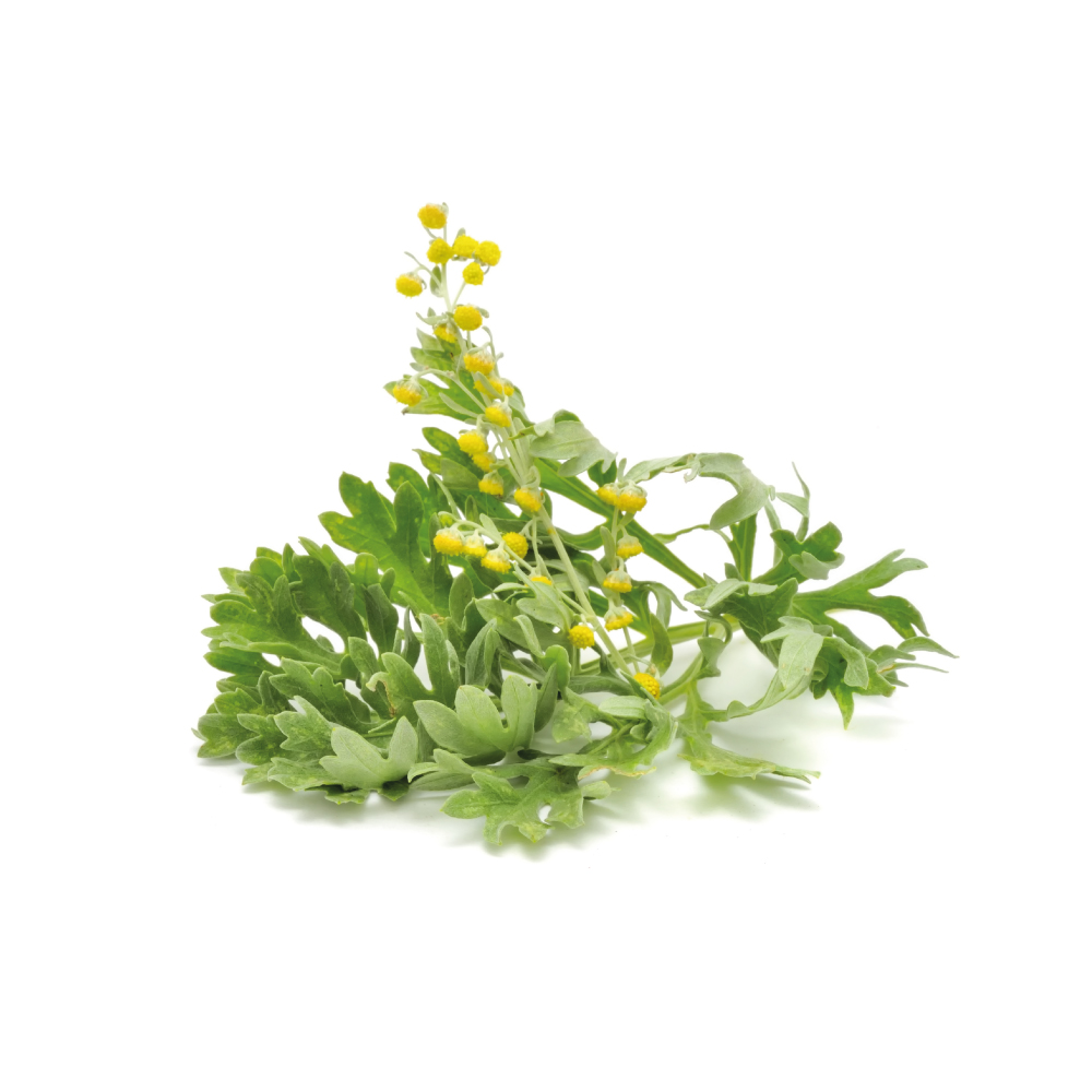 ModerateModerateWormwoodArtemisia absinthiumFlavonol glycosides of quercetin and isorhamnetin with phenolic acids scavenge radicals reliably in DPPH, FRAP, hydroxyl and nitric-oxide assays.Mechanismradical scavenging (flavonol glycosides)Read the evidenceShow lessMonograph →
The herb is reliably active in cell-free assays (DPPH, FRAP, hydroxyl- and NO-radical scavenging), attributed to flavonol glycosides of quercetin and isorhamnetin plus phenolic acids 392,
A 2025 characterisation of flowers and leaves found rich polyphenol content but only moderate in-vitro antioxidant activity, where most herbs are described as strong on the same assays.
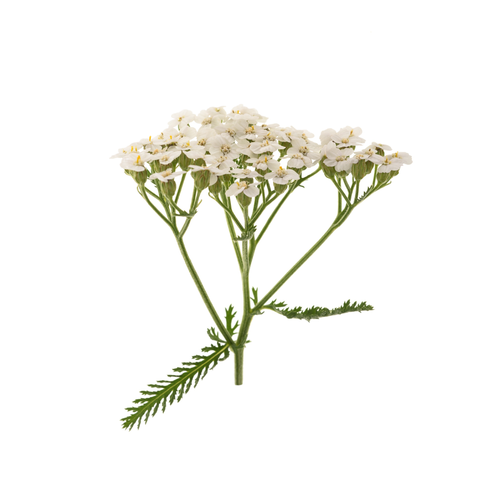 ModerateModerateYarrowAchillea millefoliumChlorogenic acid, quercetin, luteolin and apigenin give the flavonoid and phenolic-acid fraction strong, replicated radical scavenging across the standard assays.Mechanismradical scavenging; lipid-peroxidation ↓Read the evidenceShow lessMonograph →
The flavonoid and phenolic-acid fraction of chlorogenic acid, quercetin, luteolin and apigenin scavenges radicals strongly and reproducibly across standard assays 394,
The activity is a supporting mechanism beneath the hepatoprotective and anti-inflammatory actions.
The evidence is in vitro and cell-based only, with no human data.
Show 33 more herbs with a mild antioxidant action
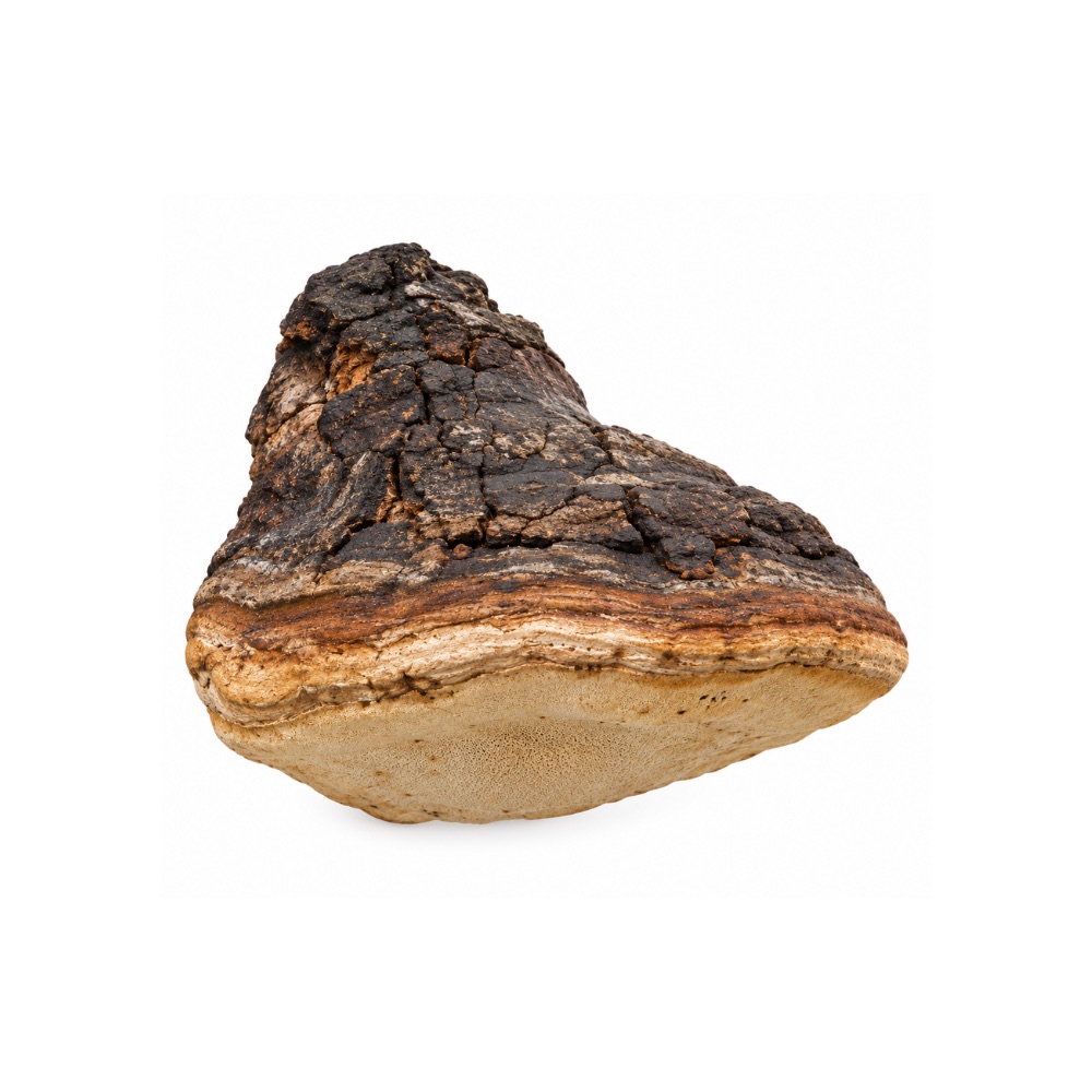 MildMildAgarikonFomitopsis officinalisPhenolics and ergosterol derivatives scavenge DPPH radicals in vitro.MechanismDPPH radical scavenging (phenolics, ergosterol derivatives)Read the evidenceShow less
In-vitro DPPH scavenging has been recorded, with the activity inferred from the phenolics and ergosterol derivatives 137,
There is no functional or in-vivo confirmation.
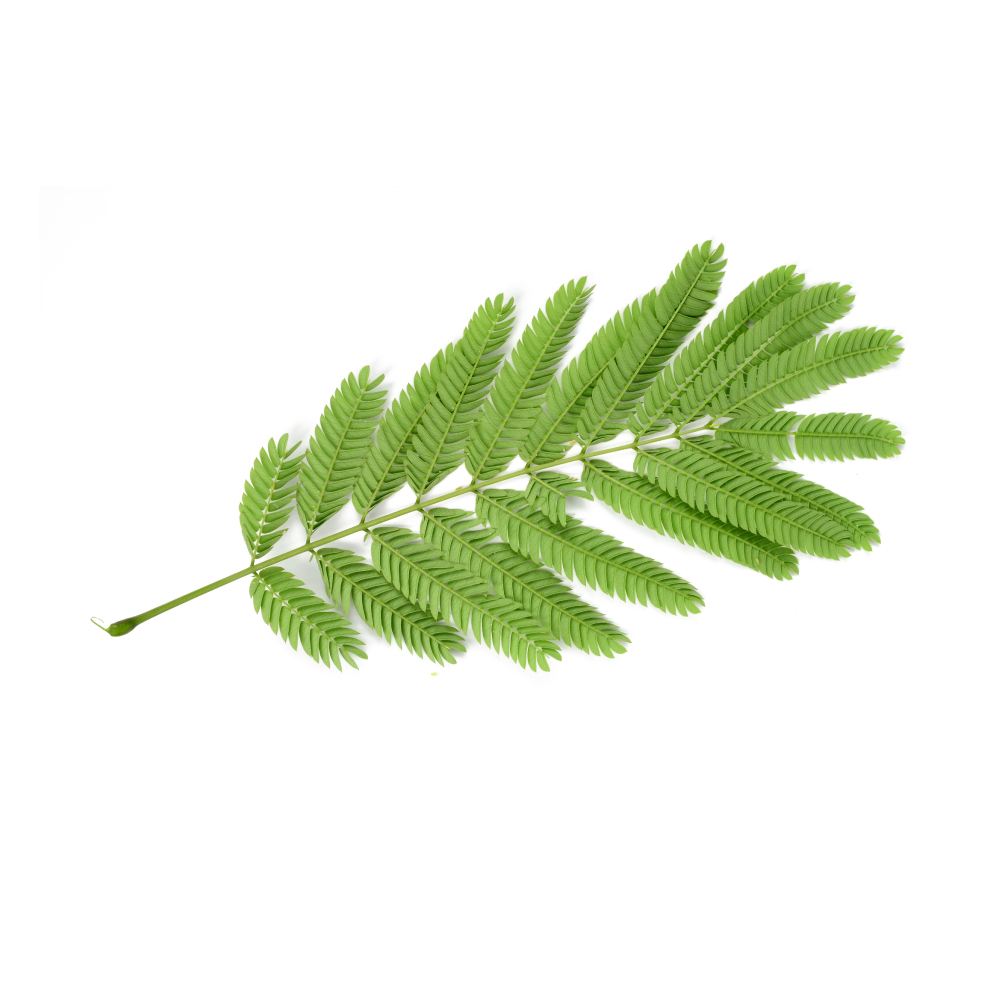 MildMildAlbizziaAlbizzia lebbeckThe stem bark's flavonols, leucoanthocyanidins and tannins protect pancreatic islets from oxidative damage in diabetic rats.MechanismROS scavenging; tissue oxidative-damage ↓ (rodent)Read the evidenceShow lessMonograph →
The stem-bark extract’s islet protection in diabetic rats is described as protection from oxidative damage 139Reference 139In vitroTarget-guided isolation, in-vitro antidiabetic, antioxidant activity and molecular docking studies of some flavonoids from Albizzia lebbeck Benth. bark — in vitro / in silicoView study →, and the one dedicated antioxidant study is an alloxan-diabetic rat model 141Reference 141AnimalAntioxidant activity of Albizzia lebbeck (Linn.) Benth. in alloxan diabetic rats — animal modelView study →.
The phytochemistry points at the source: a bark load of flavonols at about 24 mg/g plus the heartwood leucoanthocyanidins melacacidin and lebbecacidin and 7–11% tannins 140Reference 140Pharmacognostical screening and phytochemical evaluation of Albizia lebbeck Benth. heartwood — analytical study.
There are no assay values and no human markers, and the antioxidant activity is the engine under the pancreatic effect rather than a demonstrated action in its own right.
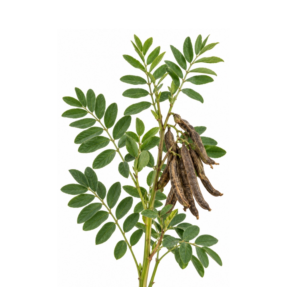 MildMildAmerican LicoriceGlycyrrhiza lepidotaIts flavonoids scavenge radicals in vitro.Mechanismflavonoid radical scavengingRead the evidenceShow less
The root’s flavonoids give standard in-vitro radical scavenging.
Nothing beyond the assay has been measured.
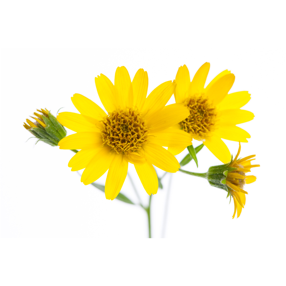 MildMildArnicaArnica montanaFlower extracts rich in quercetin glycosides scavenge radicals in standard assays and protected fibroblasts from hydrogen-peroxide damage at 10 mg/L.Mechanismdirect phenolic/flavonoid free-radical scavenging (DPPH/ORAC/TEAC); H₂O₂-induced damage ↓ in fibroblastsRead the evidenceShow lessMonograph →
Ethanolic arnica flower extracts are rich in phenolics and flavonoids and scavenge free radicals in standard assays, and at 10 mg/L the extract protected mouse NCTC fibroblasts against hydrogen-peroxide damage, preserving viability and morphology 157Reference 157In vitroEvaluation of antioxidant and cytoprotective activities of Arnica montana L. and Artemisia absinthium L. ethanolic extracts — in vitroView study →. Activity is attributed chiefly to quercetin and its glycosides isoquercitrin, astragalin and rutin.
An Artemisia absinthium extract outperformed arnica in the same assay battery 157Reference 157In vitroEvaluation of antioxidant and cytoprotective activities of Arnica montana L. and Artemisia absinthium L. ethanolic extracts — in vitroView study →.
There is no human antioxidant outcome and no plausible systemic one, since the herb only reaches intact skin, so the activity contributes to the topical skin-protective reputation.
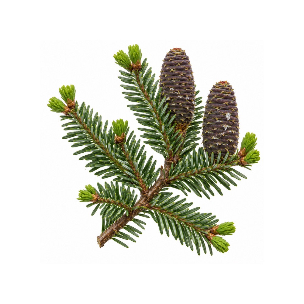 MildMildBalsam FirAbies balsameaBark extracts scavenge radicals in assay.Mechanismbark-extract radical scavengingRead the evidenceShow less
Radical-scavenging activity of the bark extracts has been recorded in standard assays 168,
The evidence is assay-level only.
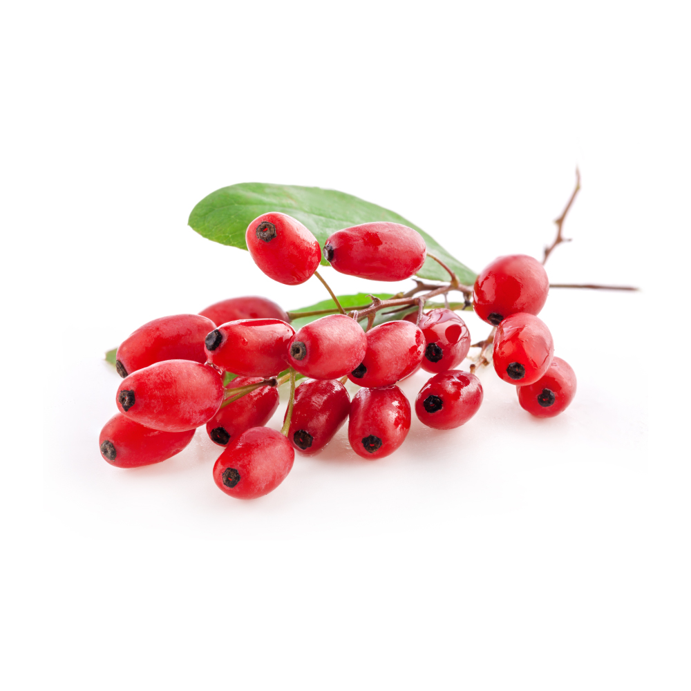 MildMildBarberryBerberis spp.The fruit anthocyanins and polyphenols scavenge radicals, not berberine, and a fruit-extract trial in diabetics raised serum total antioxidant capacity.Mechanismanthocyanin and polyphenol radical scavenging (fruit, leaf, stem); serum total antioxidant capacity ↑ (human)Read the evidenceShow lessMonograph →
Berberis vulgaris fruit, leaf and stem show measurable radical scavenging driven by anthocyanins and other polyphenols rather than by berberine 171,
The fruit-extract RCT in diabetics reported a significant rise in serum total antioxidant capacity 173Reference 173RCTThe effects of Berberis vulgaris fruit extract on serum lipoproteins, apoB, apoA-I, homocysteine, glycemic control and total antioxidant capacity in type 2 diabetic patients — randomised controlled trialView study →.
That is one serum-marker outcome in one trial, with everything else inferred from in-vitro assays and no mapping to a disease endpoint.
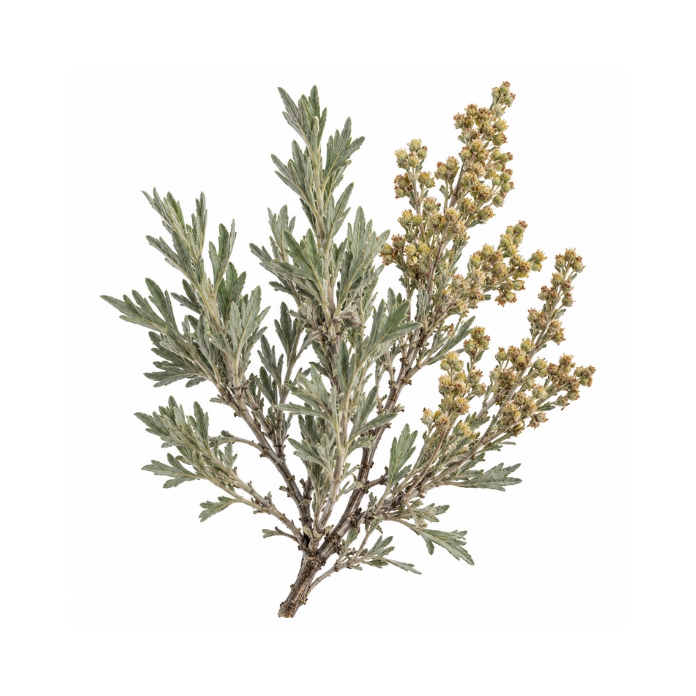 MildMildBig SagebrushArtemisia tridentataAn unusually well-catalogued flavonoid fraction, including several methylated flavonols, scavenges radicals in vitro.Mechanismflavonoid and phenolic radical scavengingRead the evidenceShow less
The flavonoid fraction is unusually well catalogued for the plant, including several methylated flavonols such as casticin, axillarin and quercetagetin trimethyl ether, and it scavenges radicals in vitro.
The evidence is assay-level throughout.
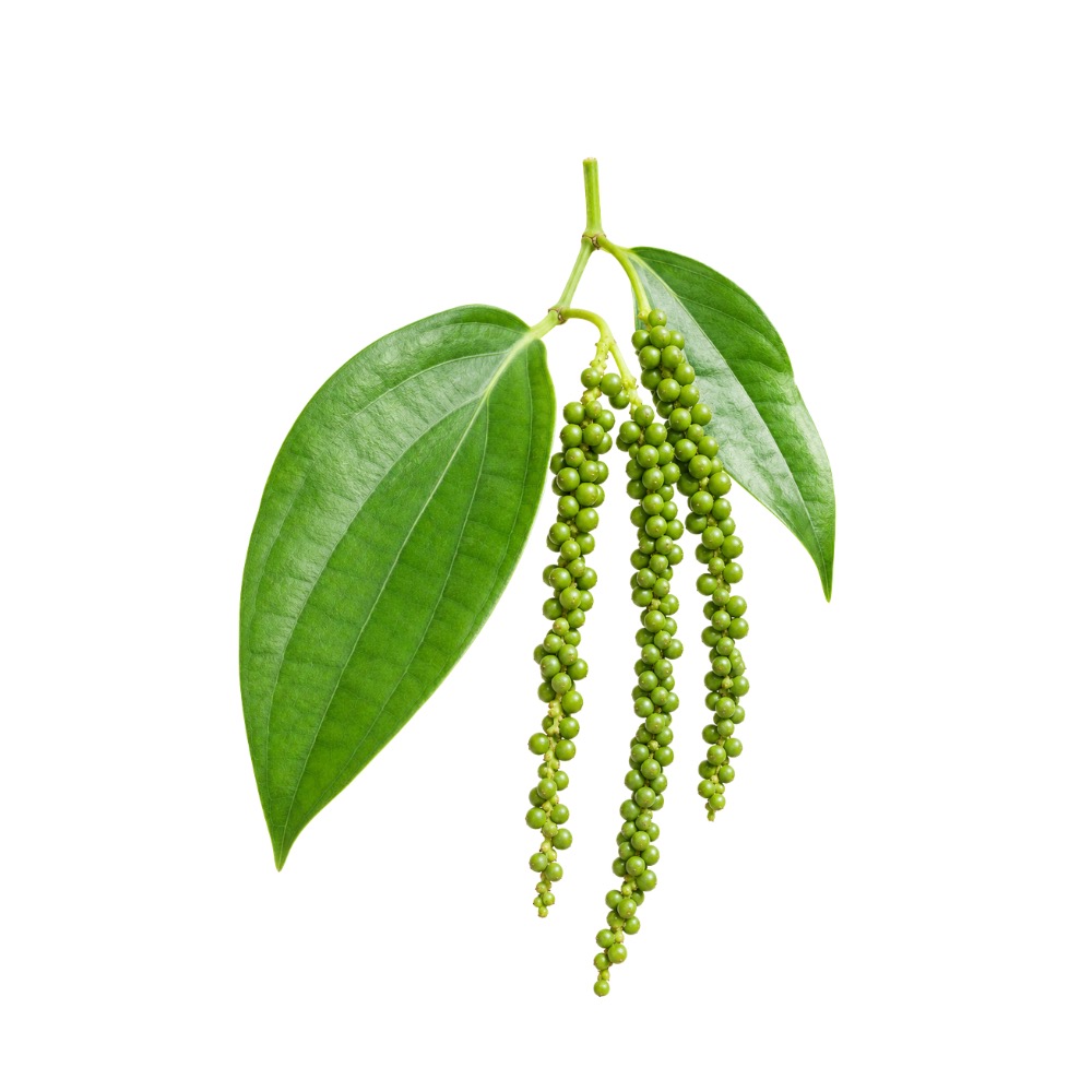 MildMildBlack PepperPiper nigrumPiperine upregulates Nrf2/HO-1 and lowers reactive-oxygen markers in cells, and reduced lipid peroxidation while preserving glutathione in mouse liver.MechanismNrf2/HO-1 ↑; lipid peroxidation ↓; glutathione preservedRead the evidenceShow lessMonograph →
In cell models piperine upregulates the Nrf2/HO-1 cytoprotective pathway and lowers reactive-oxygen markers, protecting UV-B-irradiated human keratinocytes from oxidative and inflammatory damage 184Reference 184In vitroAntioxidant and anti-inflammatory effects of piperine on UV-B-irradiated human HaCaT keratinocytes — in vitroView study →, and it reduced lipid peroxidation while preserving glutathione in mouse liver-protection models 185Reference 185In vitroEvaluation of the liver protective potential of piperine — mouse in vivo / in vitroView study →.
The action is inductive rather than scavenging.
There is no clinical antioxidant endpoint, and whole-pepper dietary intake has never been shown to change human antioxidant status.
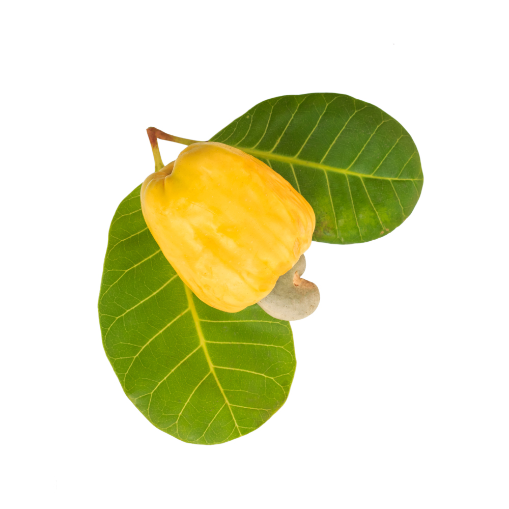 MildMildCashewAnacardium occidentaleAnacardic acid in the shell liquid inhibits xanthine oxidase, and the cashew apple carries about five times the vitamin C of citrus with carotenoids and twelve flavonol glycosides.Mechanismxanthine oxidase inhibition (anacardic acid); dietary ascorbate, carotenoid and flavonol loadRead the evidenceShow lessMonograph →
Xanthine oxidase inhibition by anacardic acid is the likely reason for the juice’s short shelf life, and the acid is listed as antioxidant among its activities 208Reference 208In vitroAnacardic acid, an inhibitor of histone acetyltransferase, suppresses NF-κB–regulated gene products and potentiates apoptosis — in vitroView study →. The cashew apple carries about five times the vitamin C of citrus plus β-carotene, β-cryptoxanthin and twelve myricetin and quercetin glycosides, the same flavonol set found in cranberry 209Reference 209Determination of the flavonoid components of cashew apple (Anacardium occidentale) by LC-DAD-ESI/MSView study →.
The shell liquid’s antimutagenic and genoprotective result 210Reference 210In vitroEvaluation of toxic, cytotoxic, mutagenic and antimutagenic activities of natural and technical cashew nut shell liquids — in vitro bioassaysView study → is adjacent support.
No antioxidant assay of the plant has been reported, so the claim rests on composition and on the enzyme mechanism.
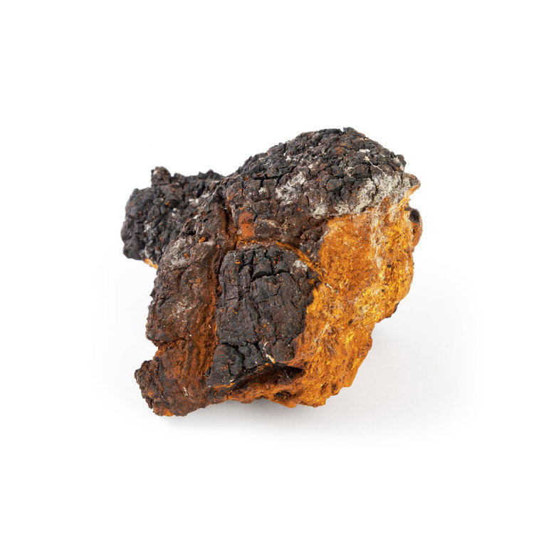 MildMildChagaInonotus obliquusA water-soluble melanin complex at 50–60% of the crude extractives and a polyphenol fraction, with one in-vivo antilipidperoxidative result in diabetic mice.Mechanismwater-soluble melanin complex; polyphenol fraction; lipid-peroxidation ↓ (in vivo, diabetic mice)Read the evidenceShow lessMonograph →
The chemistry is a water-soluble melanin complex reported at about 50–60% of crude extractives 216Reference 216Melanin complex of the fungus Inonotus obliquus — analytical study (water-soluble melanin complex, ~50–60% of crude extractives), plus polyphenols 217Reference 217Chemical characterization and biological activity of Chaga (Inonotus obliquus), a medicinal “mushroom” — analytical study (inotodiol, betulin, betulinic acid, β-glucan content; host-tree variation)View study →.
The one in-vivo endpoint is the antilipidperoxidative effect recorded alongside the blood-sugar result in diabetic mice 218Reference 218AnimalAntihyperglycemic and antilipidperoxidative effects of dry matter of culture broth of Inonotus obliquus in alloxan-diabetes mice — animal modelView study →.
Chaga is sold on ORAC scores, an assay the USDA withdrew from its own database in 2012 because tube capacity does not represent what happens in the body, and there is no dedicated antioxidant research on the fungus.
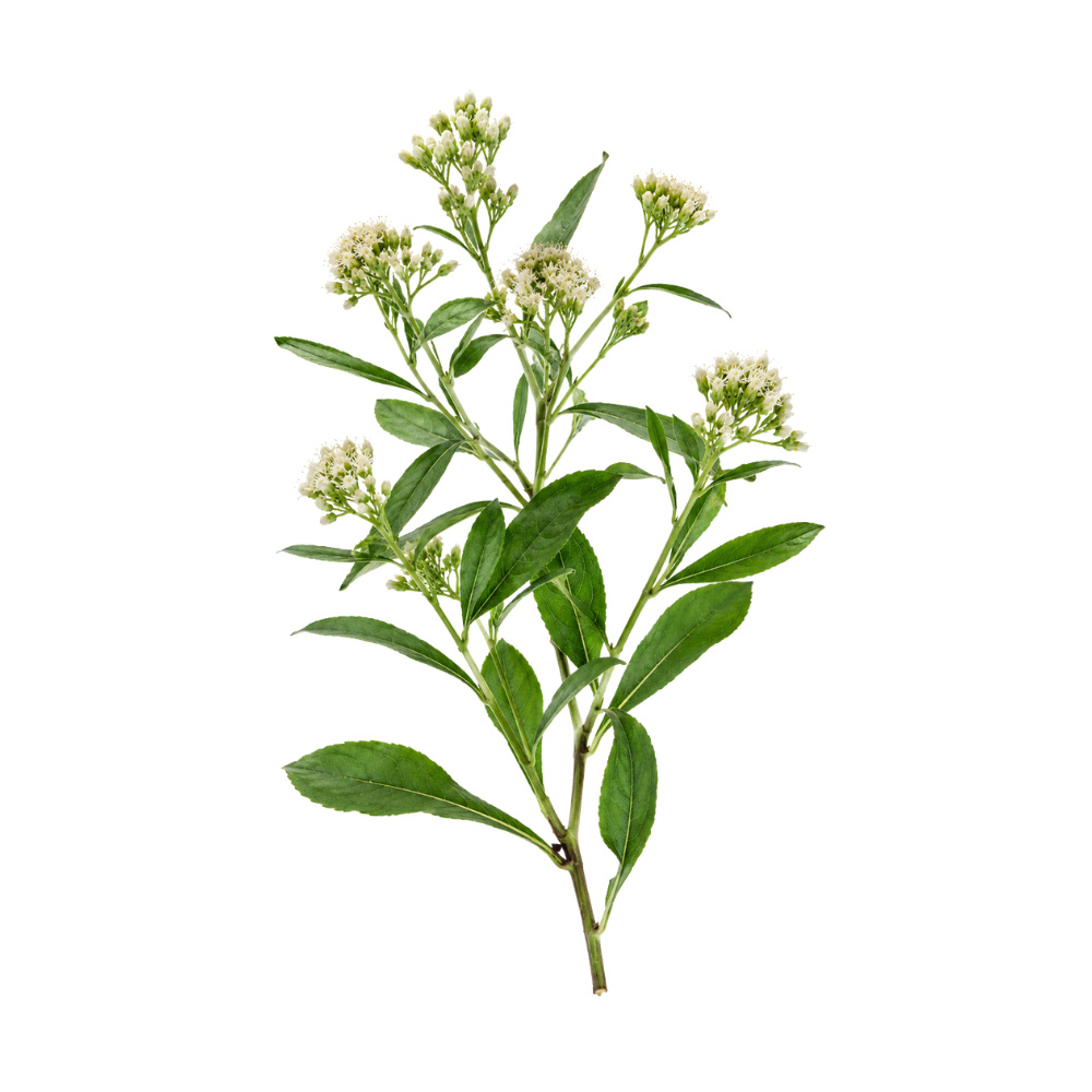 MildMildChilcaBaccharis latifoliaThe leaf extract cut stimulated ROS production in human neutrophils by about 55%, and the leaves are a documented source of quercetin and rutin.Mechanismneutrophil respiratory-burst (ROS) ↓; flavonoid content (quercetin, rutin)Read the evidenceShow lessMonograph →
In human neutrophils the leaf methanol extract inhibited PMA-stimulated ROS production by about 55% 221Reference 221Activity of plant extracts on the respiratory burst and the stress protein synthesisView study →, a cellular respiratory-burst assay that blunts oxidative signalling in immune cells rather than a chemical radical-scavenging test.
The leaves are a documented source of quercetin and rutin, whose antioxidant activity is well established but whose concentration in chilca varies with leaf type and soil 222Reference 222Flavonoid contents in leaves of Baccharis latifolia according to the type of leaf and its dependence on the physicochemical properties of soilsView study →.
There are no ORAC, DPPH, FRAP or in-vivo oxidative-stress data.
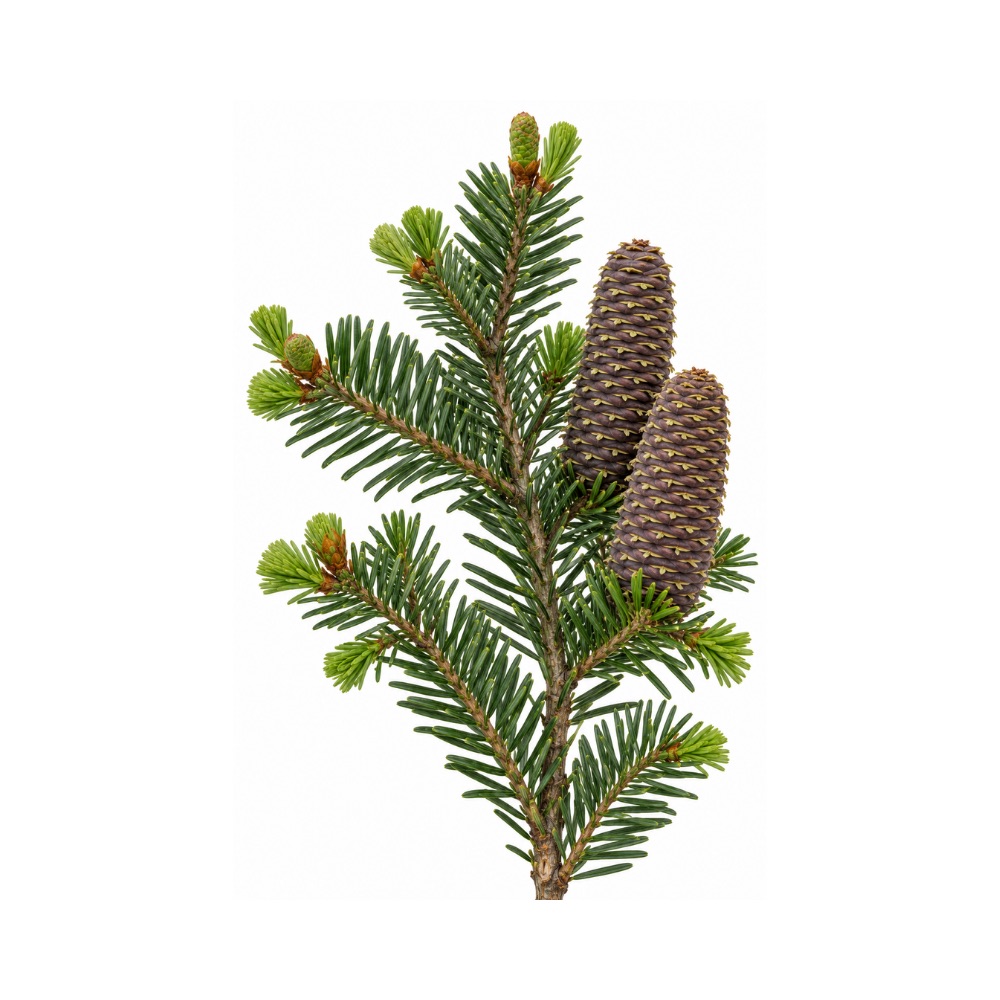 MildMildGrand FirAbies grandisIts resin acid dehydroabietic acid activates SIRT1, extends lifespan in nematodes and protects human dermal fibroblasts.Mechanismresin-acid and monoterpene radical scavenging; SIRT1 activation (dehydroabietic acid)Read the evidenceShow less
Dehydroabietic acid, a grand-fir resin acid, activates SIRT1, extends lifespan in C. elegans and protects human dermal fibroblasts 131Reference 131In vitroThe natural phytochemical dehydroabietic acid is an anti-aging reagent that mediates the direct activation of SIRT1 — in vitro / animal model (C. elegans)View study →.
The result belongs to the molecule, and it does not establish that a fir decoction is antioxidant.
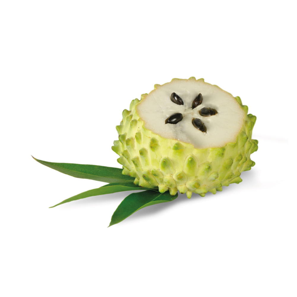 MildMildGraviolaAnnona muricataLeaf extracts scavenge radicals in vitro and shift SOD, catalase, glutathione and lipid-peroxidation markers across dozens of animal studies, the mechanism under its liver and blood-sugar work.Mechanismradical scavenging; tissue antioxidant markersRead the evidenceShow lessMonograph →
Across dozens of in-vivo studies, leaf extracts shift SOD, catalase, glutathione and lipid-peroxidation markers, and they scavenge radicals in vitro; the activity underlies much of the hepatoprotective and antidiabetic data.
Almost any polyphenol-rich botanical scores well on these assays, so the finding carries little discriminating information, and there are no human data.
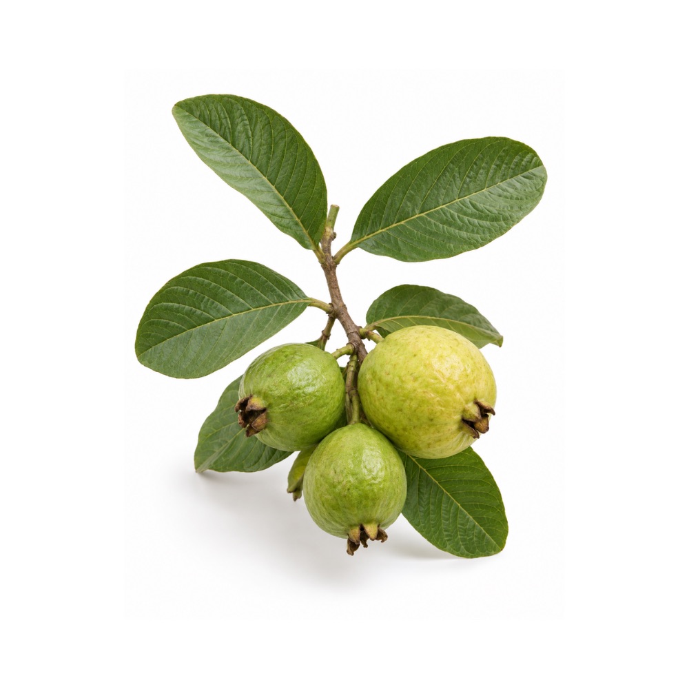 MildMildGuavaPsidium guajavaThe leaf carries 510–575 mg of phenolics per gram and scavenges DPPH radicals strongly, and the fruit holds several times the vitamin C of citrus.Mechanismphenolic radical scavenging (DPPH/FRAP), in vitroRead the evidenceShow less
Aqueous and ethanolic leaf extracts carry very high total phenolic content, about 510–575 mg gallic-acid-equivalent per gram of dried leaf, with strong DPPH radical scavenging 406Reference 406In vitroAntioxidant power of phytochemicals from Psidium guajava leaf — in vitroView study →, and leaf extracts are antimutagenic in the same assays that show the scavenging 407Reference 407In vitroAntioxidant and antimutagenic potential of Psidium guajava leaf extracts — in vitroView study →. The fruit carries several times the vitamin C of citrus.
Radical scavenging is one of the most reproducible findings on the leaf, and all of it is in vitro, with no human antioxidant-outcome data.
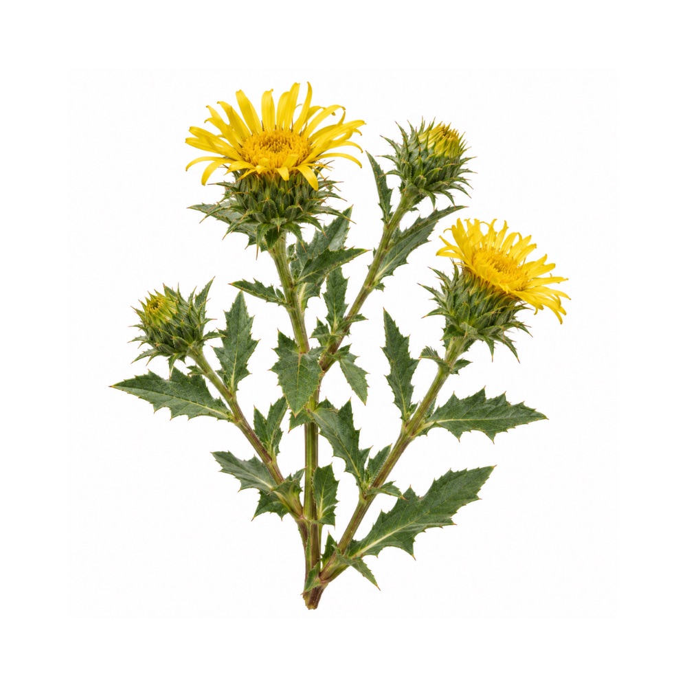 MildMildGumweedGrindelia squarrosaIts phenolic and methylated-flavonoid fraction scavenges radicals in vitro.Mechanismphenolic and methylated-flavonoid radical scavengingRead the evidenceShow less
The phenolic fraction scavenges radicals in vitro.
Nothing beyond the assay has been measured.
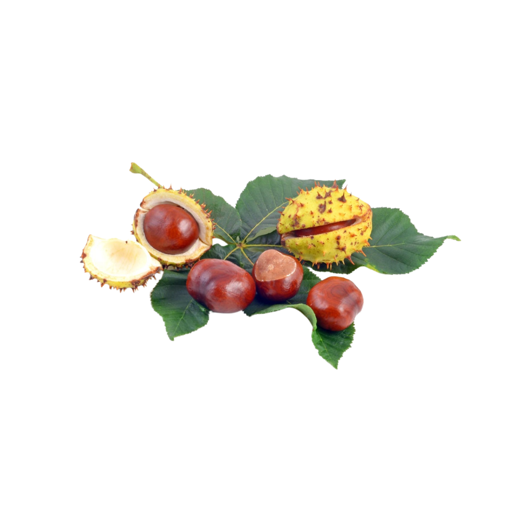 MildMildHorse ChestnutAesculus hippocastanumThe seed's rutin, quercetin and kaempferol glycosides and condensed proanthocyanidins scavenge free radicals in vitro, a minor fraction next to the saponins that carry the herb's activity.Mechanismfree-radical scavenging (flavonol glycoside + proanthocyanidin fraction)Read the evidenceShow lessMonograph →
The seed’s polyphenol fraction of rutin, quercetin and kaempferol glycosides plus condensed proanthocyanidins shows measurable free-radical scavenging capacity in vitro, varying with extraction solvent and harvest time 287,
The polyphenols are a minor fraction next to the saponins that carry the herb’s vascular activity 287,
There are no human antioxidant-status data.
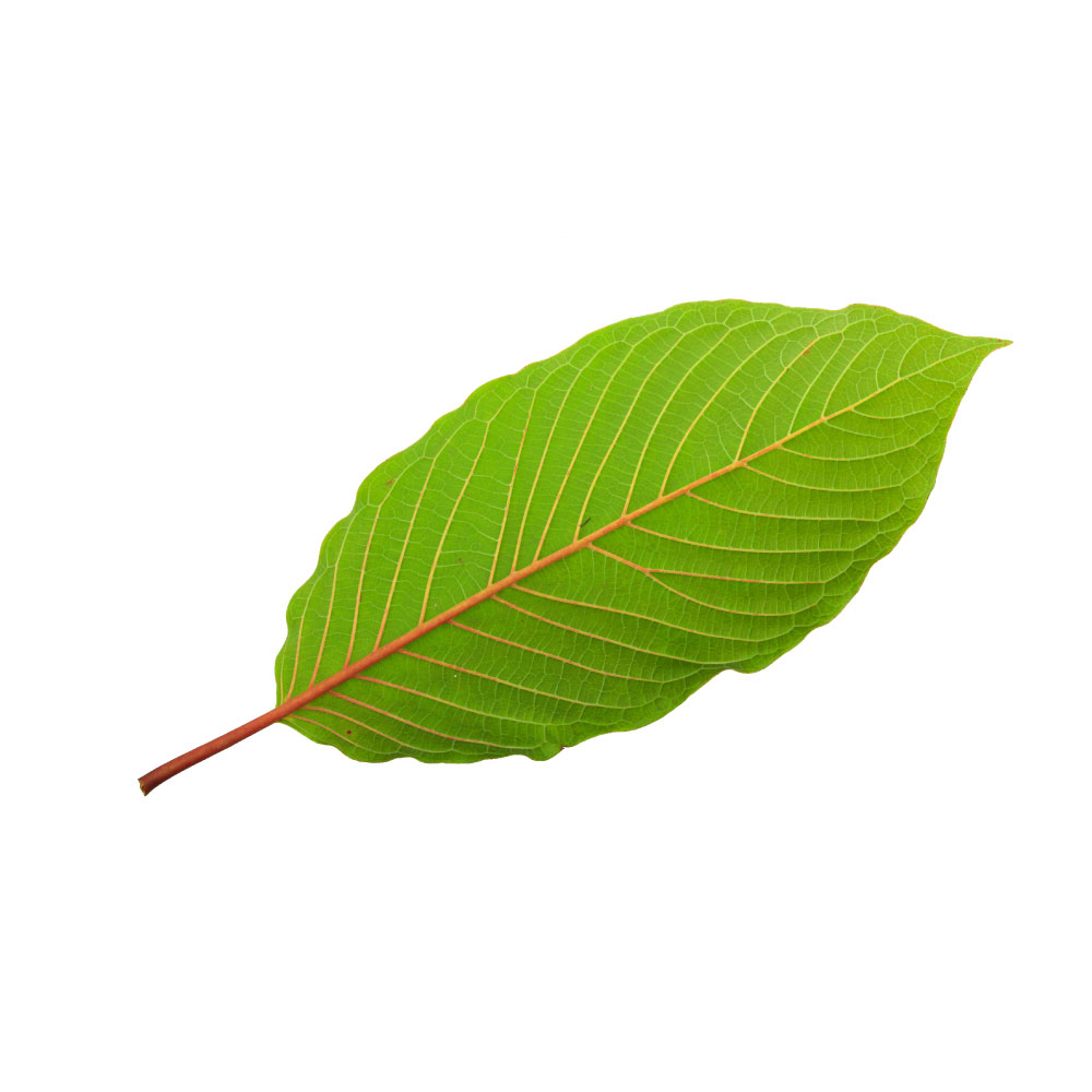 MildMildKratomMitragyna speciosaLeaf extracts scavenge DPPH radicals in proportion to phenolic content, through epicatechin and the flavonoids rather than the alkaloids.Mechanismphenolic radical scavenging (epicatechin, flavonoids)Read the evidenceShow lessMonograph →
The flavonoid-rich methanolic extract (IC50 ≈ 37 µg/mL) outperformed both aqueous and alkaloid extracts on DPPH, with scavenging tracking phenolic content 293Reference 293In vitroEvaluation of antioxidant and antibacterial activities of aqueous, methanolic and alkaloid extracts from Mitragyna speciosa leaves — in vitroView study →, while isolated mitragynine shows only moderate ABTS and DPPH scavenging 294Reference 294In vitroAntioxidant value and antiproliferative efficacy of mitragynine and a silane reduced analogue — in vitroView study →.
The capacity tracks epicatechin and other flavonoids rather than the alkaloids, so the extract that maximises the antioxidant reading is not the one people consume.
The evidence is in vitro, extract-specific and unremarkable against ordinary dietary polyphenol sources.
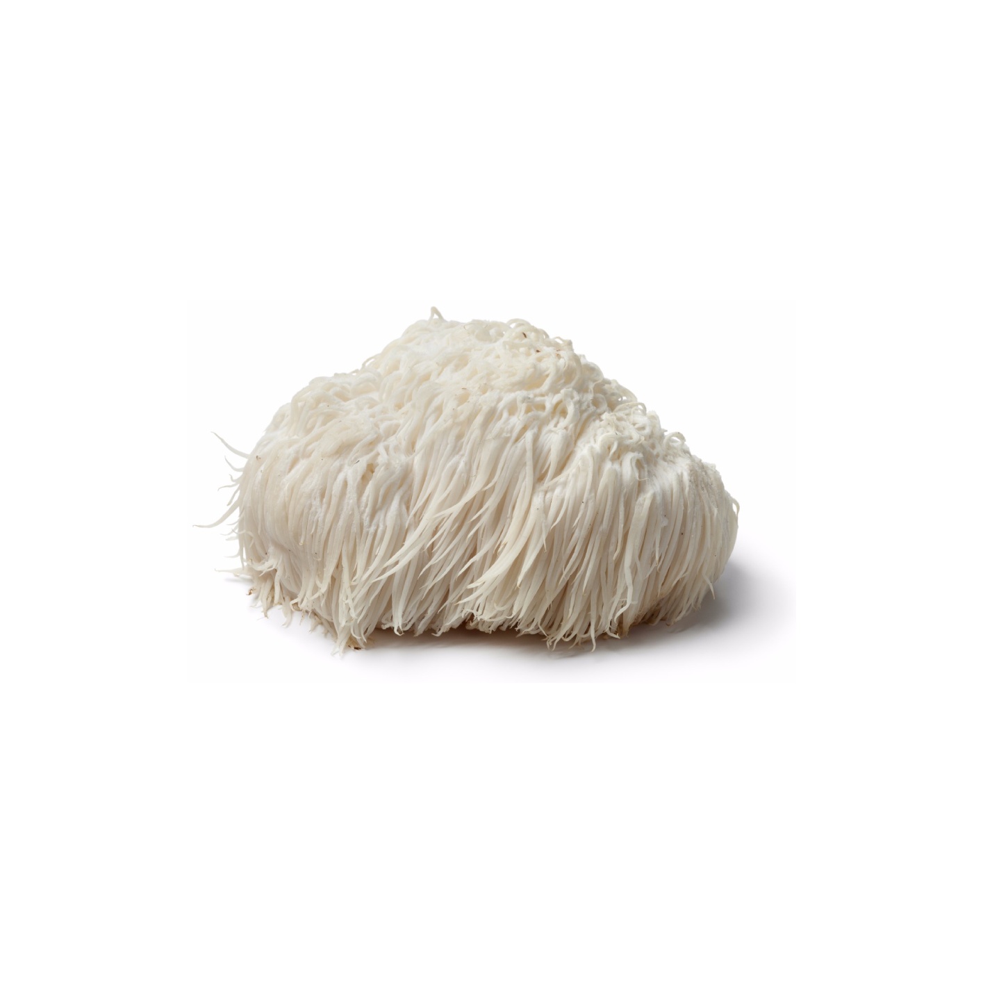 MildMildLion’s ManeHericium erinaceusMycelial polysaccharide fractions scavenge radicals in vitro and lower tissue oxidative-stress markers in mice, the connective mechanism under its neuroprotective and hepatoprotective findings.Mechanismfree-radical scavenging (chemical assay); tissue oxidative-stress markers ↓; molecular target not establishedRead the evidenceShow lessMonograph →
Mycelial endo-polysaccharide fractions showed strong in-vitro radical scavenging alongside a hepatoprotective effect in mice, with the most active fraction’s protection attributed to its antioxidant capacity 298Reference 298In vitroAntioxidant and hepatoprotective potential of endo-polysaccharides from Hericium erinaceus — in vitro and mouse studyView study →. The wider review treats antioxidant activity as the connective thread beneath the neuroprotective, hepatoprotective and anti-inflammatory findings 299Reference 299ReviewChemistry, nutrition, and health-promoting properties of Hericium erinaceus mushroom — reviewView study →.
No molecular pathway is established. The measured material is an isolated mycelial water-soluble polysaccharide fraction rather than the fruiting body most products supply, and grain-grown mycelium products carry a large starch fraction the research material does not.
It is also not the ethanol and terpenoid fraction the anti-inflammatory and cognitive work rests on.
Results vary widely with extract and fraction, and no human antioxidant endpoint has been measured.
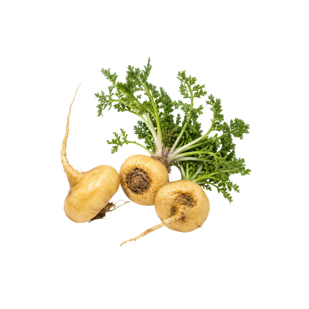 MildMildMacaLepidium meyeniiDietary maca improved antioxidant status in hypertriglyceridaemic rats, and black maca lowered brain malondialdehyde in ovariectomised mice.Mechanismlipid-peroxidation (MDA) reductionRead the evidenceShow lessMonograph →
In hypertriglyceridaemic rats 1% dietary maca improved antioxidant status alongside lipid and glucose parameters 300Reference 300AnimalThe influence of maca (Lepidium meyenii) on antioxidant status, lipid and glucose metabolism in rats — animal modelView study →, and in ovariectomised mice black maca lowered brain malondialdehyde, a lipid-peroxidation marker, in tandem with its memory benefit 301Reference 301AnimalAqueous extract of black maca (Lepidium meyenii) on memory impairment induced by ovariectomy in mice — animal modelView study →.
No human study has measured an oxidative-stress endpoint after maca, and the malondialdehyde data are black-maca-specific, so generic yellow powder is not the tested article.
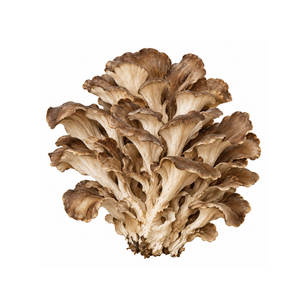 MildMildMaitakeGrifola frondosaPurified polysaccharides scavenge radicals in assay, and a phenolic-rich extract lowered ROS and extended lifespan in nematodes through DAF-16/FOXO and SKN-1/NRF2.Mechanismradical scavenging; Nrf2/SKN-1 and FOXO/DAF-16 activationRead the evidenceShow less
Standard assays record radical scavenging by purified polysaccharides 310Reference 310In vitroIsolation, purification and antioxidant activities of polysaccharides from Grifola frondosa — in vitroView study →, and a selenium-enriched polysaccharide more so 311Reference 311In vitroStructural elucidation and antioxidant activity of a novel Se-polysaccharide from Se-enriched Grifola frondosa — in vitroView study →, though that is an engineered material rather than a natural constituent.
In C. elegans a phenolic-rich extract lowered ROS and lipofuscin and extended lifespan via DAF-16/FOXO and SKN-1/NRF2 312Reference 312AnimalGrifola frondosa (Maitake) extract reduces fat accumulation and improves health span in C. elegans through the DAF-16/FOXO and SKN-1/NRF2 signalling pathways — in vivo (invertebrate model)View study →.
An invertebrate lifespan model is a long way from a human antioxidant claim, and there are no human data.
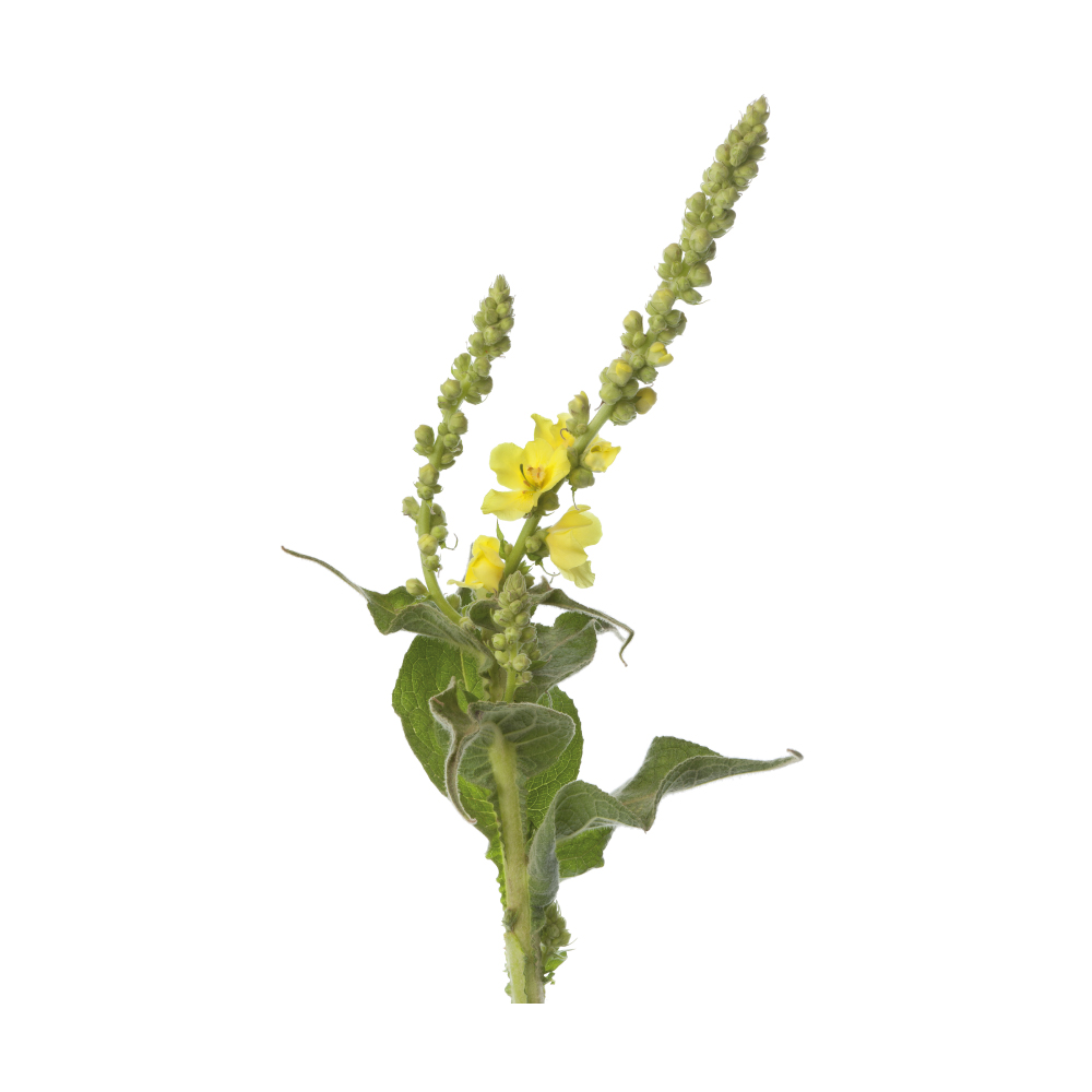 MildMildMulleinVerbascum thapsusVerbascoside is the herb's most studied phenolic, with documented antioxidant activity alongside its anti-inflammatory and wound-healing actions.Mechanismverbascoside (acteoside) radical scavenging; leucosceptoside A as its feruloyl analogueRead the evidenceShow lessMonograph →
Verbascoside, also called acteoside, is the most studied single phenolic in mullein and carries documented antioxidant activity.
It does not occur alone. Leucosceptoside A is its feruloyl analogue — the same scaffold with a methyl cap on one hydroxyl — and the two travel together across the figwort family.
The antioxidant work is constituent-level rather than herb-level, and the preparation people actually use is a mucilage-led leaf or flower infusion for the respiratory tract.
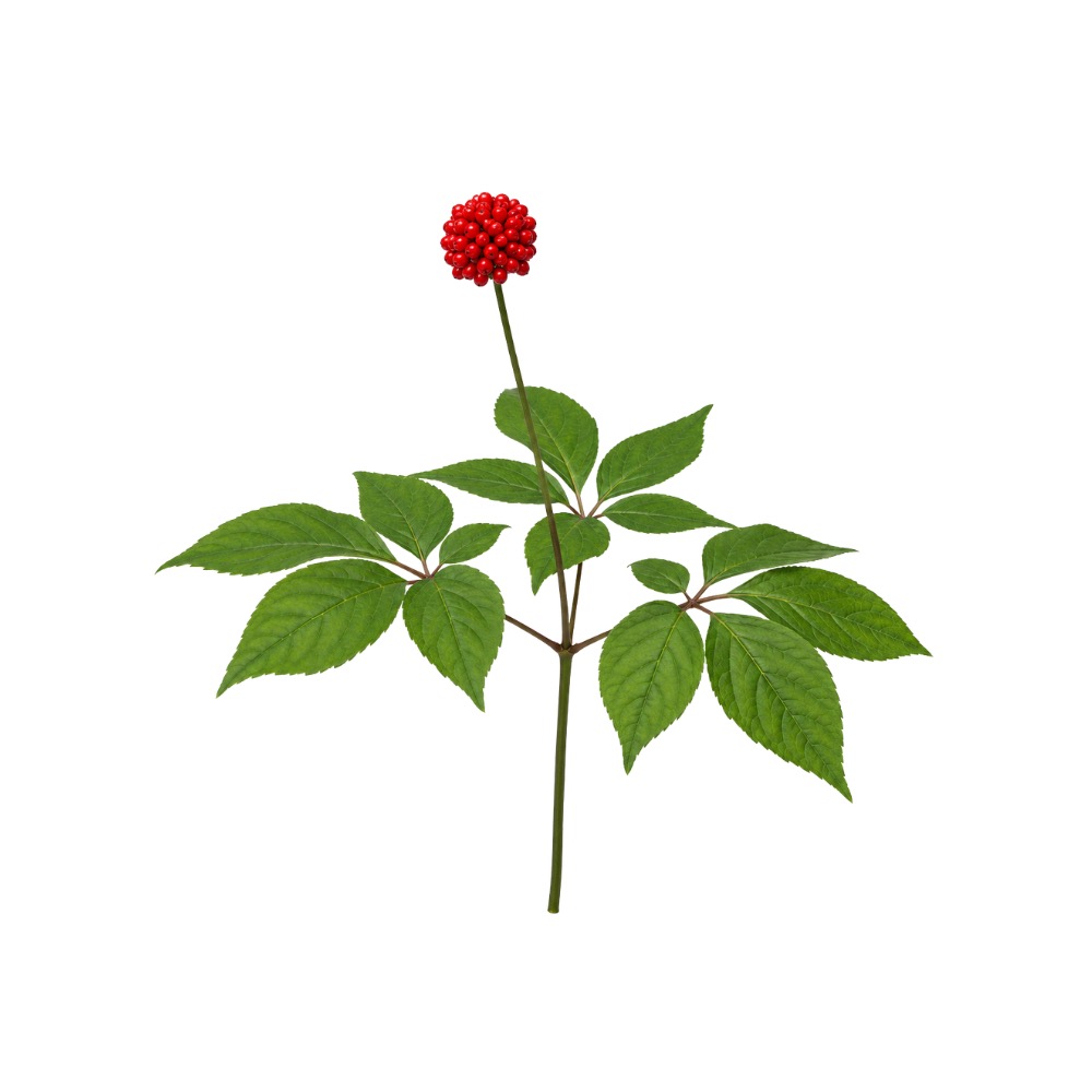 MildMildNotoginsengPanax notoginsengNotoginseng saponins raise superoxide dismutase and glutathione and lower malondialdehyde in mice, and notoginsenoside R1 enhances mitophagy against oxidative decline.MechanismSOD/GSH ↑, malondialdehyde ↓; mitophagy ↑ (notoginsenoside R1)Read the evidenceShow lessMonograph →
The saponins raised superoxide dismutase and glutathione and lowered malondialdehyde in apoE mice 326Reference 326AnimalPanax notoginseng saponins suppress RAGE/MAPK signalling and NF-κB activation in apolipoprotein-E-deficient atherosclerosis-prone mice — in vivoView study →, and in senescence-accelerated SAMP8 mice prevented oxidative stress and dementia-like decline by enhancing mitophagy 327Reference 327AnimalPanax notoginseng saponins prevent dementia and oxidative stress in brains of SAMP8 mice by enhancing mitophagy — in vivoView study →. The organ-protective effects of notoginsenoside R1 across ischaemia-reperfusion models are partly antioxidant in origin 328Reference 328Systematic reviewNotoginsenoside R1 for organ ischemia/reperfusion injury — a preclinical systematic reviewView study →.
The antioxidant activity is a secondary read-out in animal tissue rather than a measured clinical outcome, and it explains how the better-supported indications work.
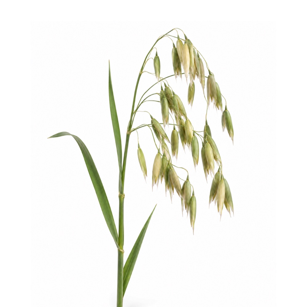 MildMildOatsAvena sativaAvenanthramides, ferulic acid and caffeic acid scavenge radicals in vitro, and eight weeks of avenanthramide-rich oat cookies raised plasma antioxidant capacity in postmenopausal women.Mechanismdirect free-radical scavenging (avenanthramides, ferulic acid, caffeic acid); plasma antioxidant capacity ↑ (human)Read the evidenceShow less
Eight weeks of avenanthramide-rich oat cookies raised plasma antioxidant capacity in postmenopausal women following downhill walking 329Reference 329RCTAvenanthramide supplementation attenuates exercise-induced inflammation in postmenopausal women — randomised controlled trialView study →, a human datum most of the field lacks.
The three common avenanthramides show measurable antioxidant capacity in vitro alongside their anti-inflammatory activity 330Reference 330In vitroIn vitro total antioxidant capacity and anti-inflammatory activity of three common oat-derived avenanthramides — in vitroView study →, with ferulic acid and caffeic acid contributing 331Reference 331ReviewMultiple Antioxidative and Bioactive Molecules of Oats (Avena sativa L.) in Human Health — reviewView study →.
The human reading is a surrogate marker inside an exercise-recovery trial designed around inflammation, the fraction tested is purified rather than dietary, and avenanthramides are present in oats at low concentration, so the activity borrows nothing from the strength of oats’ lipid-lowering evidence.
MildOregon GrapeMahonia aquifoliumIts protoberberine, aporphine and bisbenzylisoquinoline alkaloids scavenge radicals and inhibit lipoxygenase in cell-free systems.Mechanismalkaloid radical scavenging, cell-free systemsRead the evidenceShow less
The bark’s protoberberine, aporphine and bisbenzylisoquinoline alkaloids show antiradical and antioxidant activity in cell-free systems, alongside lipoxygenase inhibition 305,
The activity is cell-free only, and it is the least distinctive of the plant’s actions.
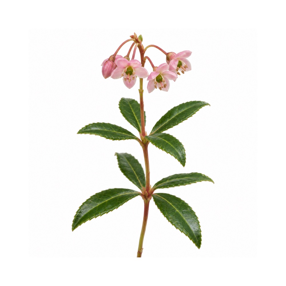 MildMildPipsissewaChimaphila umbellataIsolated chimaphilin and the flavonoids scavenge radicals in vitro.Mechanismchimaphilin and flavonoid radical scavengingRead the evidenceShow less
The activity is recorded in vitro, mostly on the isolated compound chimaphilin.
Nothing beyond the assay has been measured.
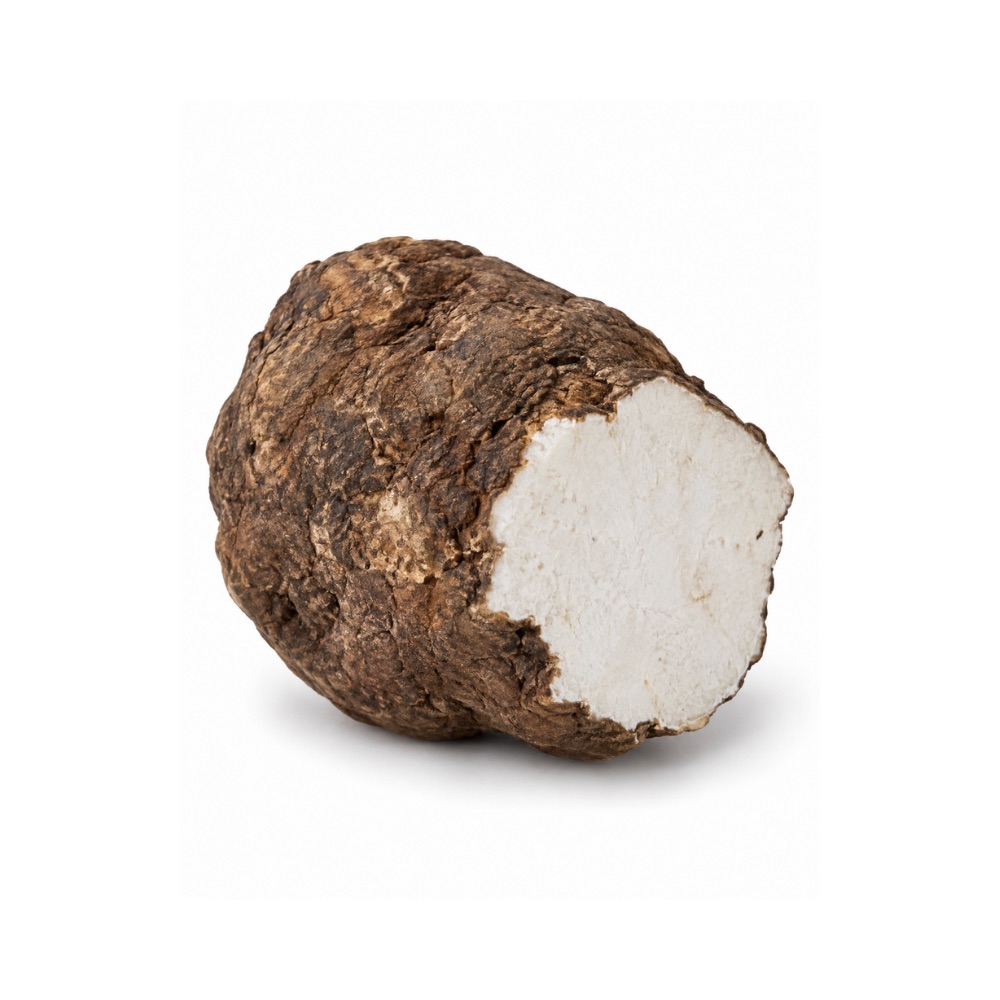 MildMildPoriaWolfiporia extensaWater-soluble polysaccharides scavenge radicals in assay, with a modest Nrf2 signal in vivo.Mechanismpolysaccharide radical scavenging; Nrf2 activationRead the evidenceShow less
Radical scavenging by the water-soluble polysaccharides varies with extraction method 391Reference 391In vitroAntioxidant property of water-soluble polysaccharides from Poria cocos Wolf using different extraction methods — in vitroView study →, and there is some in-vivo Nrf2 signal.
The activity is standard for a fungal polysaccharide, and there are no human data.
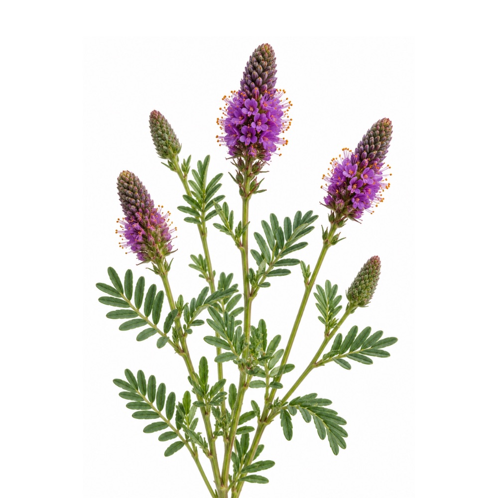 MildMildPurple Prairie CloverDalea purpureaIts stilbenes and flavan-3-ols scavenge radicals in vitro.Mechanismstilbene and flavan-3-ol radical scavengingRead the evidenceShow less
The stilbenes and condensed tannins scavenge radicals in vitro.
The activity is standard for a tannin- and stilbene-rich legume, and it has not been measured beyond the assay.
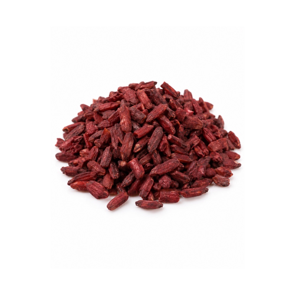 MildMildRed Yeast RiceMonascus purpureusThe yellow pigments monascin and ankaflavin enhanced anti-oxidative systems in a mouse model of alcoholic liver disease, and the extract protected proangiogenic cells from high-glucose oxidative stress through heme oxygenase-1.MechanismMonascus pigment (monascin, ankaflavin) antioxidant activity; heme oxygenase-1 induction (preclinical)Read the evidenceShow lessMonograph →
Monascin and ankaflavin enhanced anti-oxidative and anti-inflammatory systems in a mouse model of alcoholic liver disease 410Reference 410AnimalMonascin and ankaflavin of Monascus purpureus prevent alcoholic liver disease through regulating AMPK-mediated lipid metabolism and enhancing both anti-inflammatory and anti-oxidative systems — animal modelView study →, and red yeast rice protected bone-marrow-derived proangiogenic cells against high-glucose-induced senescence and oxidative stress via heme oxygenase-1 in vitro 411Reference 411In vitroRed yeast rice protects circulating bone marrow-derived proangiogenic cells against high-glucose-induced senescence and oxidative stress: the role of heme oxygenase-1 — in vitroView study →.
The activity belongs to the yellow Monascus pigments, which are not the lipid-active fraction. Monacolin K, the molecule behind every human lipid and cardiovascular result, is chemically identical to lovastatin and carries none of the antioxidant evidence.
There are no human antioxidant data.
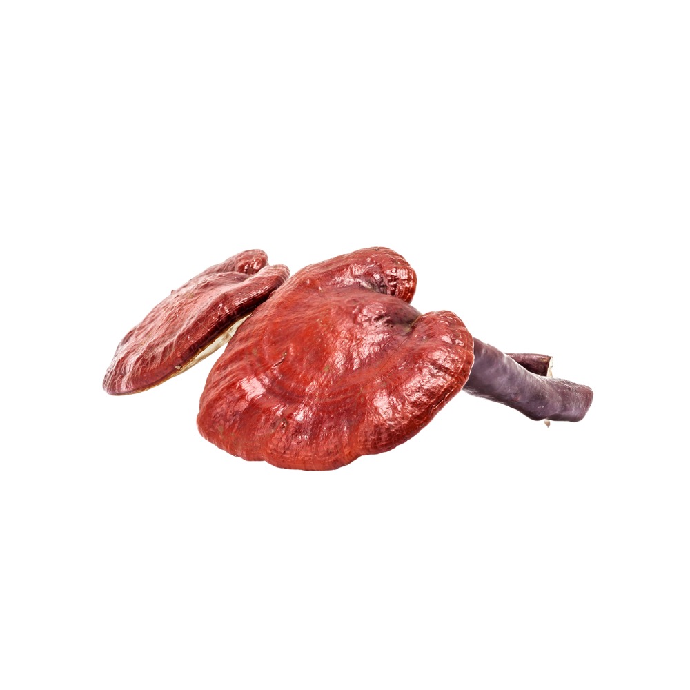 MildMildReishiGanoderma lucidumRaises plasma antioxidant capacity for a few hours after a dose, and a six-month placebo-controlled crossover of an enriched extract improved total antioxidant status.Mechanismplasma antioxidant capacity ↑ (acute, transient); intracellular glutathione ↑ and lipid peroxidation ↓ (preclinical); phenolic and triterpene contentRead the evidenceShow lessMonograph →
A controlled crossover found reishi transiently raised plasma antioxidant capacity after ingestion, but four weeks of supplementation did not consistently improve a panel of oxidative-stress, coronary-risk and DNA-damage biomarkers 342Reference 342Clinical trialGanoderma lucidum (“Lingzhi”), a Chinese medicinal mushroom: biomarker responses in a controlled human supplementation study — controlled clinical trialView study →. A later randomised, double-blind, placebo-controlled crossover of a triterpenoid- and polysaccharide-enriched extract reported improved total antioxidant status over six months 343Reference 343RCTTriterpenoids and polysaccharide peptides-enriched Ganoderma lucidum: a randomised, double-blind, placebo-controlled crossover study of antioxidation and hepatoprotective efficacy in healthy volunteers — randomised controlled trialView study →.
The two trials differ in both extract and duration, and the picture is unresolved.
Preclinically the effect is robust and reproducible: extracts raise intracellular glutathione and inhibit lipid peroxidation across many models, attributed to the phenolic and triterpene fractions.
An antioxidant effect that appears in plasma for a few hours and then fails to move a damage marker over four weeks is a weak one, and the acute rise may reflect absorbed antioxidants circulating rather than the body’s oxidative burden changing.
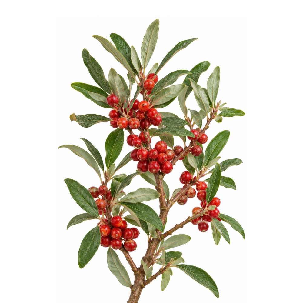 MildMildSoapberryShepherdia canadensisA documented source of lycopene and the unusual derivative methyl apo-6′-lycopenoate, carotenoids with well-characterised antioxidant activity elsewhere.Mechanismcarotenoid content (lycopene, methyl apo-6′-lycopenoate) — inferred, not assayedRead the evidenceShow less
Lycopene and the unusual derivative methyl apo-6′-lycopenoate have been isolated from the fruit 368Reference 368Major carotenoids of Shepherdia canadensis: isolation and synthesis of methyl apo-6′-lycopenoate — constituent isolationView study →, and lycopene is a well-characterised antioxidant elsewhere.
No whole-extract antioxidant assay of the plant has been performed, not even a DPPH screen, so the constituents are the whole of the basis.
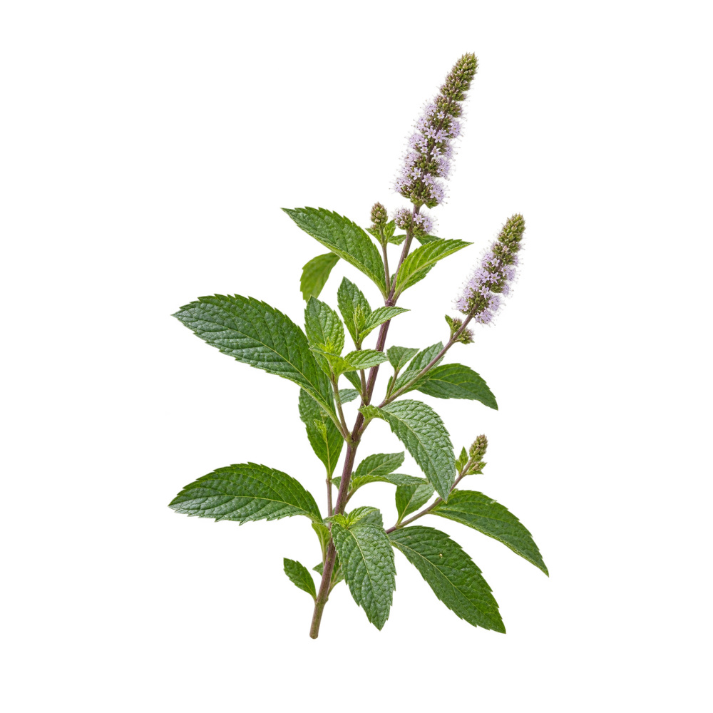 MildMildSpearmintMentha spicataRosmarinic acid carries the antioxidant side, and the standardised extract used in the cognition trials is concentrated to 14–15% of it.Mechanismrosmarinic acid radical scavenging; standardised dried extract at 14–15% rosmarinic acidRead the evidenceShow lessMonograph →
Rosmarinic acid is the phenolic behind spearmint’s antioxidant activity, alongside the carvone-dominated essential oil that defines the herb.
The preparation matters more than usual. A standardised dried extract concentrated to roughly 14–15% rosmarinic acid is the form used in every positive cognition trial, at 900 mg a day.
There is no evidence a cup of tea delivers a comparable dose.
The antioxidant work is constituent-level. What people take spearmint for is digestion.
MildMildSweet WormwoodArtemisia annuaExtracts scavenge radicals in vitro, Asian chemotypes more than European, and aqueous extract raised intestinal antioxidant capacity in broilers.Read the evidenceShow lessMonograph →
Artemisinin-bearing extracts of A. annua scavenge radicals in vitro alongside their anti-inflammatory activity 405Reference 405In vitroAnti-inflammatory, antioxidant and antimicrobial effects of artemisinin extracts from Artemisia annua L. — in vitroView study →, and the essential oil is antiradical, with Asian chemotypes more active than European ones.
In broilers, aqueous extract increased intestinal antioxidant capacity and improved gut morphology 404Reference 404AnimalArtemisia annua L. aqueous extract promotes intestine immunity and antioxidant function in broilers — animal modelView study →.
Activity is chemotype-dependent, so it varies with the material rather than being a property of the species, and there are no human data. The herb’s substantiated work is antimalarial, and even there the human evidence belongs to the isolated derivative.
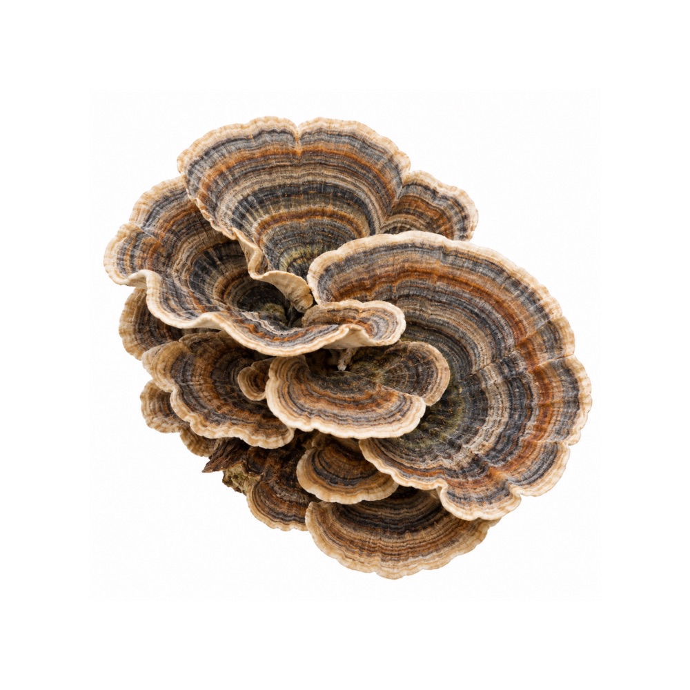 MildMildTurkey TailTrametes versicolorPolysaccharide and protein fractions scavenge DPPH, ABTS, superoxide and hydroxyl radicals in assay, with the phenolic acids present at only microgram-per-gram levels.MechanismDPPH/ABTS/superoxide/hydroxyl radical scavenging by polysaccharide and phenolic fractionsRead the evidenceShow less
Six purified polysaccharide fractions showed concentration-dependent radical scavenging, with lower-molecular-weight, higher-protein fractions most active 386Reference 386In vitroScreening and comparison of antioxidant activities of polysaccharides from Coriolus versicolor — in vitroView study →, and a protein-rich extract and its hydrolysate scavenged DPPH and ABTS 387Reference 387In vitroAntioxidant and antimicrobial properties of a protein-rich extract obtained from Trametes versicolor — in vitroView study →.
The likely small-molecule contributors are the phenolic acids, p-hydroxybenzoic at 113 µg/g, then protocatechuic acid and vanillic acid 388Reference 388In vitroAssessment of bioactive compounds and antioxidant activity of turkey tail medicinal mushroom Trametes versicolor — in vitro/analyticalView study →, concentrations that are low in absolute terms.
Every measurement is at the bench, with no human biomarker study.
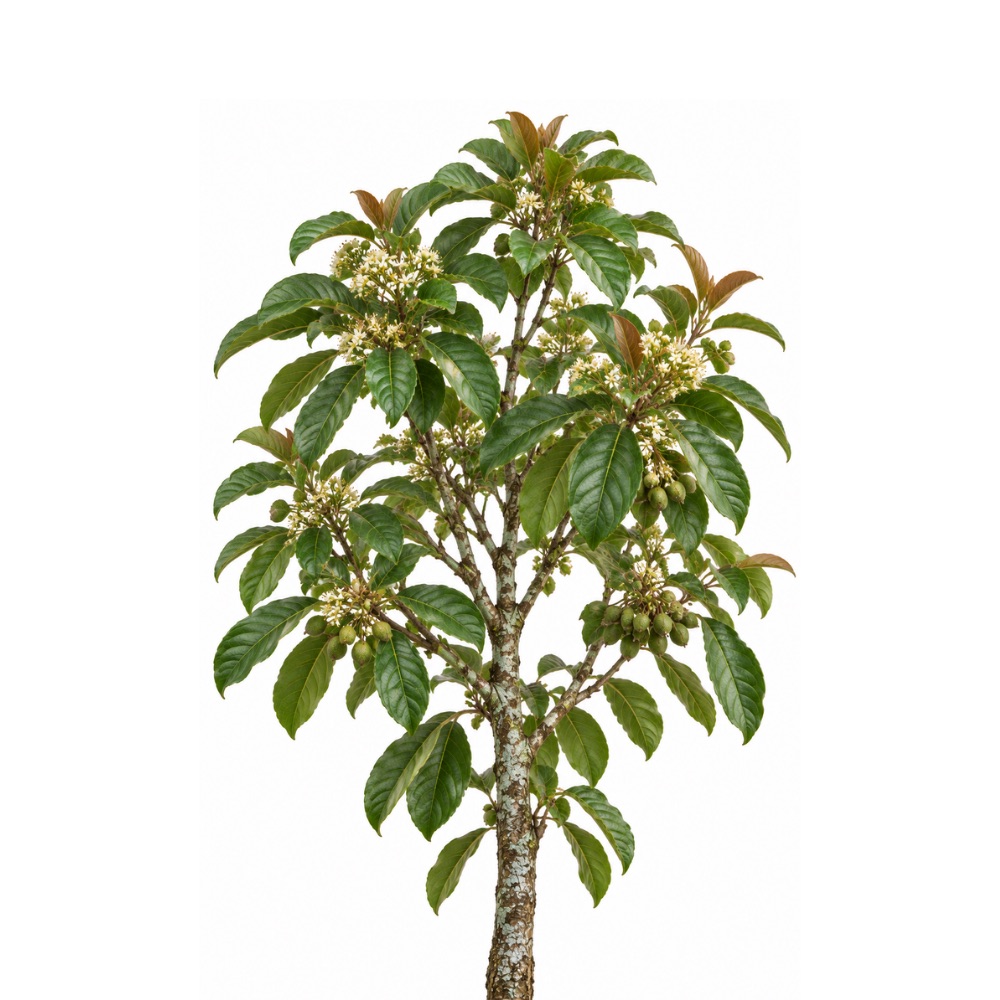 MildMildUlei IronwoodIryanthera uleiA phenol-enriched leaf fraction of eight glycosylated flavonoids and two neolignans scavenges radicals in the DPPH, ABTS and FRAP assays, and the genus carries potent lignan and tocotrienol antioxidants.Mechanismflavonoid-fraction radical scavenging (DPPH/ABTS/FRAP); lignan and tocotrienol lipid-peroxidation inhibition (genus)Read the evidenceShow less
The ethanolic leaf extract of Colombian I. ulei, enriched for phenols, contained eight glycosylated flavonoids, two free flavonoids and two neolignans, with afzelin confirmed by isolation, and scavenged radicals in the DPPH, ABTS and FRAP assays 413Reference 413In vitroLC-DAD-UV and LC-ESI-MS-based analyses, antioxidant capacity and antimicrobial activity of a polar fraction from Iryanthera ulei leaves — in vitro.
Across the genus, lignans and tocotrienols isolated from I. juruensis fruit inhibit lipid peroxidation in vitro 414Reference 414In vitroLipophyllic antioxidants from Iryanthera juruensis fruits — in vitroView study →.
All of it is cell-free chemistry, with no cellular, animal or human study of I. ulei.
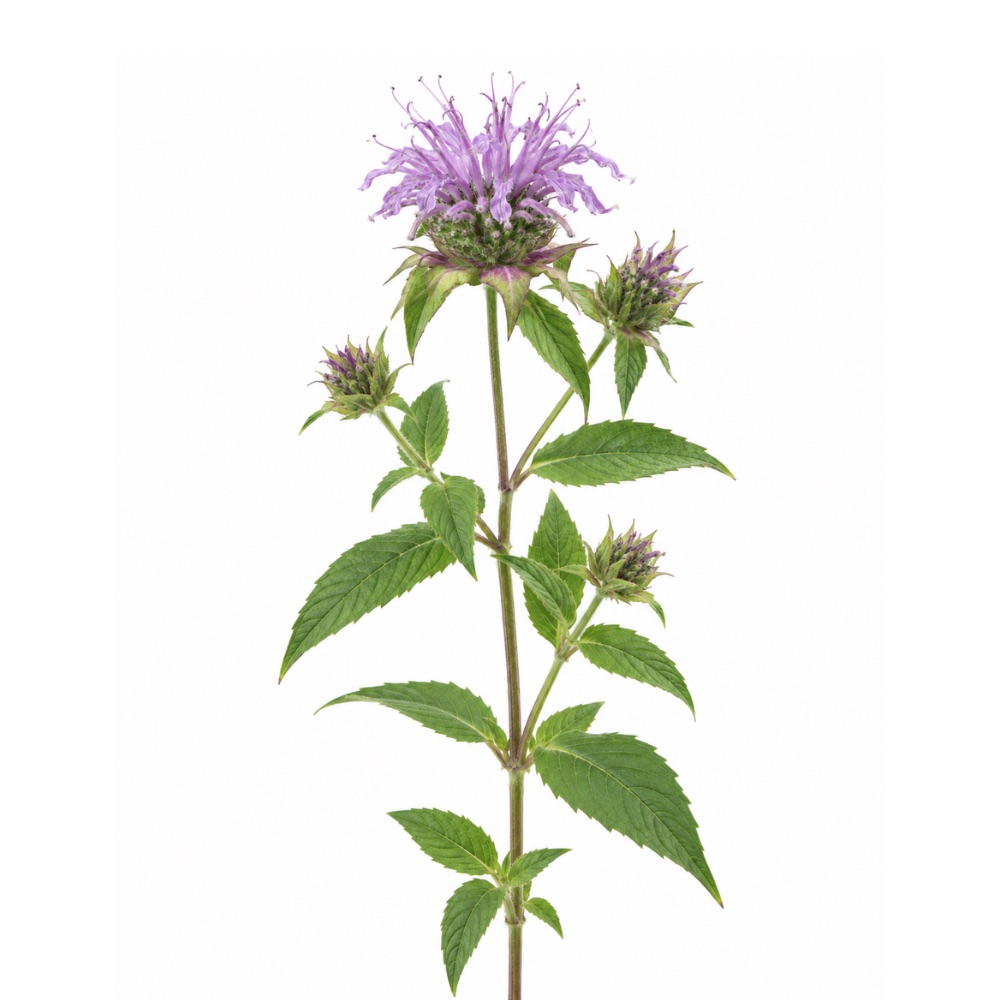 MildMildWild BergamotMonarda fistulosaRosmarinic acid and the flavonoids give it the in-vitro radical scavenging typical of the mint family.Mechanismrosmarinic acid and flavonoid radical scavengingRead the evidenceShow less
The rosmarinic acid and flavonoids of the leaf scavenge radicals in vitro.
The activity is standard for a rosmarinic-acid-rich Lamiaceae, and it has not been measured beyond the assay.
No herbs match these filters.
Also Tagged
15 more herbs carry an antioxidant action.
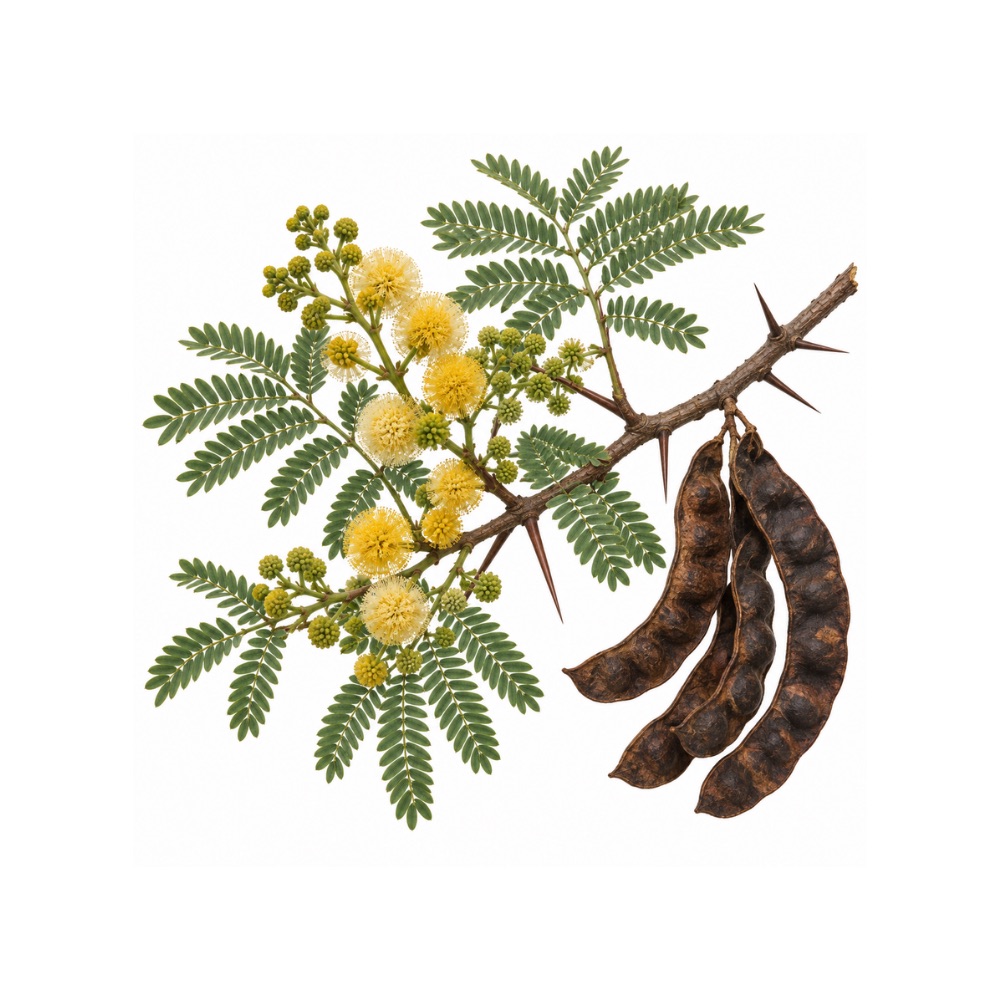
Babul
Vachellia nilotica
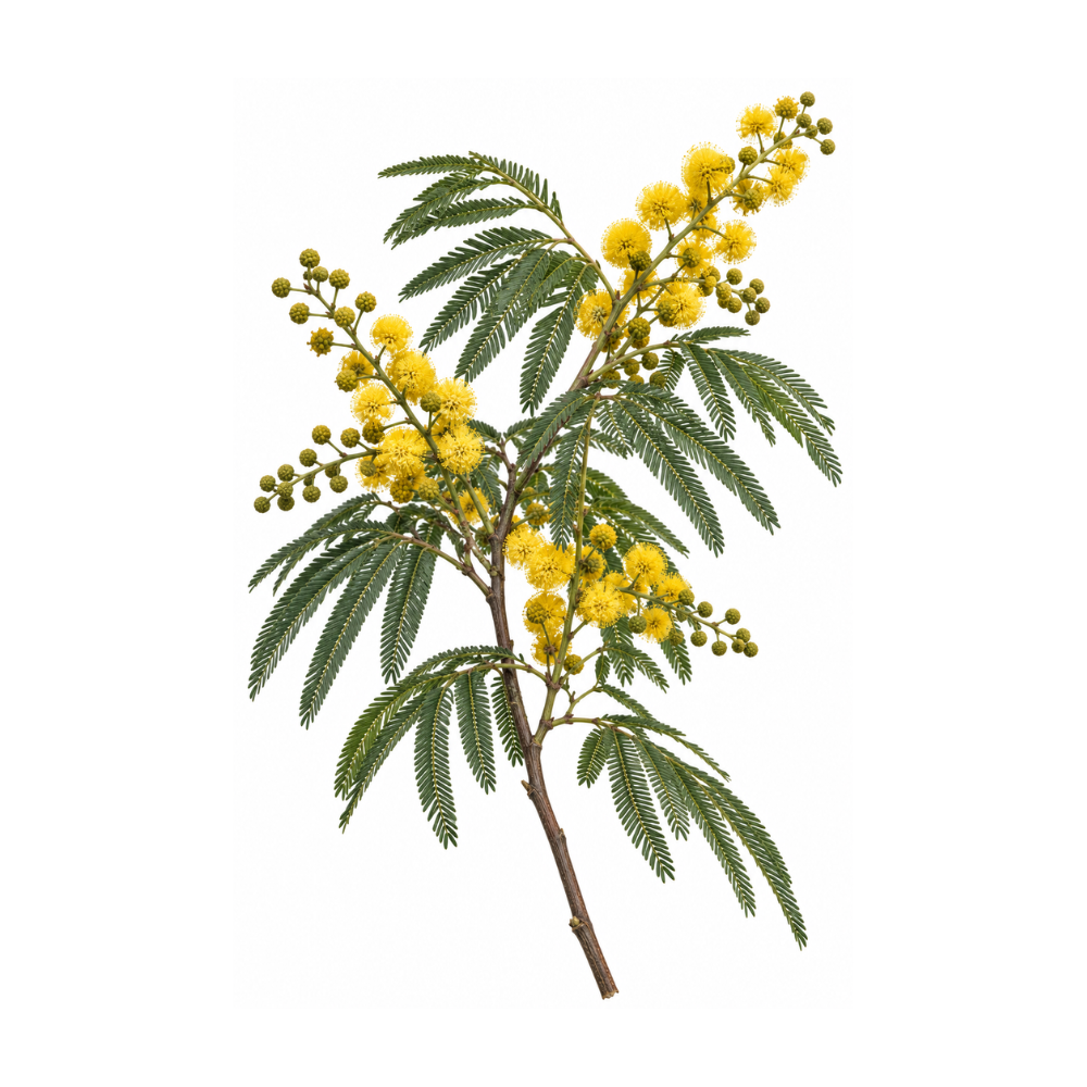
Black Wattle
Acacia mearnsii
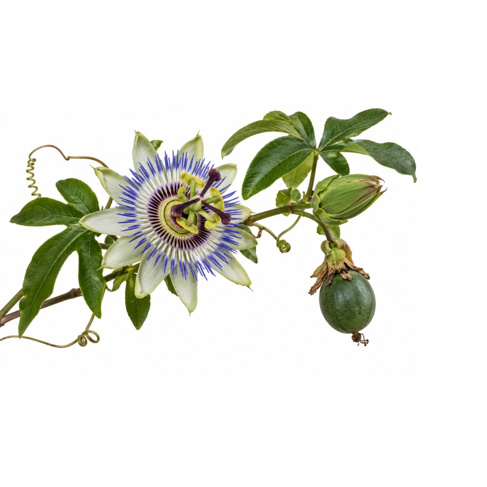
Blue Passionflower
Passiflora caerulea
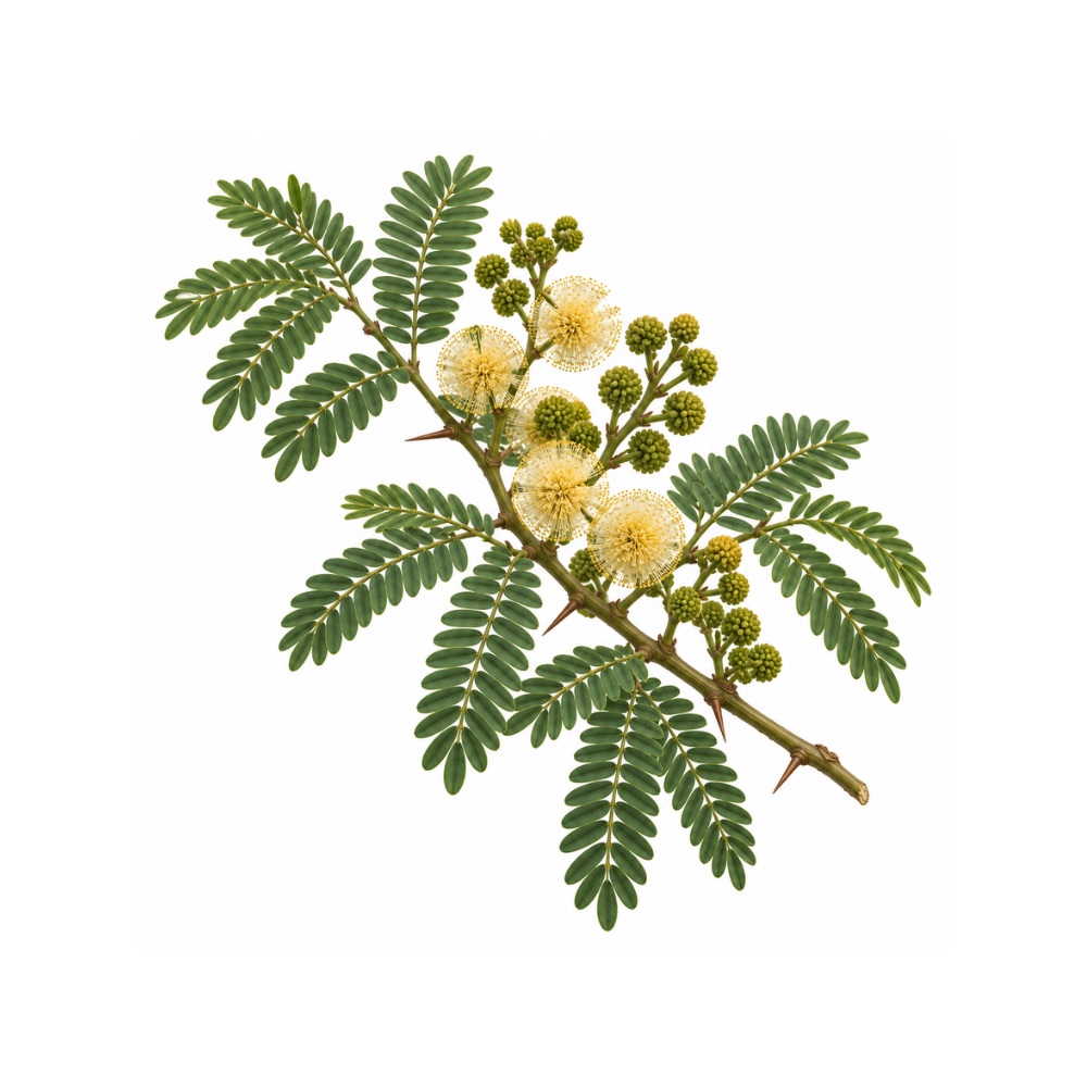
Catechu
Senegalia catechu
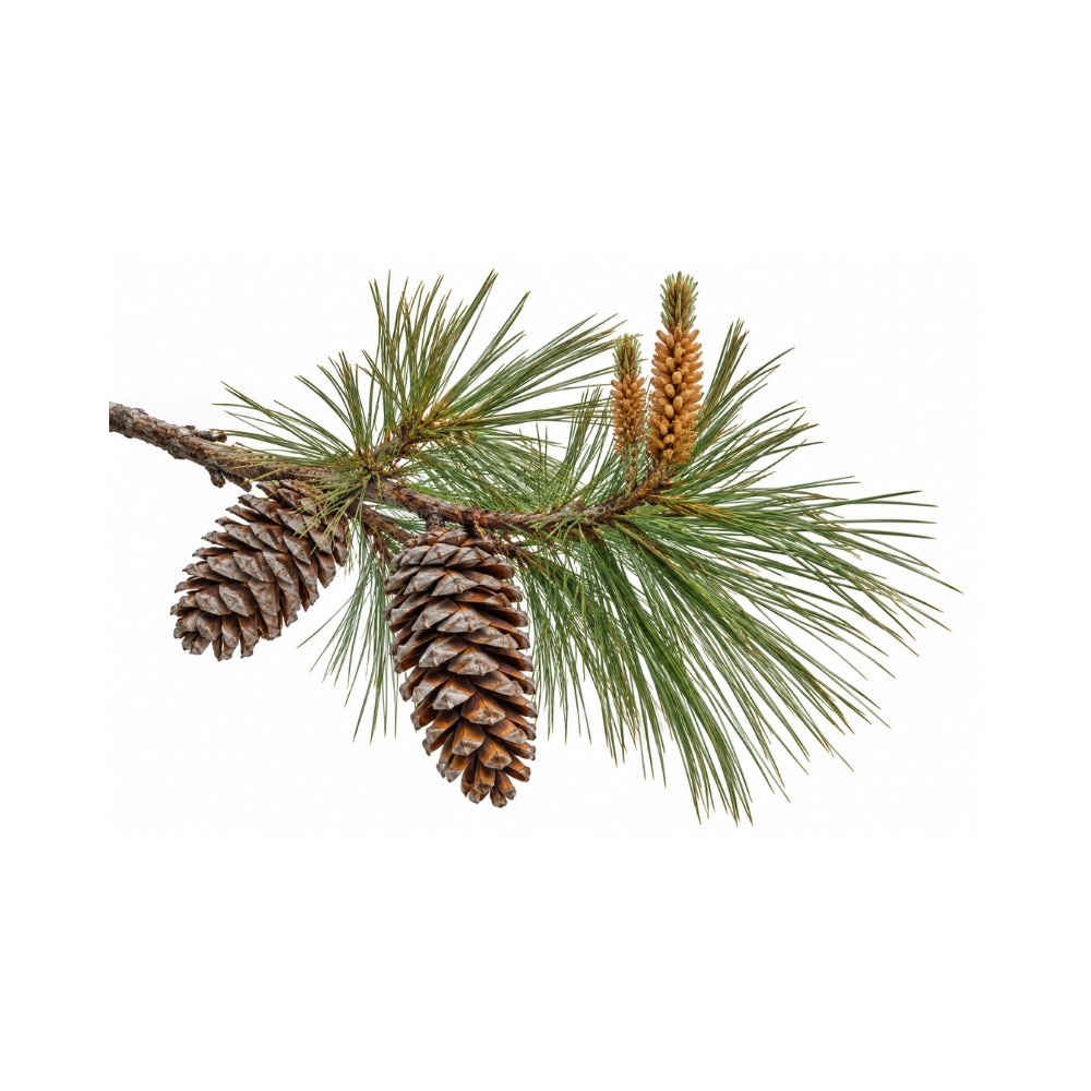
Chir Pine
Pinus roxburghii
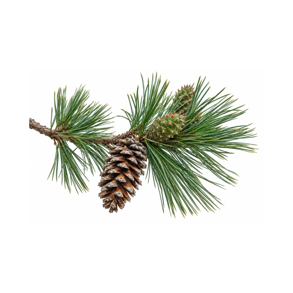
Korean Pine
Pinus koraiensis
Korean Red Pine
Pinus densiflora
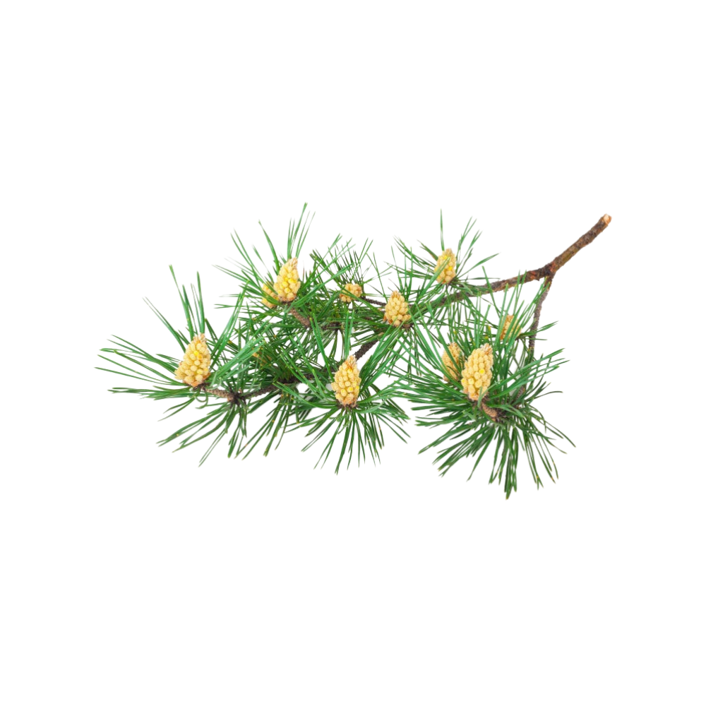
Maritime Pine
Pinus pinaster
Masson Pine
Pinus massoniana
Monterey Pine
Pinus radiata
Mugo Pine
Pinus mugo
Passion Fruit
Passiflora edulis
Pine
Pinus sylvestris
Stinking Passionflower
Passiflora foetida
Winged-Stem Passionflower
Passiflora alata
Safety
- No antioxidant herb improves with a higher dose; high-dose antioxidant supplements have failed in trials where the same compounds eaten as food looked protective.
- High-dose antioxidants blunt the adaptations to endurance training, which are signalled by the radicals exercise generates.
- Concentrated extracts are a different article from the plant, and high-dose catechin extracts carry a rare hepatotoxicity signal that the brewed leaf does not.
- Tannin-rich antioxidant herbs bind dietary iron; take them about two hours apart from iron supplements and from oral medication, and with care in iron deficiency and pregnancy.
- Antioxidant supplements can interfere with chemotherapy and radiotherapy, which work partly through oxidative damage, so tell the oncology team.
- A scavenging assay describes the preparation studied, and a number for a concentrated extract says nothing about a cup of the infusion.
- Antioxidant activity does not cancel a cancer signal: very hot yerba maté and guayusa are linked to oesophageal cancer, and sugary guarana drinks were pro-oxidant in mice.
References
- Villamil-Galindo E, Van de Velde F, Piagentini AM (2021). Strawberry agro-industrial by-products as a source of bioactive compounds — effect of cultivar on the phenolic profile and the antioxidant capacity. Bioresources and Bioprocessing. https://pubmed.ncbi.nlm.nih.gov/38650292/
- Nowicka A, Kucharska AZ, Sokół-Łętowska A, Fecka I (2019). Comparison of polyphenol content and antioxidant capacity of strawberry fruit from 90 cultivars of Fragaria × ananassa Duch. Food Chemistry. https://pubmed.ncbi.nlm.nih.gov/30174053/
- Kim SB, Hwang SH, Suh HW, Lim SS (2017). Phytochemical analysis of Agrimonia pilosa Ledeb, its antioxidant activity and aldose reductase inhibitory potential — in vitro and ex vivo. International Journal of Molecular Sciences. https://pubmed.ncbi.nlm.nih.gov/28208627/
- Bazylko A, Piwowarski JP, Filipek A, Bonarewicz J, Tomczyk M (2013). In vitro antioxidant and anti-inflammatory activities of extracts from Potentilla recta and its main ellagitannin, agrimoniin. Journal of Ethnopharmacology. https://pubmed.ncbi.nlm.nih.gov/23811215/
- Fedotcheva TA, Sheichenko OP, Fedotcheva NI (2021). New properties and mitochondrial targets of polyphenol agrimoniin as a natural anticancer and preventive agent — in vitro. Pharmaceutics. https://pubmed.ncbi.nlm.nih.gov/34959369/
- Aruoma OI, Halliwell B, Aeschbach R, Löligers J. (1992). Antioxidant and pro-oxidant properties of active rosemary constituents: carnosol and carnosic acid — in vitro. Xenobiotica. https://pubmed.ncbi.nlm.nih.gov/1378672/
- Loussouarn M, Krieger-Liszkay A, Svilar L, Bily A, Birtić S, Havaux M. (2017). Carnosic acid and carnosol, two major antioxidants of rosemary, act through different mechanisms — in vitro and in planta. Plant Physiology. https://pubmed.ncbi.nlm.nih.gov/28916593/
- Satoh T, Kosaka K, Itoh K, Kobayashi A, Yamamoto M, Shimojo Y, et al. (2008). Carnosic acid, a catechol-type electrophilic compound, protects neurons both in vitro and in vivo through activation of the Keap1/Nrf2 pathway via S-alkylation of targeted cysteines on Keap1 — mice. Journal of Neurochemistry. https://pubmed.ncbi.nlm.nih.gov/17995931/
- EFSA Panel on Food Additives and Nutrient Sources added to Food (ANS). (2018). Refined exposure assessment of extracts of rosemary (E 392) from its use as food additive — review, including the Regulation (EU) No 231/2012 and JECFA specifications. EFSA Journal. https://doi.org/10.2903/j.efsa.2018.5373
- European Commission. (2012). Commission Regulation (EU) No 231/2012 laying down specifications for food additives listed in Annexes II and III to Regulation (EC) No 1333/2008 — monograph. Official Journal of the European Union. https://eur-lex.europa.eu/eli/reg/2012/231/oj
- Li X, Zhang Q, Hou N, et al. (2019). Carnosol as a Nrf2 activator improves endothelial barrier function through antioxidative mechanisms. International Journal of Molecular Sciences, 20(4), 880. https://pubmed.ncbi.nlm.nih.gov/30781644/
- Takahashi T, Tabuchi T, Tamaki Y, Kosaka K, Takikawa Y, Satoh T. (2009). Carnosic acid and carnosol inhibit adipocyte differentiation in mouse 3T3-L1 cells through induction of phase 2 enzymes and activation of glutathione metabolism. Biochemical and Biophysical Research Communications, 382(3), 549–554. https://pubmed.ncbi.nlm.nih.gov/19289108/
- Singletary KW. (1996). Rosemary extract and carnosol stimulate rat liver glutathione-S-transferase and quinone reductase activities. Cancer Letters, 100(1–2), 139–144. https://pubmed.ncbi.nlm.nih.gov/8620433/
- Fidelis M, et al. (2020). Camu-camu seed (Myrciaria dubia) — from side stream to an antioxidant, antihyperglycemic, antiproliferative, antimicrobial, antihemolytic, anti-inflammatory, and antihypertensive ingredient. Food Chemistry. https://pubmed.ncbi.nlm.nih.gov/31816536/
- Fidelis M, et al. (2020). Response surface optimization of phenolic compounds from jabuticaba (Myrciaria cauliflora [Mart.] O.Berg) seeds: antioxidant, antimicrobial, antihyperglycemic, antihypertensive and cytotoxic assessments. Food and Chemical Toxicology. https://pubmed.ncbi.nlm.nih.gov/32450285/
- Adiamo OQ, et al. (2024). In-vitro colonic fermentation of Kakadu plum (Terminalia ferdinandiana) fruit powder: microbial biotransformation of phenolic compounds and cytotoxicity. Food Chemistry. https://pubmed.ncbi.nlm.nih.gov/38555694/
- Frešer F, et al. (2026). Ellagitannins as Al(III) chelators: determined binding sites with a predictive model. Spectrochimica Acta Part A: Molecular and Biomolecular Spectroscopy. https://pubmed.ncbi.nlm.nih.gov/42242147/
- Messaoudene M, Pidgeon R, Richard C, et al. (2022). A natural polyphenol exerts antitumor activity and circumvents anti-PD-1 resistance through effects on the gut microbiota. Cancer Discovery. https://pubmed.ncbi.nlm.nih.gov/35031549/
- Nguyen TL, Rusten A, Bugge MS, Malterud KE, Diallo D, Paulsen BS, Wangensteen H (2016). Flavonoids, gallotannins and ellagitannins in Syzygium guineense and the traditional use among Malian healers — in vitro. Journal of Ethnopharmacology. https://pubmed.ncbi.nlm.nih.gov/27647014/
- Kähkönen M, Kylli P, Ollilainen V, Salminen JP, Heinonen M (2012). Antioxidant activity of isolated ellagitannins from red raspberries and cloudberries — in vitro. Journal of Agricultural and Food Chemistry. https://pubmed.ncbi.nlm.nih.gov/22229937/
- Dah-Nouvlessounon D, Chokki M, Hoteyi IMS, et al. (2023). Pharmacological property and cytotoxic effect showing antiproliferative potency in human melanoma cell lines (A375) of Combretum racemosum leaf and root extracts used in Benin. Antioxidants. https://pubmed.ncbi.nlm.nih.gov/38247456/
- Piwowarski JP, Granica S, Stefańska J, Kiss AK (2016). Differences in metabolism of ellagitannins by human gut microbiota ex vivo cultures. Journal of Natural Products. https://pubmed.ncbi.nlm.nih.gov/28006907/
- Daniel EM, Ratnayake S, Kinstle T, Stoner GD (1991). The effects of pH and rat intestinal contents on the liberation of ellagic acid from purified and crude ellagitannins — in vitro. Journal of Natural Products. https://pubmed.ncbi.nlm.nih.gov/1791480/
- Chen CH, Liu TZ, Kuo TC, Lu FJ, Chen YC, Chang-Chien YW, Lin CC (2004). Casuarinin protects cultured MDCK cells from hydrogen peroxide-induced oxidative stress and DNA oxidative damage. Planta Medica, 70(11), 1022-1026. https://pubmed.ncbi.nlm.nih.gov/15549656/
- Kim EB, Lee EK, Son SY, et al. (2022). Antiacne and anti-inflammatory effects of phenolic compounds from Quercus acutissima. Evidence-Based Complementary and Alternative Medicine, 2022, 9078475. https://pubmed.ncbi.nlm.nih.gov/36624865/
- Perera HDSM, Perera AS, Samarasekera JKRR, et al. (2025). Investigating the antioxidant, pro-inflammatory enzyme inhibitory, neuroprotective, and antineuroinflammatory bioactives of Osbeckia octandra using in vitro and in silico approaches. ACS Omega, 10(45), 53775-53790. https://pubmed.ncbi.nlm.nih.gov/41280792/
- Al-Sayed E, Michel HE, Khattab MA, El-Shazly M, Singab AN (2020). Protective role of casuarinin from Melaleuca leucadendra against ethanol-induced gastric ulcer in rats. Planta Medica, 86(1), 32-44. https://pubmed.ncbi.nlm.nih.gov/31689719/
- Song H, et al. (2023). Cryptotanshinone alleviates lipopolysaccharide and cigarette smoke-induced chronic obstructive pulmonary disease in mice via the Keap1/Nrf2 axis — mouse in vivo study. Biomedicine & Pharmacotherapy. https://pubmed.ncbi.nlm.nih.gov/37399718/
- Nagappan A, Kim JH, et al. (2019). Cryptotanshinone from the Salvia miltiorrhiza Bunge attenuates ethanol-induced liver injury by activation of AMPK/SIRT1 and Nrf2 signaling pathways — mouse in vivo study. International Journal of Molecular Sciences. https://pubmed.ncbi.nlm.nih.gov/31906014/
- Gao N, Huang Z, et al. (2025). Cryptotanshinone alleviates cerebral ischemia reperfusion injury by regulating ferroptosis through the PI3K/AKT/Nrf2 and SLC7A11/GPX4 signaling pathway — rodent in vivo study. Journal of Ethnopharmacology. https://pubmed.ncbi.nlm.nih.gov/40222690/
- Wang Q, Liu Y, et al. (2022). Cryptotanshinone ameliorates MPP⁺-induced oxidative stress and apoptosis of SH-SY5Y neuroblastoma cells: the role of STAT3 in Parkinson’s disease — in vitro study. Metabolic Brain Disease. https://pubmed.ncbi.nlm.nih.gov/35396628/
- Zhao GR, Zhang HM, Ye TX, et al. (2008). Characterization of the radical scavenging and antioxidant activities of danshensu and salvianolic acid B. Food and Chemical Toxicology, 46(1), 73–81. https://pubmed.ncbi.nlm.nih.gov/17719161/
- Guo C, Ma Z, Tao X, et al. (2025). Therapeutic time window of sodium danshensu on cerebral ischemia and its mechanism of inhibiting oxidative stress and ferroptosis through the Nrf2 pathway. Brain Research Bulletin, 227, 111396. https://pubmed.ncbi.nlm.nih.gov/40403934/
- Yu C, Dong H, Wang Q, et al. (2021). Danshensu attenuates cisplatin-induced nephrotoxicity through activation of the Nrf2 pathway and inhibition of NF-κB. Biomedicine & Pharmacotherapy, 142, 111995. https://pubmed.ncbi.nlm.nih.gov/34435595/
- Zhang J, Zhang Q, Liu G, Zhang N. (2019). Therapeutic potentials and mechanisms of the Chinese traditional medicine Danshensu. European Journal of Pharmacology, 864, 172710. https://pubmed.ncbi.nlm.nih.gov/31586468/
- Zhou X, Chan SW, Tseng HL, et al. (2012). Danshensu is the major marker for the antioxidant and vasorelaxation effects of danshen (Salvia miltiorrhiza) water-extracts produced by different heat water-extractions. Phytomedicine, 19(14), 1263–1269. https://pubmed.ncbi.nlm.nih.gov/23026310/
- Lu B, Xu Y, Xu L, Cong X, Yin L, Li H, Peng J (2012). Mechanism investigation of dioscin against CCl4-induced acute liver damage in mice. Environmental Toxicology and Pharmacology. https://pubmed.ncbi.nlm.nih.gov/22516057/
- Xu T, Zheng L, Xu L, Yin L, Qi Y, Xu Y, Han X, Peng J (2014). Protective effects of dioscin against alcohol-induced liver injury. Archives of Toxicology. Correction published 2019. https://pubmed.ncbi.nlm.nih.gov/24146112/
- Qiao Y, Xu L, Tao X, Yin L, Qi Y, Xu Y, Han X, Tang Z, Ma X, Liu K, Peng J (2018). Protective effects of dioscin against fructose-induced renal damage via adjusting Sirt3-mediated oxidative stress, fibrosis, lipid metabolism and inflammation. Toxicology Letters. https://pubmed.ncbi.nlm.nih.gov/29197622/
- Wang S, Zheng Y, Jin S, Fu Y, Liu Y (2022). Dioscin protects against cisplatin-induced acute kidney injury by reducing ferroptosis and apoptosis through activating Nrf2/HO-1 signaling. Antioxidants. https://pubmed.ncbi.nlm.nih.gov/36552651/
- Leng J, et al. (2020). Neuroprotective effect of diosgenin in a mouse model of diabetic peripheral neuropathy involves the Nrf2/HO-1 pathway — mouse in vivo study. BMC Complementary Medicine and Therapies. https://pubmed.ncbi.nlm.nih.gov/32336289/
- Haraguchi H, Saito T, Okamura N, Yagi A (1995). Inhibition of lipid peroxidation and superoxide generation by diterpenoids from Rosmarinus officinalis. Planta Medica. https://pubmed.ncbi.nlm.nih.gov/7480180/
- Zeng HH, Tu PF, Zhou K, Wang H, Wang BH, Lu JF (2001). Antioxidant properties of phenolic diterpenes from Rosmarinus officinalis. Acta Pharmacologica Sinica. https://pubmed.ncbi.nlm.nih.gov/11749806/
- Miura K, Kikuzaki H, Nakatani N (2002). Antioxidant activity of chemical components from sage (Salvia officinalis L.) and thyme (Thymus vulgaris L.) measured by the oil stability index method. Journal of Agricultural and Food Chemistry. https://pubmed.ncbi.nlm.nih.gov/11902922/
- Nakatani N, Inatani R (1984). Two antioxidative diterpenes from rosemary (Rosmarinus officinalis L.) and a revised structure for rosmanol. Agricultural and Biological Chemistry 48(8):2081–2085. https://doi.org/10.1271/bbb1961.48.2081
- Fischedick JT, Standiford M, Johnson DA, Johnson JA (2013). Structure activity relationship of phenolic diterpenes from Salvia officinalis as activators of the nuclear factor E2-related factor 2 pathway. Bioorganic & Medicinal Chemistry. https://pubmed.ncbi.nlm.nih.gov/23507152/
- Llurba Montesino N, Kaiser M, Mäser P, Schmidt TJ (2021). Salvia officinalis L.: antitrypanosomal activity and active constituents against Trypanosoma brucei rhodesiense. Molecules. https://pubmed.ncbi.nlm.nih.gov/34072147/
- Lin SY, et al. (2008). Antioxidant, anti-semicarbazide-sensitive amine oxidase, and anti-hypertensive activities of geraniin isolated from Phyllanthus urinaria. Food and Chemical Toxicology. https://pubmed.ncbi.nlm.nih.gov/18495318/
- Kumaran A, Karunakaran RJ (2006). Nitric oxide radical scavenging active components from Phyllanthus emblica L. Plant Foods for Human Nutrition. https://pubmed.ncbi.nlm.nih.gov/16688481/
- Chung APYS, et al. (2018). Geraniin protects high-fat diet-induced oxidative stress in Sprague Dawley rats. Frontiers in Nutrition. https://pubmed.ncbi.nlm.nih.gov/29616223/
- Ito H (2011). Metabolites of the ellagitannin geraniin and their antioxidant activities. Planta Medica. https://pubmed.ncbi.nlm.nih.gov/21294073/
- Janarthanam VA, Issac PK, Guru A, et al. (2025). In vitro and in silico evaluation of the antioxidant and anticancer potential of hamamelitannin from Hamamelis virginiana — in vitro/in silico. Cell Biochem Biophys. https://pubmed.ncbi.nlm.nih.gov/40549288/
- Masaki H, Atsumi T, Sakurai H. (1995). Protective activity of hamamelitannin on cell damage induced by superoxide anion radicals in murine dermal fibroblasts — in vitro. Biol Pharm Bull. https://pubmed.ncbi.nlm.nih.gov/7735252/
- Masaki H, Atsumi T, Sakurai H. (1995). Protective activity of hamamelitannin on cell damage of murine skin fibroblasts induced by UVB irradiation — in vitro. J Dermatol Sci. https://pubmed.ncbi.nlm.nih.gov/7577835/
- Touriño S, Lizárraga D, Carreras A, et al. (2008). Highly galloylated tannin fractions from witch hazel (Hamamelis virginiana) bark: electron transfer capacity, in vitro antioxidant activity, and effects on skin-related cells — in vitro. Chem Res Toxicol. https://pubmed.ncbi.nlm.nih.gov/18311930/
- Kanazawa K, Sakakibara H (2000). High content of dopamine, a strong antioxidant, in Cavendish banana. Journal of Agricultural and Food Chemistry. https://pubmed.ncbi.nlm.nih.gov/10725161/
- Liu Q, et al. (2020). Functions of dopamine in plants: a review. Plant Signaling & Behavior. https://pubmed.ncbi.nlm.nih.gov/33040671/
- Kulma A, Szopa J (2007). Catecholamines are active compounds in plants. Plant Science. https://doi.org/10.1016/j.plantsci.2006.10.013
- Eisenhofer G, Coughtrie MW, Goldstein DS (1999). Dopamine sulphate: an enigma resolved. Clinical and Experimental Pharmacology and Physiology (Supplement). https://pubmed.ncbi.nlm.nih.gov/10386253/
- Higashino H, Karatsu A, Masuda T (2024). Catalytic antioxidant activity of two diterpenoid polyphenols of rosemary, carnosol, and isorosmanol, against lipid oxidation in the presence of cysteine thiol. Journal of Agricultural and Food Chemistry 72(4):2193–2201. https://pubmed.ncbi.nlm.nih.gov/38254316/
- Hamasaki H, Masuda T (2025). Enhancement of antioxidant activity of isorosmanol in aqueous micellar solution by a cysteine thiol. ACS Food Science & Technology. https://doi.org/10.1021/acsfoodscitech.5c00619
- Berger A, Sarniguet J, Hugon G, Cubedo N, Morel S, Chabi B, Vernus B, Bonnieu A, Brun A, Matecki S, Rossel M, Carnac G (2025). Advances in identifying functional groups in carnosol analogues to address their efficacy in skeletal muscle function. Biomedicine & Pharmacotherapy 190:118397. https://pubmed.ncbi.nlm.nih.gov/40753935/
- Kashchenko NI, Olennikov DN, Chirikova NK (2021). Metabolites of Siberian raspberries — LC-MS profile, seasonal variation, antioxidant activity and thermal stability of Rubus matsumuranus phenolome. Plants, 10(11), 2317. https://pubmed.ncbi.nlm.nih.gov/34834680/
- Tomás-Barberán FA, García-Villalba R, González-Sarrías A, Selma MV, Espín JC (2014). Ellagic acid metabolism by human gut microbiota — consistent observation of three urolithin phenotypes in intervention trials, independent of food source, age and health status. Journal of Agricultural and Food Chemistry, 62(28), 6535-6538. https://pubmed.ncbi.nlm.nih.gov/24976365/
- Cortés-Martín A, Selma MV, Espín JC, García-Villalba R (2018). The gut microbiota urolithin metabotypes revisited — the human metabolism of ellagic acid is mainly determined by aging. Food & Function, 9(8), 4100-4106. https://pubmed.ncbi.nlm.nih.gov/30004553/
- Mullen W, McGinn J, Lean ME, MacLean MR, Gardner P, Duthie GG, Yokota T, Crozier A (2002). Ellagitannins, flavonoids, and other phenolics in red raspberries and their contribution to antioxidant capacity and vasorelaxation properties. Journal of Agricultural and Food Chemistry, 50(18), 5191-5196. https://pubmed.ncbi.nlm.nih.gov/12188628/
- Liu JY, Hou YL, Cao R, et al. (2017). Protodioscin ameliorates oxidative stress, inflammation and histology outcome in complete Freund’s adjuvant induced arthritis rats — [animal model]. Apoptosis. https://pubmed.ncbi.nlm.nih.gov/28916869/
- Gil MI, Tomás-Barberán FA, Hess-Pierce B, Holcroft DM, Kader AA (2000). Antioxidant activity of pomegranate juice and its relationship with phenolic composition and processing. Journal of Agricultural and Food Chemistry, 48(10), 4581–4589. https://pubmed.ncbi.nlm.nih.gov/11052704/
- Aguilar-Zárate P, Wong-Paz JE, Michel M, et al. (2017). Characterisation of pomegranate-husk polyphenols and semi-preparative fractionation of punicalagin. Phytochemical Analysis, 28(5), 433–438. https://pubmed.ncbi.nlm.nih.gov/28543801/
- Navale GR, Chauhan R, Saini S, Roy P, Ghosh K (2023). Effect of cycloastragenol and punicalagin on PrP(106–126) and Aβ(25–35) oligomerization and fibrillization. Biophysical Chemistry, 302, 107108. https://pubmed.ncbi.nlm.nih.gov/37734278/
- Chen B, Tuuli MG, Longtine MS, et al. (2012). Pomegranate juice and punicalagin attenuate oxidative stress and apoptosis in human placenta and in human placental trophoblasts. American Journal of Physiology — Endocrinology and Metabolism, 302(9), E1142–E1152. https://pubmed.ncbi.nlm.nih.gov/22374759/
- Cerdá B, Espín JC, Parra S, Martínez P, Tomás-Barberán FA (2004). The potent in vitro antioxidant ellagitannins from pomegranate juice are metabolised into bioavailable but poor antioxidant hydroxy-6H-dibenzopyran-6-one derivatives by the colonic microflora of healthy humans. European Journal of Nutrition, 43(4), 205–220. https://pubmed.ncbi.nlm.nih.gov/15309440/
- Londhe JS, Devasagayam TP, Foo LY, Ghaskadbi SS (2008). Antioxidant activity of some polyphenol constituents of the medicinal plant Phyllanthus amarus Linn. Redox Report. https://pubmed.ncbi.nlm.nih.gov/18796238/
- Londhe JS, Devasagayam TP, Foo LY, Ghaskadbi SS (2009). Radioprotective properties of polyphenols from Phyllanthus amarus Linn. Journal of Radiation Research. https://pubmed.ncbi.nlm.nih.gov/19461166/
- Mihaylova R, Elincheva V, Momekov G, Simeonova R (2025). Unlocking the therapeutic potential of ellagitannins: a comprehensive review of key representatives. Molecules. https://pubmed.ncbi.nlm.nih.gov/41302388/
- Escuder B, Torres R, Lissi E, Labbé C, Faini F. (2002). Antioxidant capacity of abietanes from Sphacele salviae — in vitro. Natural Product Letters. https://pubmed.ncbi.nlm.nih.gov/12168765/
- Etsassala NGER, Adeloye AO, El-Halawany A, Hussein AA, Iwuoha EI. (2019). Investigation of in-vitro antioxidant and electrochemical activities of isolated compounds from Salvia chamelaeagnea extract. Antioxidants. https://pubmed.ncbi.nlm.nih.gov/31013747/
- Etsassala NGER, Badmus JA, Marnewick JL, Iwuoha EI, Nchu F, Hussein AA. (2020). Alpha-glucosidase and alpha-amylase inhibitory activities, molecular docking, and antioxidant capacities of Salvia aurita constituents — in vitro. Antioxidants. https://pubmed.ncbi.nlm.nih.gov/33228164/
- Cheng AC, Lee MF, Tsai ML, Lai CS, Lee JH, Ho CT, Pan MH. (2011). Rosmanol potently induces apoptosis through both the mitochondrial apoptotic pathway and death receptor pathway in human colon adenocarcinoma COLO 205 cells — in vitro. Food and Chemical Toxicology. https://pubmed.ncbi.nlm.nih.gov/21112365/
- Nakatani N, Inatani R. (1981). Structure of rosmanol, a new antioxidant from rosemary (Rosmarinus officinalis L.). Agricultural and Biological Chemistry 45(10):2385–2386. https://doi.org/10.1271/bbb1961.45.2385
- Ho JH, Hong CY. (2011). Salvianolic acids — small compounds with multiple mechanisms for cardiovascular protection. Journal of Biomedical Science, 18(1), 30. https://pubmed.ncbi.nlm.nih.gov/21569331/
- Du GH, Qiu Y, Tian YE, Zhang JT. (1995). Prevention of galactose-induced cataractogenesis in rats by salvianolic acid A [in Chinese]. Yao Xue Xue Bao (Acta Pharmaceutica Sinica), 30(8), 561–566. https://pubmed.ncbi.nlm.nih.gov/8571774/
- Zhou GJ, Wang W, Xie XM, et al. (2014). Post-harvest induced production of salvianolic acids and significant promotion of antioxidant properties in roots of Salvia miltiorrhiza (Danshen). Molecules, 19(6), 7207–7222. https://pubmed.ncbi.nlm.nih.gov/24886944/
- Sun J, Song J, Zhang W, et al. (2018). Some pharmacokinetic parameters of salvianolic acid A following single-dose oral administration to rats. Pharmaceutical Biology, 56(1), 399–406. https://pubmed.ncbi.nlm.nih.gov/30122142/
- He G, et al. (2023). Salvianolic acid B — a review of pharmacological effects, safety, combination therapy, new dosage forms and novel drug delivery routes. Pharmaceutics, 15(9), 2235. https://pubmed.ncbi.nlm.nih.gov/37765204/
- Shao Y, Zhai L, Jiang S, Sun F, Liu X. (2025). Salvianolic acid B — a promising cardioprotective agent. Frontiers in Immunology, 16, 1681358. https://pubmed.ncbi.nlm.nih.gov/41357244/
- Wu YT, Chen YF, Hsieh YJ, Jaw I, Shiao MS, Tsai TH. (2006). Bioavailability of salvianolic acid B in conscious and freely moving rats. International Journal of Pharmaceutics, 326(1–2), 25–31. https://pubmed.ncbi.nlm.nih.gov/16905282/
- Gao DY, Han LM, Zhang LH, Fang XL, Wang JX. (2009). Bioavailability of salvianolic acid B and effect on blood viscosities after oral administration of salvianolic acids in beagle dogs. Archives of Pharmacal Research, 32(5), 773–779. https://pubmed.ncbi.nlm.nih.gov/19471893/
- Wu Y, Xu S, Tian XY (2020). The effect of salvianolic acid on vascular protection and possible mechanisms. Oxidative Medicine and Cellular Longevity, 2020, 5472096. https://pubmed.ncbi.nlm.nih.gov/33062143/
- González-Barrio R, Borges G, Mullen W, Crozier A (2010). Bioavailability of anthocyanins and ellagitannins following consumption of raspberries by healthy human volunteers and subjects with an ileostomy. Journal of Agricultural and Food Chemistry. https://pubmed.ncbi.nlm.nih.gov/20218618/
- Dobani S, et al. (2021). Ex vivo fecal fermentation of human ileal fluid collected after raspberry consumption modifies (poly)phenolics and modulates genoprotective effects in colonic epithelial cells. Redox Biology. https://pubmed.ncbi.nlm.nih.gov/33486151/
- Choi YH (2023). Reduction of high glucose-induced oxidative injury in human retinal pigment epithelial cells by sarsasapogenin through inhibition of ROS generation and inactivation of NF-κB/NLRP3 inflammasome pathway. Genes & Genomics. https://pubmed.ncbi.nlm.nih.gov/37354257/
- Wang F, Zhang F, Lin B, Xiao W, Wang X, Wang N (2025). Sarsasapogenin stimulates angiogenesis and osteogenesis coupling to treat estrogen deficiency-induced osteoporosis by activating the GPX4/SLIT3/ROBO1 axis. Phytomedicine. https://pubmed.ncbi.nlm.nih.gov/39637471/
- Mandlik DS, Mandlik SK, Patel S (2021). Protective effect of sarsasapogenin in TNBS induced ulcerative colitis in rats associated with downregulation of pro-inflammatory mediators and oxidative stress. Immunopharmacology and Immunotoxicology. https://pubmed.ncbi.nlm.nih.gov/34338577/
- Smita SS, Raj Sammi S, Laxman TS, Bhatta RS, Pandey R (2017). Shatavarin IV elicits lifespan extension and alleviates Parkinsonism in Caenorhabditis elegans. Free Radical Research (animal model). https://pubmed.ncbi.nlm.nih.gov/29069955/
- Smita SS, Trivedi S, Pandey T, Trivedi M, Pandey R (2020). A bioactive compound Shatavarin IV-mediated longevity as revealed by dietary restriction-induced autophagy in Caenorhabditis elegans. Biogerontology (animal model). https://pubmed.ncbi.nlm.nih.gov/32888154/
- Das D, Sarkar D, Bose Dasgupta S, Banik SP, Bagchi D (2026). Shatavarin IV, a bioactive constituent of Asparagus racemosus, exerts antioxidant and anti-inflammatory effects in LPS-treated cultured SH-SY5Y cells via TrkB-BDNF axis. Journal of the American Nutrition Association (in vitro). https://pubmed.ncbi.nlm.nih.gov/41494017/
- Ke L, Zhong C, Chen Z, Zheng Z, Li S, Chen B, Wu Q, Yao H (2023). Tanshinone I: pharmacological activities, molecular mechanisms against diseases and future perspectives. Phytomedicine. https://pubmed.ncbi.nlm.nih.gov/36608501/
- Huang X, Jin L, Deng H, Wu D, Shen QK, Quan ZS, Zhang CH, Guo HY (2022). Research and development of natural product tanshinone I: pharmacology, total synthesis, and structure modifications. Frontiers in Pharmacology. https://pubmed.ncbi.nlm.nih.gov/35903340/
- Li Y, Fan Y, Su H, Wang Q, Li GF, Hu Y, Jiang J, Tan B, Qiu F (2019). Metabolic characteristics of tanshinone I in human liver microsomes and S9 subcellular fractions. Xenobiotica. https://pubmed.ncbi.nlm.nih.gov/29357726/
- Guo R, Li L, Su J, et al. (2020). Pharmacological activity and mechanism of tanshinone IIA in related diseases — review. Drug Design, Development and Therapy. https://pubmed.ncbi.nlm.nih.gov/33192051/
- Zhang X, Jiang H, Zhang L, et al. (2024). Efficacy of tanshinone IIA in rat models with myocardial ischemia-reperfusion injury — systematic mini-review and meta-analysis of 15 studies. PeerJ. https://pubmed.ncbi.nlm.nih.gov/39161965/
- Xie X, Xu Y, Zhou X, Su P, Jiang X, Jin Z (2023). The protective effect of tanshinone IIA on cerebral ischemic injury in animal models — systematic review and meta-analysis of 13 studies. Journal of Ethnopharmacology. https://pubmed.ncbi.nlm.nih.gov/37400004/
- Hao H, Wang G, Cui N, Li J, Xie L, Ding Z (2007). Identification of a novel intestinal first pass metabolic pathway: NQO1 mediated quinone reduction and subsequent glucuronidation — in vivo and in vitro metabolism study. Current Drug Metabolism. https://pubmed.ncbi.nlm.nih.gov/17305492/
- Yin J, Hwang IH, Lee MW (2019). Anti-acne vulgaris effect including skin barrier improvement and 5α-reductase inhibition by tellimagrandin I from Carpinus tschonoskii. BMC Complementary and Alternative Medicine. https://pubmed.ncbi.nlm.nih.gov/31752827/
- Fernandes AS, de Melo Bisneto AV, Silva LS, Bailão EFLC, Cardoso CG, Carneiro CC, da Costa Santos S, Chen-Chen L (2024). Tellimagrandin-I and camptothin A exhibit chemopreventive effects in Salmonella enterica serovar Typhimurium strains and human lymphocytes. Journal of Toxicology and Environmental Health, Part A. https://pubmed.ncbi.nlm.nih.gov/38073488/
- Yi ZC, Liu YZ, Li HX, Wang Z (2009). Chebulinic acid and tellimagrandin I inhibit DNA strand breaks by hydroquinone/Cu(II) and H2O2/Cu(II), but potentiate DNA strand breaks by H2O2/Fe(II). Toxicology in Vitro. https://pubmed.ncbi.nlm.nih.gov/19328845/
- Yi ZC, Liu YZ, Li HX, Wang Z (2009). Prooxidant action of chebulinic acid and tellimagrandin I — causing copper-dependent DNA strand breaks. Toxicology in Vitro. https://pubmed.ncbi.nlm.nih.gov/19344683/
- Wu YY, et al. (2011). Evaluation of the antioxidant effects of four main theaflavin derivatives through chemiluminescence and DNA damage analyses. Journal of Zhejiang University Science B. https://pubmed.ncbi.nlm.nih.gov/21887850/
- Feng Q, et al. (2002). Black tea polyphenols, theaflavins, prevent cellular DNA damage by inhibiting oxidative stress and suppressing cytochrome P450 1A1 in cell cultures. Journal of Agricultural and Food Chemistry. https://pubmed.ncbi.nlm.nih.gov/11754570/
- Mulder TP, et al. (2001). Analysis of theaflavins in biological fluids using liquid chromatography-electrospray mass spectrometry. Journal of Chromatography B. https://pubmed.ncbi.nlm.nih.gov/11530986/
- Arent SM, Senso M, Golem DL, McKeever KH (2010). The effects of theaflavin-enriched black tea extract on muscle soreness, oxidative stress, inflammation, and endocrine responses to acute anaerobic interval training: a randomized, double-blind, crossover study. Journal of the International Society of Sports Nutrition, 7, 11. https://pubmed.ncbi.nlm.nih.gov/20178632/
- Babich H, Gottesman RT, Liebling EJ, Schuck AG (2008). Theaflavin-3-gallate and theaflavin-3’-gallate, polyphenols in black tea with prooxidant properties. Basic & Clinical Pharmacology & Toxicology, 103(1), 66-74. https://pubmed.ncbi.nlm.nih.gov/18346048/
- Zhang S, Li H, Yang SJ (2010). Tribulosin protects rat hearts from ischemia/reperfusion injury — isolated Langendorff-perfused rat heart, animal model. Acta Pharmacologica Sinica. https://pubmed.ncbi.nlm.nih.gov/20453871/
- Limboo A, Pradhan D, Singh AL, Srivastava S (2026). Pharmacopoeial standardisation and bioactive metabolite-based identification of elite sources of Tribulus terrestris L. from Rajasthan and adjoining regions, India. Natural Product Research. https://doi.org/10.1080/14786419.2026.2653209
- Piwowarski JP, Granica S, Kiss AK (2014). Influence of gut microbiota-derived ellagitannins’ metabolites urolithins on pro-inflammatory activities of human neutrophils — urolithin A cut stimulated ROS by 42.6–53.7% at 1 µM. Planta Medica, 80(11), 887–895. https://pubmed.ncbi.nlm.nih.gov/24995502/
- Singh R, Chandrashekharappa S, Bodduluri SR, et al. (2019). Enhancement of the gut barrier integrity by a microbial metabolite through the Nrf2 pathway — urolithin A and UAS03 via AhR–Nrf2 and tight-junction proteins. Nature Communications, 10(1), 89. https://pubmed.ncbi.nlm.nih.gov/30626868/
- Zhao H, Zhu H, Yun H, Liu J, Song G, Teng J, Zou D, Lu N, Liu C (2024). Assessment of urolithin A effects on muscle endurance, strength, inflammation, oxidative stress and protein metabolism in male athletes with resistance training — an 8-week randomized, double-blind, placebo-controlled study (n = 20). Journal of the International Society of Sports Nutrition, 21(1), 2419388. https://pubmed.ncbi.nlm.nih.gov/39487653/
- Monsalve Acevedo A, Sanctuary C, Aitken RJ, Wilkins A, Harrison N, Naughton M (2025). Effects of urolithin A supplementation on performance and antioxidant status in academy soccer players during preseason — a pilot randomised controlled trial. Frontiers in Nutrition, 12, 1674446. https://pubmed.ncbi.nlm.nih.gov/41245402/
- Ávila-Gálvez MÁ, Giménez-Bastida JA, González-Sarrías A, Espín JC (2019). Tissue deconjugation of urolithin A glucuronide to free urolithin A in systemic inflammation. Food & Function, 10(6), 3135–3141. https://pubmed.ncbi.nlm.nih.gov/31041969/
- Lee G, Park JS, Lee EJ, Ahn JH, Kim HS (2019). Anti-inflammatory and antioxidant mechanisms of urolithin B in activated microglia. Phytomedicine, 55, 50–57. https://pubmed.ncbi.nlm.nih.gov/30668443/
- Zheng D, Liu Z, Zhou Y, et al. (2020). Urolithin B, a gut microbiota metabolite, protects against myocardial ischemia/reperfusion injury via p62/Keap1/Nrf2 signaling pathway. Pharmacological Research, 153, 104655. https://pubmed.ncbi.nlm.nih.gov/31996327/
- Qu Z, An H, Feng M, Huang W, Wang D, Zhang Z, Yan L (2022). Urolithin B suppresses osteoclastogenesis via inhibiting RANKL-induced signalling pathways and attenuating ROS activities. Journal of Cellular and Molecular Medicine, 26(16), 4428–4439. https://pubmed.ncbi.nlm.nih.gov/35781786/
- Chen P, Chen F, Lei J, Wang G, Zhou B (2021). The gut microbiota metabolite urolithin B improves cognitive deficits by inhibiting Cyt C-mediated apoptosis and promoting the survival of neurons through the PI3K pathway in aging mice. Frontiers in Pharmacology, 12, 768097. https://pubmed.ncbi.nlm.nih.gov/34867396/
- Kaneshima T, Myoda T, Nakata M, Fujimori T, Toeda K, Nishizawa M (2016). Antioxidant activity of C-glycosidic ellagitannins from the seeds and peel of camu-camu (Myrciaria dubia) — in vitro. LWT – Food Science and Technology, 69:76–81. https://doi.org/10.1016/j.lwt.2016.01.024
- Willemann JR, Escher GB, Kaneshima T, Furtado MM, Sant’Ana AS, Vieira do Carmo MA, Azevedo L, Granato D (2020). Response surface optimization of phenolic compounds extraction from camu-camu (Myrciaria dubia) seed coat based on chemical properties and bioactivity — in vitro. Journal of Food Science. https://pubmed.ncbi.nlm.nih.gov/32645216/
- Fidelis M, de Oliveira SM, Sousa Santos J, Bragueto Escher G, Silva Rocha R, Gomes Cruz A, Araújo Vieira do Carmo M, Azevedo L, Kaneshima T, Oh WY, Shahidi F, Granato D (2020). From byproduct to a functional ingredient: camu-camu (Myrciaria dubia) seed extract as an antioxidant agent in a yogurt model — in vitro. Journal of Dairy Science. https://pubmed.ncbi.nlm.nih.gov/31759605/
- Ren, Y., DeRose, K., Li, L., et al. (2023). Vincamine, from an antioxidant and a cerebral vasodilator to its anticancer potential — review. Bioorganic & Medicinal Chemistry. https://pubmed.ncbi.nlm.nih.gov/37579526/
- Abouelhamd, A., et al. (2025). Vincamine induces cytoprotective autophagy via regulation of AMPK/mTOR signalling in gentamicin-induced hepatotoxicity and nephrotoxicity in rats — animal model. Scientific Reports. https://pubmed.ncbi.nlm.nih.gov/40542078/
- Abouelhamd, A., et al. (2026). Mechanistic insights into vincamine’s cardio-protection against gentamicin toxicity: crosstalk between Nrf2/HO-1 and related pathways — animal model. Journal of Applied Toxicology. https://pubmed.ncbi.nlm.nih.gov/41387276/
- Kim, J., et al. (2015). The natural phytochemical dehydroabietic acid is an anti-aging reagent that mediates the direct activation of SIRT1 — in vitro / animal model (C. elegans). Molecular and Cellular Endocrinology. https://pubmed.ncbi.nlm.nih.gov/25976661/
- Bafna, A., & Mishra, S. (2010). Antioxidant and immunomodulatory activity of the alkaloidal fraction of Cissampelos pareira Linn — in vitro and in vivo (mice). Scientia Pharmaceutica. https://pubmed.ncbi.nlm.nih.gov/21179368/
- Thim-Uam A, Thepmalee C, Chaiwangyen W, et al. (2025). Phytochemical screening and in vitro antioxidant and anticancer evaluation of stem and leaf extracts of Cissampelos pareira L. — in vitro. Mediators of Inflammation. https://pubmed.ncbi.nlm.nih.gov/40689394/
- Ali, N. E., Kaddam, L. A., Alkarib, S. Y., et al. (2020). Gum Arabic Augmented Total Antioxidant Capacity and Reduced C-Reactive Protein among Haemodialysis Patients in a Phase II Trial — randomised trial. Int J Nephrol. https://pubmed.ncbi.nlm.nih.gov/32328307/
- Ali, B. H., Al Za’abi, M., Al Suleimani, Y., et al. (2020). Gum arabic reduces inflammation, oxidative and nitrosative stress in the gastrointestinal tract of mice with chronic kidney disease — mouse, in vivo. Naunyn Schmiedebergs Arch Pharmacol. https://pubmed.ncbi.nlm.nih.gov/32157347/
- Ogawa, S., & Yazaki, Y. (2018). Tannins from Acacia mearnsii De Wild. Bark: Tannin Determination and Biological Activities — review, in vitro. Molecules. https://pubmed.ncbi.nlm.nih.gov/29621196/
- Fijałkowska, A., Muszyńska, B., Sułkowska-Ziaja, K., et al. (2020). Medicinal potential of mycelium and fruiting bodies of an arboreal mushroom Fomitopsis officinalis in therapy of lifestyle diseases — [in vitro]. Scientific Reports. https://pubmed.ncbi.nlm.nih.gov/33208786/
- Altannavch, N., Zhou, X., Khan, M. A., et al. (2022). Anti-oxidant and anticancerous effect of Fomitopsis officinalis mushroom on hepatocellular carcinoma cells in vitro through the NF-κB pathway — [in vitro]. Anti-Cancer Agents in Medicinal Chemistry. https://pubmed.ncbi.nlm.nih.gov/34102992/
- Kumar, P., et al. (2014). Target-guided isolation, in-vitro antidiabetic, antioxidant activity and molecular docking studies of some flavonoids from Albizzia lebbeck Benth. bark — in vitro / in silico. BMC Complementary and Alternative Medicine. https://pubmed.ncbi.nlm.nih.gov/24886138/
- Yadav, S. S., Galib, Prajapati, P. K., & Harisha, C. R. (2011). Pharmacognostical screening and phytochemical evaluation of Albizia lebbeck Benth. heartwood — analytical study. Asian Journal of Biomedical and Pharmaceutical Sciences, 1(5), 1–6.
- Resmi, C. R., Venukumar, M. R., Latha, M. S. (2006). Antioxidant activity of Albizzia lebbeck (Linn.) Benth. in alloxan diabetic rats — animal model. Indian Journal of Physiology and Pharmacology. https://pubmed.ncbi.nlm.nih.gov/17193903/
- Durhan, Yalçın, Çavuşoğlu, et al. (2022). Amaranthus lividus leaf extract radical scavenging and phytochemical profile — in vitro. Scientific Reports. https://doi.org/10.1038/s41598-022-08421-8
- Sarker, U., & Oba, S. (2020). Nutrients, minerals, pigments, phytochemicals and radical-scavenging activity in Amaranthus blitum — analytical characterisation of 16 genotypes. Scientific Reports. https://doi.org/10.1038/s41598-020-59848-w
- Yilmaz-Ozden, T., Can, A., Karatug, A., et al. (2016). Water extract of A. lividus protects rat kidney against CCl₄ injury — animal study. Toxicology and Industrial Health. https://pubmed.ncbi.nlm.nih.gov/25415872/
- Amornrit, Santiyanont, et al. (2016). Methanol extracts of A. lividus and A. tricolor protect SH-SY5Y neuroblastoma cells against H₂O₂ — in vitro. Genetics and Molecular Research. https://doi.org/10.4238/gmr.15027562
- Zeashan, H., et al. (2009). Hepatoprotective and antioxidant activity of Amaranthus spinosus against CCl4 induced toxicity — in vitro. Journal of Ethnopharmacology. https://doi.org/10.1016/j.jep.2009.05.010
- Zeashan, H., et al. (2008). Hepatoprotective activity of Amaranthus spinosus in experimental animals — animal study. Food and Chemical Toxicology. https://doi.org/10.1016/j.fct.2008.08.013
- Salawu, Akindahunsi (2007). Ethanolic extracts of tropical vegetables including amaranth reduce serum AST/ALT after CCl₄ damage — animal study. Journal of Medicinal Food. https://doi.org/10.1089/jmf.2006.212
- Usharani, P., et al. (2019). Evaluation of the effects of a standardized aqueous extract of Phyllanthus emblica fruits on endothelial dysfunction, oxidative stress, systemic inflammation and lipid profile in subjects with metabolic syndrome: a randomised, double-blind, placebo-controlled clinical study. BMC Complementary and Alternative Medicine. https://pubmed.ncbi.nlm.nih.gov/31060549/
- Khanna, S., et al. (2015). Supplementation of a standardized extract from Phyllanthus emblica improves cardiovascular risk factors and platelet aggregation in overweight/class-1 obese adults. Journal of Medicinal Food. https://pubmed.ncbi.nlm.nih.gov/25756303/
- Chen, T. S., et al. (2009). Supplementation of Emblica officinalis (Amla) extract reduces oxidative stress in uremic patients. American Journal of Chinese Medicine. https://pubmed.ncbi.nlm.nih.gov/19222108/
- Vishwanatha, U., et al. (2016). Effect of Amalaki rasayana on DNA damage and repair in randomized aged human individuals. Journal of Ethnopharmacology. https://pubmed.ncbi.nlm.nih.gov/27364038/
- Lewis, C. R., et al. (2026). Geroprotection through modulation of heart rate variability, oxidative enzymes, tissue integrity and gene expression by Amalaki Rasayana — rat in vivo model. Journal of Ayurveda and Integrative Medicine. https://pubmed.ncbi.nlm.nih.gov/42172813/
- Amalraj, A., & Gopi, S. (2017). Medicinal properties of Terminalia arjuna (Roxb.) Wight & Arn. — a review. Journal of Traditional and Complementary Medicine, 7(1), 65–78. https://pubmed.ncbi.nlm.nih.gov/28053890/
- Maulik, S. K., Wilson, V., Seth, S., et al. (2016). Clinical efficacy of water extract of stem bark of Terminalia arjuna in patients of chronic heart failure — a double-blind, randomised controlled trial. Phytomedicine, 23(11), 1211–1219. https://pubmed.ncbi.nlm.nih.gov/26988798/
- Shukla, S. K., Sharma, S. B., & Singh, U. R. (2015). Terminalia arjuna augments cardioprotection via antioxidant and antiapoptotic cascade in isoproterenol-induced cardiotoxicity in rats — animal model. Indian Journal of Experimental Biology, 53(12), 810–818. https://pubmed.ncbi.nlm.nih.gov/26742326/
- Craciunescu, O., Constantin, D., Gaspar, A., et al. (2012). Evaluation of antioxidant and cytoprotective activities of Arnica montana L. and Artemisia absinthium L. ethanolic extracts — in vitro. Chemistry Central Journal. https://pubmed.ncbi.nlm.nih.gov/22958433/
- Salekzamani S, Ebrahimi-Mameghani M, Rezazadeh K. (2019). The antioxidant activity of artichoke (Cynara scolymus) — systematic review and meta-analysis of animal studies. Phytotherapy Research. https://pubmed.ncbi.nlm.nih.gov/30345589/
- Zhang, J., et al. (2020). Astragaloside IV derived from Astragalus membranaceus — a research review on the pharmacological effects. Advances in Pharmacology (review). https://pubmed.ncbi.nlm.nih.gov/32089240/
- Zhang, Q., Liu, J., Duan, H., et al. (2021). Activation of Nrf2/HO-1 signaling — an important molecular mechanism of herbal medicine in the treatment of atherosclerosis (review, includes astragalus constituents). Journal of Advanced Research. https://pubmed.ncbi.nlm.nih.gov/35024180/
- Hu, H.C., et al. (2022). Anti-inflammatory and antioxidant activity of astragalus polysaccharide in ulcerative colitis — a systematic review and meta-analysis of animal studies. Frontiers in Pharmacology. https://pubmed.ncbi.nlm.nih.gov/36532736/
- Silveira Rabelo, A. C., & Caldeira Costa, D. (2018). A review of biological and pharmacological activities of Baccharis trimera — review. Chemico-Biological Interactions, 296, 65–75. https://pubmed.ncbi.nlm.nih.gov/30240600/
- Pádua, B. C., Silva, L. D., Rossoni, J. V., et al. (2010). Antioxidant properties of Baccharis trimera in the neutrophils of Fisher rats — in vitro and in vivo (rat). Journal of Ethnopharmacology, 129(3), 381–386. https://pubmed.ncbi.nlm.nih.gov/20430095/
- Pádua, B. C., Rossoni Júnior, J. V., de Brito Magalhães, C. L., et al. (2014). Protective effect of Baccharis trimera extract on acute hepatic injury in a model of inflammation induced by acetaminophen — in vivo (rat). Mediators of Inflammation, 2014, 196598. https://doi.org/10.1155/2014/196598
- Pádua, B. C., Rossoni Júnior, J. V., de Brito Magalhães, C. L., et al. (2013). Baccharis trimera improves the antioxidant defense system and inhibits iNOS and NADPH oxidase expression in a rat model of inflammation — in vivo (rat). Current Pharmaceutical Biotechnology, 14(11), 975–984. https://pubmed.ncbi.nlm.nih.gov/24372242/
- Raja SB et al. (2011). Isolation and partial characterisation of a novel lectin from Aegle marmelos fruit and its effect on adherence and invasion of Shigellae to HT29 cells — [in vitro]. PLoS ONE, 6(1), e16231. https://pubmed.ncbi.nlm.nih.gov/21283697/
- Sabu MC, & Kuttan R (2004). Antidiabetic activity of Aegle marmelos and its relationship with its antioxidant properties — [animal]. Indian Journal of Physiology and Pharmacology, 48(1), 81-8. https://pubmed.ncbi.nlm.nih.gov/15270373/
- Legault, J., Dahl, W., Debiton, E., Pichette, A., & Madelmont, J. C. (2003). Antitumor activity of balsam fir oil: production of reactive oxygen species induced by alpha-humulene as possible mechanism of action — in vitro. Planta Medica. https://pubmed.ncbi.nlm.nih.gov/12802719/
- Wang, Cáceres, Hossain, et al. (2019). The antioxidant and enzyme inhibitory activity of balsam fir (Abies balsamea (L.) Mill.) bark solvent extracts and pyrolysis oil — in vitro. Waste and Biomass Valorization. https://doi.org/10.1007/s12649-018-00551-3
- Stanciauskaite, M., Marksa, M., Babickaite, L., Majiene, D., & Ramanauskiene, K. (2021). Comparison of ethanolic and aqueous Populus balsamifera L. bud extracts by different extraction methods: chemical composition, antioxidant and antibacterial activities — [in vitro]. Pharmaceuticals (Basel). https://pubmed.ncbi.nlm.nih.gov/34681242/
- Och, A. et al. (2023). Evaluation of the antioxidant and anti-lipoxygenase activity of Berberis vulgaris L. leaves, fruits and stem and their LC-MS/MS polyphenolic profile — in vitro. Antioxidants. https://pubmed.ncbi.nlm.nih.gov/37508005/
- Aliakbarlu, J. et al. (2018). Effect of extraction conditions on antioxidant activity of barberry (Berberis vulgaris) fruit — in vitro. Veterinary Research Forum. https://pubmed.ncbi.nlm.nih.gov/30713616/
- Shidfar, F. et al. (2012). The effects of Berberis vulgaris fruit extract on serum lipoproteins, apoB, apoA-I, homocysteine, glycemic control and total antioxidant capacity in type 2 diabetic patients — randomised controlled trial. Iranian Journal of Pharmaceutical Research. https://pubmed.ncbi.nlm.nih.gov/24250489/
- Chávez-Bustos, E. A., Morales-González, A., Anguiano-Robledo, L., et al. (2022). Bauhinia forficata Link — antioxidant, genoprotective, and hypoglycemic activity in a murine model — animal study. Plants. https://pubmed.ncbi.nlm.nih.gov/36432781/
- Franco, R. R., Mota Alves, V. H., Ribeiro Zabisky, L. F., et al. (2020). Antidiabetic potential of Bauhinia forficata Link leaves — a non-cytotoxic source of lipase and glycoside-hydrolase inhibitors with antioxidant and antiglycation properties — in vitro study. Biomedicine & Pharmacotherapy. https://pubmed.ncbi.nlm.nih.gov/31877553/
- Peroza, L. R., Busanello, A., Leal, C. Q., et al. (2013). Bauhinia forficata prevents vacuous chewing movements induced by haloperidol in rats and has antioxidant potential in vitro — animal/in vitro study. Neurochemical Research. https://pubmed.ncbi.nlm.nih.gov/23377855/
- Miceli, N., Buongiorno, L. P., Celi, M. G., et al. (2016). Role of the flavonoid-rich fraction in the antioxidant and cytotoxic activities of Bauhinia forficata Link (Fabaceae) leaves extract — in vitro study. Natural Product Research. https://pubmed.ncbi.nlm.nih.gov/26118983/
- Gliozzi, M., Walker, R., Muscoli, S., et al. (2013). Bergamot polyphenolic fraction enhances rosuvastatin-induced effect on LDL-cholesterol, LOX-1 expression and protein kinase B phosphorylation. International Journal of Cardiology. https://pubmed.ncbi.nlm.nih.gov/24239156/
- Mollace, R., Macrì, R., Serra, M., et al. (2025). The antioxidant power of bergamot polyphenolic fraction Gold potentiates the effects of L-citrulline in athlete performance and vasodilation. Nutrients. https://pubmed.ncbi.nlm.nih.gov/40218864/
- Fogacci, F., Rizzoli, E., Giovannini, M., et al. (2022). Effect of dietary supplementation with Eufortyn Colesterolo Plus on serum lipids, endothelial reactivity and NAFLD indices (ANEMONE study). Nutrients. https://pubmed.ncbi.nlm.nih.gov/35631240/
- Seidel, V., Bailleul, F., Libot, F., & Tillequin, F. (1997). A phenylpropanoid glycoside from Ballota nigra — phytochemical. Phytochemistry. https://pubmed.ncbi.nlm.nih.gov/9041717/
- Seidel, V., Bailleul, F., & Tillequin, F. (1996). Phenylpropanoid glycosides from Ballota nigra — in vitro / phytochemical. Planta Medica. https://pubmed.ncbi.nlm.nih.gov/17252437/
- Seidel, V., Verholle, M., Malard, Y., et al. (2000). Phenylpropanoids from Ballota nigra L. inhibit in vitro LDL peroxidation — in vitro. Phytotherapy Research. https://pubmed.ncbi.nlm.nih.gov/10685104/
- Jaisin, Y., et al. (2020). Antioxidant and anti-inflammatory effects of piperine on UV-B-irradiated human HaCaT keratinocytes — in vitro. Life Sci. https://pubmed.ncbi.nlm.nih.gov/33091445/
- Koul, I. B., & Kapil, A. (1993). Evaluation of the liver protective potential of piperine — mouse in vivo / in vitro. Planta Medica. https://pubmed.ncbi.nlm.nih.gov/8255933/
- Lanhers, M. C., et al. (1991). Hepatoprotective and anti-inflammatory effects of a traditional medicinal plant of Chile, Peumus boldus — animal in vivo and in vitro. Planta Medica. https://pubmed.ncbi.nlm.nih.gov/1891491/
- Fernández, J., et al. (2009). Effect of boldo (Peumus boldus Molina) infusion on lipoperoxidation induced by cisplatin in mice liver — animal in vivo. Phytotherapy Research. https://pubmed.ncbi.nlm.nih.gov/19145575/
- Sundararajan, R., et al. (2006). Cytisus scoparius link — a natural antioxidant — in vitro. BMC Complementary and Alternative Medicine. https://pubmed.ncbi.nlm.nih.gov/16542432/
- Raja, S., et al. (2007). Antioxidant effect of Cytisus scoparius against carbon tetrachloride treated liver injury — animal model, in vivo. Journal of Ethnopharmacology. https://pubmed.ncbi.nlm.nih.gov/16930896/
- Nirmal, J., Babu, C. S., Harisudhan, T., et al. (2008). Evaluation of behavioural and antioxidant activity of Cytisus scoparius in rats exposed to chronic unpredictable mild stress — animal model, in vivo. BMC Complementary and Alternative Medicine. https://pubmed.ncbi.nlm.nih.gov/18435835/
- González, N., Ribeiro, D., Fernandes, E., et al. (2013). Potential use of Cytisus scoparius extracts in topical applications for skin protection against oxidative damage — in vitro. Journal of Photochemistry and Photobiology B. https://pubmed.ncbi.nlm.nih.gov/23751539/
- Wang, D., et al. (2014). In vitro and in vivo antioxidant activity of a fructan from the roots of Arctium lappa L. — in vitro and animal. International Journal of Biological Macromolecules. https://pubmed.ncbi.nlm.nih.gov/24508920/
- Li, Y., et al. (2019). Extraction and antioxidant activities of polysaccharides from roots of Arctium lappa L. — animal model. International Journal of Biological Macromolecules. https://pubmed.ncbi.nlm.nih.gov/30439439/
- Maghsoumi-Norouzabad, L., et al. (2016). Effects of Arctium lappa L. (Burdock) root tea on inflammatory status and oxidative stress in patients with knee osteoarthritis — randomised controlled trial. International Journal of Rheumatic Diseases. https://pubmed.ncbi.nlm.nih.gov/25350500/
- Baharum Z, Akim AM, et al. (2014). In vitro antioxidant and antiproliferative activities of methanolic plant part extracts of Theobroma cacao. Molecules. https://pubmed.ncbi.nlm.nih.gov/25389662/
- Oboh G, Ademosun AO, et al. (2014). In vitro studies on the antioxidant property and inhibition of alpha-amylase, alpha-glucosidase, and angiotensin-I-converting enzyme by polyphenol-rich extracts from cocoa (Theobroma cacao) bean. Patholog Res Int. https://pubmed.ncbi.nlm.nih.gov/25295218/
- Lin X, Zhang I, et al. (2016). Cocoa flavanol intake and biomarkers for cardiometabolic health: a systematic review and meta-analysis of randomised controlled trials. J Nutr. https://pubmed.ncbi.nlm.nih.gov/27683874/
- E. O Afoakwa, E. Ofosu-Ansah, A. S. Budu, H. Mensah-Brown, J. F. Takrama. (2015). Roasting Effects on Phenolic content and Free-Radical Scavenging Activities of Pulp Pre-Conditioned and Fermented cocoa (Theobroma cacao) Beans. African Journal of Food, Agriculture, Nutrition and Development. Vol 15. No, 1.
- Taylor L. The Healing Power of Rainforest Herbs: A Guide to Understanding and Using Herbal Medicinals. Garden City Park, NY: Square One Publishers; 2005. (Ethnobotanical/secondary source — used for traditional-use, harvesting and comparative-composition context only.)
- Fracassetti D, Costa C, Moulay L, Tomás-Barberán FA. Ellagic acid derivatives, ellagitannins, proanthocyanidins and other phenolics, vitamin C and antioxidant capacity of two powder products from camu-camu fruit (Myrciaria dubia). Food Chemistry. 2013;139(1–4):578–588. https://pubmed.ncbi.nlm.nih.gov/23561148/
- Zanatta CF, Cuevas E, Bobbio FO, Winterhalter P, Mercadante AZ. Determination of anthocyanins from camu-camu (Myrciaria dubia) by HPLC-PDA, HPLC-MS and NMR. Journal of Agricultural and Food Chemistry. 2005;53(24):9531–9535. https://pubmed.ncbi.nlm.nih.gov/16302773/
- Langley PC, et al. Antioxidant and associated capacities of camu camu (Myrciaria dubia): a systematic review. Journal of Alternative and Complementary Medicine. 2015;21(1):8–14. https://pubmed.ncbi.nlm.nih.gov/25275221/
- Inoue T, et al. Tropical fruit camu-camu (Myrciaria dubia) has anti-oxidative and anti-inflammatory properties. Journal of Cardiology. 2008. https://pubmed.ncbi.nlm.nih.gov/18922386/
- Do, N. Q., Zheng, S., Park, B., et al. (2021). Camu-camu fruit extract inhibits oxidative stress and inflammatory responses by regulating NFAT and Nrf2 signalling in high-glucose-induced human keratinocytes — in vitro. Molecules. https://pubmed.ncbi.nlm.nih.gov/34073317/
- da Silva FC, et al. Antigenotoxic effect of acute, subacute and chronic treatments with camu-camu (Myrciaria dubia) juice on mice blood cells. Food and Chemical Toxicology. 2012;50(7):2275–2281. https://pubmed.ncbi.nlm.nih.gov/22542553/
- da Silva FC, et al. Antigenotoxic and antimutagenic effects of Myrciaria dubia juice in mice submitted to ethanol treatment. Journal of Toxicology and Environmental Health, Part A. 2019;82(17):956–968. https://pubmed.ncbi.nlm.nih.gov/31570063/
- Ellinger S, et al. Bolus consumption of a specifically designed fruit juice rich in anthocyanins and ascorbic acid did not influence markers of antioxidative defense in healthy humans. Journal of Agricultural and Food Chemistry. 2012;60(45):11292–11300. https://pubmed.ncbi.nlm.nih.gov/23072538/
- Sung, B., et al. (2008). Anacardic acid, an inhibitor of histone acetyltransferase, suppresses NF-κB–regulated gene products and potentiates apoptosis — in vitro. Blood. https://pubmed.ncbi.nlm.nih.gov/18349320/
- de Brito, E. S., et al. (2007). Determination of the flavonoid components of cashew apple (Anacardium occidentale) by LC-DAD-ESI/MS. Food Chemistry. https://pubmed.ncbi.nlm.nih.gov/25544795/
- Leite, A. de S., et al. (2015). Evaluation of toxic, cytotoxic, mutagenic and antimutagenic activities of natural and technical cashew nut shell liquids — in vitro bioassays. BioMed Research International. https://pubmed.ncbi.nlm.nih.gov/25861638/
- Pilarski, R., Zieliński, H., Ciesiołka, D., & Gulewicz, K. (2006). Antioxidant activity of ethanolic and aqueous extracts of Uncaria tomentosa — in vitro. Journal of Ethnopharmacology. https://pubmed.ncbi.nlm.nih.gov/16202551/
- Piscoya, J., Rodriguez, Z., Bustamante, S. A., Okuhama, N. N., Miller, M. J., & Sandoval, M. (2001). Efficacy and safety of freeze-dried cat’s claw in osteoarthritis of the knee — randomised controlled trial. Inflammation Research. https://pubmed.ncbi.nlm.nih.gov/11603848/
- Sandoval, M., Okuhama, N. N., Zhang, X. J., et al. (2002). Anti-inflammatory and antioxidant activities of cat’s claw are independent of their alkaloid content — in vitro. Phytomedicine. https://pubmed.ncbi.nlm.nih.gov/12120814/
- Dreifuss, A. A., Bastos-Pereira, A. L., Fabossi, I. A., et al. (2013). Uncaria tomentosa exerts extensive anti-neoplastic effects against the Walker-256 tumour by modulating oxidative stress and not by alkaloid activity — animal model. PLoS ONE. https://pubmed.ncbi.nlm.nih.gov/23408945/
- Dreifuss, A. A., Bastos-Pereira, A. L., Ávila, T. V., et al. (2010). Antitumoral and antioxidant effects of a hydroalcoholic extract of cat’s claw — animal model. Journal of Ethnopharmacology. https://pubmed.ncbi.nlm.nih.gov/20435132/
- Babitskaya, V. G., Scherba, V. V., & Ikonnikova, N. V. (2000). Melanin complex of the fungus Inonotus obliquus — analytical study (water-soluble melanin complex, ~50–60% of crude extractives). Applied Biochemistry and Microbiology, 36(4), 377–381.
- Glamočlija, J., Ćirić, A., Nikolić, M., et al. (2015). Chemical characterization and biological activity of Chaga (Inonotus obliquus), a medicinal “mushroom” — analytical study (inotodiol, betulin, betulinic acid, β-glucan content; host-tree variation). Journal of Ethnopharmacology. https://pubmed.ncbi.nlm.nih.gov/25576897/
- Sun, J. E., Ao, Z. H., Lu, Z. M., et al. (2008). Antihyperglycemic and antilipidperoxidative effects of dry matter of culture broth of Inonotus obliquus in alloxan-diabetes mice — animal model. Journal of Ethnopharmacology. https://pubmed.ncbi.nlm.nih.gov/18434051/
- Colpo, E., et al. (2014). Antioxidant effects of Phyllanthus niruri tea on healthy subjects — randomized crossover trial. Asian Pac J Trop Med. https://pubmed.ncbi.nlm.nih.gov/24461523/
- Giribabu, N., et al. (2014). Aqueous extract of Phyllanthus niruri leaves displays in vitro antioxidant activity and prevents oxidative stress in diabetic rat kidney — in vitro and animal model. Evid Based Complement Alternat Med. https://pubmed.ncbi.nlm.nih.gov/24991228/
- Pérez-García F, Marín E, Adzet T, Cañigueral S. Activity of plant extracts on the respiratory burst and the stress protein synthesis. Phytomedicine. 2001;8(1):31–38. https://pubmed.ncbi.nlm.nih.gov/11292237/
- Enríquez S, Quispe RE, Amurrio P, Peñaranda JC, Calle A, Orsag V, Almanza GR. Flavonoid contents in leaves of Baccharis latifolia according to the type of leaf and its dependence on the physicochemical properties of soils. Rev Bol Quim. 2018;35(5):152–160. https://www.redalyc.org/journal/4263/426358213004/html/
- Valarezo E, et al. (2025). Antioxidant Application of Clove (Syzygium aromaticum) Essential Oil in Meat and Meat Products: A Systematic Review — [review]. Plants. https://pubmed.ncbi.nlm.nih.gov/40647967/
- Abdelmuhsin AA, et al. (2025). Clove (Syzygium aromaticum) Pods: Revealing Their Antioxidant Potential via GC-MS Analysis and Computational Insights — [in vitro]. Pharmaceuticals. https://pubmed.ncbi.nlm.nih.gov/40283940/
- Barboza JN, et al. (2018). An Overview on the Anti-inflammatory Potential and Antioxidant Profile of Eugenol — [review]. Oxidative Medicine and Cellular Longevity. https://pubmed.ncbi.nlm.nih.gov/30425782/
- Grosso, G., Godos, J., Galvano, F., & Giovannucci, E. L. (2017). Coffee, caffeine, and health outcomes: an umbrella review — systematic review. Annu Rev Nutr. https://pubmed.ncbi.nlm.nih.gov/28826374/
- Ludwig, I. A., Clifford, M. N., Lean, M. E., et al. (2014). Coffee: biochemistry and potential impact on health — review. Food Funct. https://pubmed.ncbi.nlm.nih.gov/24671262/
- McCune, L. M., & Johns, T. (2002). Antioxidant activity in medicinal plants associated with the symptoms of diabetes mellitus used by the indigenous peoples of the North American boreal forest — in vitro. Journal of Ethnopharmacology. https://pubmed.ncbi.nlm.nih.gov/12241996/
- Yang, H., et al. (2019). Effect of Manitoba-grown red-osier dogwood extracts on recovering Caco-2 cells from H₂O₂-induced oxidative damage — in vitro. Antioxidants (Basel). https://pubmed.ncbi.nlm.nih.gov/31357693/
- Apea-Bah, F. B., Head, D., Scales, R., Bazylo, R., & Beta, T. (2020). Hydrothermal extraction, a promising method for concentrating phenolic antioxidants from red osier dogwood leaves and stems — in vitro (analytical). Heliyon. https://pubmed.ncbi.nlm.nih.gov/33083615/
- Forman, V., et al. (2025). Phenolic compounds profiling of nine dogwood species (Cornus L.) leaves — in vitro (analytical). PeerJ. https://pubmed.ncbi.nlm.nih.gov/40511380/
- Jiang, X., et al. (2019). Red-osier dogwood extracts prevent inflammatory responses in Caco-2 cells and a Caco-2 BBe1/EA.hy926 co-culture model — in vitro. Antioxidants (Basel). https://pubmed.ncbi.nlm.nih.gov/31557846/
- Kumari, A., et al. (2008). Screening of antioxidant potential of selected barks of Indian medicinal plants — in vitro. Biomedical and Environmental Sciences. https://pubmed.ncbi.nlm.nih.gov/18478975/
- Kumar, D. G., et al. (2012). Modulatory effects of Crataeva nurvala bark against testosterone- and NMU-induced oxidative damage in prostate — animal model. Pharmacognosy Magazine. https://pubmed.ncbi.nlm.nih.gov/24082632/
- Jahan, I., et al. (2023). Crataeva nurvala bark extract modulates oxidative-stress gene expression and restores antioxidant enzymes in kidney and heart of 2K1C rats — animal model. Evidence-Based Complementary and Alternative Medicine. https://pubmed.ncbi.nlm.nih.gov/37593003/
- Popescu Stegarus, D. I., Frum, A., Dobrea, C. M., et al. (2023). Comparative antioxidant and antimicrobial activities of several conifer needles and bark extracts — in vitro (DPPH, FRAP, ABTS; MIC). Pharmaceutics. https://pubmed.ncbi.nlm.nih.gov/38258063/
- Brennan, M., Fritsch, C., Cosgun, S., et al. (2020). Yield and compositions of bark phenolic extractives from three commercially significant softwoods show intra- and inter-specific variation — analytical. Plant Physiology and Biochemistry. https://pubmed.ncbi.nlm.nih.gov/32798903/
- Mbakidi-Ngouaby, H., Pinault, E., Gloaguen, V., et al. (2018). Profiling and seasonal variation of chemical constituents from Pseudotsuga menziesii wood — analytical (LC-MS/MS). Industrial Crops and Products. https://doi.org/10.1016/j.indcrop.2018.02.069
- Stafford, H. A., Lester, H. H. (1981). Proanthocyanidins and potential precursors in needles of Douglas fir and in cell suspension cultures — in vitro / analytical. Plant Physiology. https://pubmed.ncbi.nlm.nih.gov/16662047/
- Gevrenova, R., Zengin, G., Ozturk, G., & Zheleva-Dimitrova, D. (2025). Exploring the phytochemical profile and biological insights of Epilobium angustifolium L. herb — LC-Orbitrap profiling, antioxidant and enzyme-inhibition assays (in vitro). Plants. https://pubmed.ncbi.nlm.nih.gov/39942977/
- Simeonova, R., Mihaylova, R., Gevrenova, R., et al. (2025). Ulceroprotective effects of Epilobium angustifolium extract in DSS-induced colitis in mice (in vivo). Current Issues in Molecular Biology. https://pubmed.ncbi.nlm.nih.gov/40699843/
- Mei, Z., Hong, Y., Yang, H., et al. (2021). Huperzine A protects against traumatic brain injury through anti-oxidative effects via the Nrf2-ARE pathway — mouse in vivo. Iran J Basic Med Sci. https://pubmed.ncbi.nlm.nih.gov/35096305/
- Qian ZM, Ke Y. (2014). Huperzine A: is it an effective disease-modifying drug for Alzheimer’s disease? — review. Front Aging Neurosci. https://pubmed.ncbi.nlm.nih.gov/25191267/
- Dang TK, Hong SM, Dao VT, et al. (2023). Neuroprotective effects of total alkaloids fraction of Huperzia serrata on scopolamine-induced neurodegenerative animals — mouse in vivo. Phytother Res. https://pubmed.ncbi.nlm.nih.gov/36065796/
- Wang Y, Zhao H, Lin C, et al. (2016). Forsythiaside A inhibits LPS-induced neuroinflammation in microglia via NF-κB suppression and Nrf2/HO-1 activation. Neurochemical Research, 41. https://pubmed.ncbi.nlm.nih.gov/26498935/
- Kim YE, Bak SB, Kim MJ, et al. (2023). Forsythiaside A protects against CCl₄-induced liver injury via AMPK/Nrf2 (mouse model). International Journal of Molecular Sciences. https://www.ncbi.nlm.nih.gov/pmc/articles/PMC10707188/
- Wei C, Fan J, Sun X, Yao J, Guo Y, Zhou B, et al. (2020). Acetyl-11-keto-β-boswellic acid ameliorates cognitive deficits and reduces amyloid-β levels in APPswe/PS1dE9 mice through antioxidant and anti-inflammatory pathways — [animal model]. Free Radic Biol Med, 150, 96-108. https://pubmed.ncbi.nlm.nih.gov/32109514/
- Zhou C, Wang Y, Zhang Q, Zhou G, Ma X, Jiang X, et al. (2023). Acetyl-11-Keto-Beta-Boswellic Acid Activates the Nrf2/HO-1 Signaling Pathway in Schwann Cells to Reduce Oxidative Stress and Promote Sciatic Nerve Injury Repair — [animal model]. Planta Med, 89(15), 1468-1482. https://pubmed.ncbi.nlm.nih.gov/37541437/
- Acuña UM, Atha DE, Ma J, Nee MH, Kennelly EJ. (2002). Antioxidant capacities of ten edible North American plants — in vitro (DPPH). Phytotherapy Research. https://pubmed.ncbi.nlm.nih.gov/11807968/
- Ferguson A, Carvalho E, Gourlay G, Walker V, Martens S, Salminen JP, Constabel CP. (2018). Phytochemical analysis of salal berry (Gaultheria shallon Pursh.), a traditionally-consumed fruit from western North America with exceptionally high proanthocyanidin content — in vitro. Phytochemistry. https://pubmed.ncbi.nlm.nih.gov/29353157/
- Rjabi S et al. (2025). Antioxidant and anti-inflammatory effects of ginger supplementation in adults: a GRADE-assessed systematic review and dose-response meta-analysis of randomised controlled trials — meta-analysis. Inflammopharmacology. https://pubmed.ncbi.nlm.nih.gov/41123858/
- Jalali M et al. (2020). The effects of ginger supplementation on markers of inflammatory and oxidative stress: a systematic review and meta-analysis of clinical trials — meta-analysis. Phytotherapy Research. https://pubmed.ncbi.nlm.nih.gov/32147845/
- Maitra I, et al. (1995). Peroxyl radical scavenging activity of Ginkgo biloba extract EGb 761 — in vitro study. Biochemical pharmacology. https://pubmed.ncbi.nlm.nih.gov/7786306/
- Bastianetto S, et al. (2000). The Ginkgo biloba extract (EGb 761) protects hippocampal neurons against cell death induced by beta-amyloid — in vitro study. The European journal of neuroscience. https://pubmed.ncbi.nlm.nih.gov/10886329/
- Droy-Lefaix MT, et al. (1995). Antioxidant effect of a Ginkgo biloba extract (EGb 761) on the retina — animal model study. International journal of tissue reactions. https://pubmed.ncbi.nlm.nih.gov/8867648/
- Shukla, A., et al. (1999). Asiaticoside-induced elevation of antioxidant levels in healing wounds — animal (rat) model. Phytotherapy Research. https://pubmed.ncbi.nlm.nih.gov/10189951/
- Matthews, D. G., et al. (2019). Centella asiatica improves memory and promotes antioxidative NRF2 signaling in 5XFAD mice — animal model. Antioxidants. https://pubmed.ncbi.nlm.nih.gov/31817977/
- Gray, N. E., et al. (2018). Centella asiatica attenuates hippocampal mitochondrial dysfunction and improves memory and executive function in β-amyloid overexpressing mice — animal model. Molecular and Cellular Neuroscience. https://pubmed.ncbi.nlm.nih.gov/30253196/
- Orhan, I. E. (2012). Centella asiatica (L.) Urban: From traditional medicine to modern medicine with neuroprotective potential — review. Evidence-Based Complementary and Alternative Medicine. https://pubmed.ncbi.nlm.nih.gov/22666298/
- Baek, H. I., et al. (2026). Long-term supplementation with Centella asiatica extract (CA-HE50) on macular pigment optical density — randomised, double-blind, placebo-controlled trial. Nutrients. https://pubmed.ncbi.nlm.nih.gov/41901080/
- Foshati, S., et al. (2021). The effect of grape seed extract supplementation on oxidative stress and inflammation: a systematic review and meta-analysis of controlled trials. International Journal of Clinical Practice. https://pubmed.ncbi.nlm.nih.gov/34107109/
- Zhang, H., et al. (2016). The impact of grape seed extract treatment on blood pressure changes: a meta-analysis of 16 randomized controlled trials. Medicine (Baltimore). https://pubmed.ncbi.nlm.nih.gov/27537554/
- Feringa, H. H., et al. (2011). The effect of grape seed extract on cardiovascular risk markers: a meta-analysis of randomized controlled trials. Journal of the American Dietetic Association. https://pubmed.ncbi.nlm.nih.gov/21802563/
- Foshati, S., et al. (2022). The effect of grape (Vitis vinifera) seed extract supplementation on flow-mediated dilation, blood pressure and heart rate: a systematic review and meta-analysis with duration- and dose-response analysis. Pharmacological Research. https://pubmed.ncbi.nlm.nih.gov/34798267/
- Basile, A., et al. (2005). Antibacterial and antioxidant activities of ethanol extract from Paullinia cupana — in vitro. Journal of Ethnopharmacology. https://pubmed.ncbi.nlm.nih.gov/16040216/
- Yamaguti-Sasaki, E., et al. (2007). Antioxidant capacity and in vitro prevention of dental plaque by extracts and condensed tannins of Paullinia cupana — in vitro. Molecules. https://pubmed.ncbi.nlm.nih.gov/17960098/
- Machado, K. N., et al. (2021). TNF-α inhibition, antioxidant effects and chemical analysis of guaraná seed powder — in vitro. Food Chemistry. https://pubmed.ncbi.nlm.nih.gov/33799249/
- Schimpl, F. C., et al. (2013). Guarana: revisiting a highly caffeinated plant from the Amazon — review. Journal of Ethnopharmacology. https://pubmed.ncbi.nlm.nih.gov/23981847/
- Portella, R. L., et al. (2013). Guaraná effects on LDL oxidation in elderly people — in vitro and in vivo study. Lipids in Health and Disease. https://pubmed.ncbi.nlm.nih.gov/23391102/
- Teixeira, C. D., et al. (2025). Preventive guarana powder mitigates acute paracetamol-induced hepatotoxicity in rats — animal model. Toxicology Reports. https://pubmed.ncbi.nlm.nih.gov/39989980/
- Chisté, L. A., et al. (2019). Worsening of oxidative stress, DNA damage and atherosclerotic lesions with guarana soft drinks in LDLr-knockout mice — animal model. Oxidative Medicine and Cellular Longevity. https://pubmed.ncbi.nlm.nih.gov/31281596/
- Pardau, M. D., et al. (2017). Antioxidant and anti-inflammatory properties of Ilex guayusa tea preparations: a comparison to Camellia sinensis teas — in vitro comparative study. Food & Function. https://pubmed.ncbi.nlm.nih.gov/29134218/
- García-Ruiz, A., et al. (2017). Guayusa (Ilex guayusa L.) new tea: phenolic and carotenoid composition and antioxidant capacity — in vitro. Journal of the Science of Food and Agriculture. https://pubmed.ncbi.nlm.nih.gov/28188617/
- Kelebek, H., et al. (2024). Exploring the Impact of Infusion Parameters and In Vitro Digestion on the Phenolic Profile and Antioxidant Capacity of Guayusa — in vitro. Foods. https://pubmed.ncbi.nlm.nih.gov/38472807/
- Chóez-Guaranda, I., et al. (2022). Effect of solvent-solvent partition on antioxidant activity and GC-MS profile of guayusa — in vitro. Natural Product Research. https://pubmed.ncbi.nlm.nih.gov/33538193/
- Arteaga-Crespo, Y., et al. (2020). Optimisation of ultrasound-assisted extraction of phenolic antioxidants from Ilex guayusa — in vitro. Heliyon. https://pubmed.ncbi.nlm.nih.gov/31909247/
- Cadena-Carrera, S., et al. (2023). Green-based methods to obtain bioactive compounds from guayusa (caffeine 2.27% of raw leaf) — in vitro. Natural Product Research. https://pubmed.ncbi.nlm.nih.gov/36370059/
- Villacís-Chiriboga, J., et al. (2018). Changes in phytochemical composition, bioactivity and in vitro digestibility of guayusa leaves in different ripening stages — in vitro. Journal of the Science of Food and Agriculture. https://pubmed.ncbi.nlm.nih.gov/28906554/
- de Los Monteros-Silva, N. E., et al. (2025). Unlocking the biochemical profile of Ilex guayusa: geographical and ontogenetic factors, antioxidant and α-glucosidase inhibition — in vitro. Molecules. https://pubmed.ncbi.nlm.nih.gov/41097306/
- Brixius, K., Willms, S., Napp, A., et al. (2006). Crataegus special extract WS 1442 induces an endothelium-dependent, NO-mediated vasorelaxation via eNOS-phosphorylation at serine 1177 — in vitro. Cardiovascular Drugs and Therapy. https://pubmed.ncbi.nlm.nih.gov/16779533/
- Mejía, J. J., et al. (2023). Color, antioxidant capacity and flavonoid composition in Hibiscus rosa-sinensis cultivars — analytical (UHPLC-MS). Molecules, 28(4), 1779. https://doi.org/10.3390/molecules28041779
- Khan, Z. A., et al. (2014). Antioxidant and antibacterial activities of Hibiscus rosa-sinensis Linn. flower extracts — in vitro. Pak J Pharm Sci. https://pubmed.ncbi.nlm.nih.gov/24811803/
- Kumar, V., et al. (2012). Antidyslipidemic and antioxidant activity of Hibiscus rosa-sinensis root extract in alloxan-induced diabetic rats — in vivo. Indian J Clin Biochem. https://doi.org/10.1007/s12291-012-0223-x
- Leelapornpisid, P., et al. (2015). Potential of native Thai aromatic plant extracts (including holy basil oil) in antiwrinkle body creams: antioxidant assays — in vitro. J Cosmet Sci, 66(6), 373–388. https://pubmed.ncbi.nlm.nih.gov/26665978/
- Cai Z, Wang C, Zou L, et al. (2019). Comparative constituent profiling of Lonicera japonica flower bud vs stem/leaf. https://www.ncbi.nlm.nih.gov/pmc/articles/PMC6572465/
- Zhang X, Yu X, Sun X, et al. (2024). LC-MS identification of 86 components across Lonicera japonica parts (25 flavonoids, 24 phenolic acids, 24 iridoid glycosides, 9 amino acids). https://www.ncbi.nlm.nih.gov/pmc/articles/PMC11225679/
- Kukula-Koch, W., Kędzierski, B., Głowniak, K., et al. (2015). Influence of extrahent on antioxidant capacity of Aesculus hippocastanum seeds — in vitro. Natural Product Research. https://pubmed.ncbi.nlm.nih.gov/25109836/
- Impact of harvest time of Aesculus hippocastanum seeds on the composition, antioxidant capacity and total phenolic content (2016). Industrial Crops and Products. (Total seed aescin reported in the range ~0.82–4.16% depending on harvest time.)
- Dudek-Makuch, M., & Studzińska-Sroka, E. (2015). Horse chestnut – efficacy and safety in chronic venous insufficiency: an overview — review. Revista Brasileira de Farmacognosia, 25(5), 533–541.
- Guo, Y. et al. (2026). Optimisation of extraction and antioxidant activities of resveratrol from Polygonum cuspidatum — in vitro. Molecules. https://pubmed.ncbi.nlm.nih.gov/41683469/
- Wang, H. L. et al. (2026). Resveratrol-enriched Polygonum cuspidatum extract enhances functional bioactivity — in vitro. Nutrients. https://pubmed.ncbi.nlm.nih.gov/42356251/
- Wu, Y. Q. et al. (2025). Nrf2-mediated inhibition of ferroptosis contributes to amelioration of atherosclerosis by polydatin — ApoE-knockout mouse, in vivo. Toxicology and Applied Pharmacology. https://pubmed.ncbi.nlm.nih.gov/40886948/
- Parthasarathy, S., et al. (2009). Evaluation of antioxidant and antibacterial activities of aqueous, methanolic and alkaloid extracts from Mitragyna speciosa leaves — in vitro. Molecules. https://pubmed.ncbi.nlm.nih.gov/19924042/
- Goh, T. B., et al. (2014). Antioxidant value and antiproliferative efficacy of mitragynine and a silane reduced analogue — in vitro. Asian Pac J Cancer Prev. https://pubmed.ncbi.nlm.nih.gov/25081682/
- Lung MY et al. (2013). Antioxidant potential and antioxidant compounds of extracts from the medicinal sulphur polypore, Laetiporus sulphureus in submerged cultures — in vitro study. Int J Med Mushrooms. https://pubmed.ncbi.nlm.nih.gov/24266380/
- Klaus A et al. (2013). The edible mushroom Laetiporus sulphureus as potential source of natural antioxidants — in vitro study. Int J Food Sci Nutr. https://pubmed.ncbi.nlm.nih.gov/23324060/
- Petrović J et al. (2014). Study on chemical, bioactive and food-preserving properties of Laetiporus sulphureus — in vitro study. Food Funct. https://pubmed.ncbi.nlm.nih.gov/24810655/
- Zhang, Z., et al. (2012). Antioxidant and hepatoprotective potential of endo-polysaccharides from Hericium erinaceus — in vitro and mouse study. Int J Biol Macromol. https://pubmed.ncbi.nlm.nih.gov/22982810/
- Friedman, M. (2015). Chemistry, nutrition, and health-promoting properties of Hericium erinaceus mushroom — review. J Agric Food Chem. https://pubmed.ncbi.nlm.nih.gov/26244378/
- Vecera, R., et al. (2007). The influence of maca (Lepidium meyenii) on antioxidant status, lipid and glucose metabolism in rats — animal model. Plant Foods Hum Nutr. https://pubmed.ncbi.nlm.nih.gov/17333395/
- Rubio, J., et al. (2011). Aqueous extract of black maca (Lepidium meyenii) on memory impairment induced by ovariectomy in mice — animal model. Evid Based Complement Alternat Med. https://pubmed.ncbi.nlm.nih.gov/18955369/
- Murakami, Y., Kawata, A., Seki, Y., Koh, T., et al. (2012). Comparative inhibitory effects of magnolol, honokiol, eugenol and bis-eugenol on cyclooxygenase-2 expression and nuclear factor-kappa B activation in RAW264.7 macrophage-like cells — in vitro. In Vivo, 26(6), 941–950. https://pubmed.ncbi.nlm.nih.gov/23160676/
- Niu, L., Hou, Y., & Bai, G. (2021). The rich pharmacological activities of Magnolia officinalis and secondary effects based on significant intestinal contributions — review. Journal of Ethnopharmacology, 281, 114524. https://doi.org/10.1016/j.jep.2021.114524
- Lin, Y., Li, Y., Zeng, Y., et al. (2021). Pharmacology, toxicity, bioavailability, and formulation of magnolol: an update — review. Frontiers in Pharmacology, 12, 632767. https://doi.org/10.3389/fphar.2021.632767
- Müller K, et al. (1994). The antipsoriatic Mahonia aquifolium and its active constituents; I. Pro- and antioxidant properties and inhibition of 5-lipoxygenase — in vitro. Planta Med. https://pubmed.ncbi.nlm.nih.gov/7997469/
- Bezáková L, et al. (1996). Lipoxygenase inhibition and antioxidant properties of bisbenzylisoquinoline alkaloids isolated from Mahonia aquifolium — in vitro. Pharmazie. https://pubmed.ncbi.nlm.nih.gov/8941945/
- Misík V, et al. (1995). Lipoxygenase inhibition and antioxidant properties of protoberberine and aporphine alkaloids isolated from Mahonia aquifolium — in vitro. Planta Med. https://pubmed.ncbi.nlm.nih.gov/7480190/
- Rackova L, et al. (2007). Free radical scavenging activity and lipoxygenase inhibition of Mahonia aquifolium extract and isoquinoline alkaloids — in vitro. J Inflamm (Lond). https://pubmed.ncbi.nlm.nih.gov/17634120/
- Racková L, et al. (2004). Antiradical and antioxidant activities of alkaloids isolated from Mahonia aquifolium — structural aspects — in vitro. Bioorg Med Chem. https://pubmed.ncbi.nlm.nih.gov/15358297/
- Chen, G.-T., et al. (2012). Isolation, purification and antioxidant activities of polysaccharides from Grifola frondosa — in vitro. Carbohydrate Polymers. https://pubmed.ncbi.nlm.nih.gov/24750604/
- Mao, G., et al. (2017). Structural elucidation and antioxidant activity of a novel Se-polysaccharide from Se-enriched Grifola frondosa — in vitro. Carbohydrate Polymers. https://pubmed.ncbi.nlm.nih.gov/28189245/
- Aranaz, P., et al. (2021). Grifola frondosa (Maitake) extract reduces fat accumulation and improves health span in C. elegans through the DAF-16/FOXO and SKN-1/NRF2 signalling pathways — in vivo (invertebrate model). Nutrients. https://pubmed.ncbi.nlm.nih.gov/34836223/
- Samardžić, S. (2018). Antioxidant, anti-inflammatory and gastroprotective activity of Filipendula ulmaria and Filipendula vulgaris — ex vivo and animal model. J Ethnopharmacol, 213, 132–137. https://pubmed.ncbi.nlm.nih.gov/29132911/
- Sukhikh, S. (2022). Study of the Antioxidant Properties of Filipendula ulmaria and Alnus glutinosa — in vitro. Plants, 11(18), 2415. https://pubmed.ncbi.nlm.nih.gov/36145820/
- Savina, T. (2023). Variation in Phenolic Compounds, Antioxidant and Antibacterial Activities of Extracts from Different Plant Organs of Meadowsweet — in vitro. Molecules, 28(8), 3512. https://pubmed.ncbi.nlm.nih.gov/37110746/
- Sroka, Z. (2001). Phenolic extracts from meadowsweet and hawthorn flowers have antioxidative properties — in vitro. Z Naturforsch C, 56(9-10), 739–744. https://pubmed.ncbi.nlm.nih.gov/11724378/
- Katanić, J. (2017). Filipendula ulmaria extracts attenuate cisplatin-induced liver and kidney oxidative stress in rats — in vivo. Food Chem Toxicol, 99, 86–102. https://pubmed.ncbi.nlm.nih.gov/27871982/
- Andonova, T. (2024). DNA-Protective, Antioxidant and Anti-Carcinogenic Potential of Meadowsweet (Filipendula ulmaria) Dry Tincture — in vitro. Antioxidants, 13(10), 1200. https://pubmed.ncbi.nlm.nih.gov/39456454/
- Miraj S, Rafieian-Kopaei, Kiani S. (2017). Melissa officinalis L: a review study with an antioxidant prospective — review. Journal of Evidence-Based Complementary & Alternative Medicine. https://pubmed.ncbi.nlm.nih.gov/27620926/
- Javid AZ, Haybar H, Dehghan P, et al. (2018). The effects of Melissa officinalis in chronic stable angina on serum biomarkers of oxidative stress, inflammation and lipid profile — randomised placebo-controlled trial. Asia Pacific Journal of Clinical Nutrition. https://pubmed.ncbi.nlm.nih.gov/30045422/
- Aghemo A, Alekseeva OP, Angelico F, et al. (2022). Role of silymarin as antioxidant in clinical management of chronic liver diseases: a narrative review — review. Ann Med. https://pubmed.ncbi.nlm.nih.gov/35635048/
- Miguez MP, Anundi I, Sainz-Pardo LA, Lindros KO. (1994). Hepatoprotective mechanism of silymarin: no evidence for involvement of cytochrome P450 2E1 — in vitro / animal mechanistic study. Chem Biol Interact. https://pubmed.ncbi.nlm.nih.gov/8194125/
- Talebi M, Talebi M, et al. (2021). Biological and therapeutic activities of thymoquinone — focus on the Nrf2 signaling pathway — review. Phytother Res. https://pubmed.ncbi.nlm.nih.gov/33051921/
- Montazeri RS, Fatahi S, et al. (2021). The effect of Nigella sativa on biomarkers of inflammation and oxidative stress — systematic review and meta-analysis of randomized controlled trials. J Food Biochem. https://pubmed.ncbi.nlm.nih.gov/33559935/
- Zaki SM, Waggas DS. (2022). Protective effect of Nigella sativa against 5-fluorouracil-induced liver damage in rats — animal study. Nutr Cancer. https://pubmed.ncbi.nlm.nih.gov/34963383/
- Dou, L., et al. (2012). Panax notoginseng saponins suppress RAGE/MAPK signalling and NF-κB activation in apolipoprotein-E-deficient atherosclerosis-prone mice — in vivo. Cellular Physiology and Biochemistry. https://pubmed.ncbi.nlm.nih.gov/22613987/
- Yang, Y., et al. (2024). Panax notoginseng saponins prevent dementia and oxidative stress in brains of SAMP8 mice by enhancing mitophagy — in vivo. BMC Complementary Medicine and Therapies. https://pubmed.ncbi.nlm.nih.gov/38575939/
- Tong, Q., et al. (2019). Notoginsenoside R1 for organ ischemia/reperfusion injury — a preclinical systematic review. Frontiers in Pharmacology. https://pubmed.ncbi.nlm.nih.gov/31680976/
- Koenig, R. (2014). Avenanthramide supplementation attenuates exercise-induced inflammation in postmenopausal women — randomised controlled trial. Nutr J, 13, 21. https://pubmed.ncbi.nlm.nih.gov/24645793/
- Yang, J. (2014). In vitro total antioxidant capacity and anti-inflammatory activity of three common oat-derived avenanthramides — in vitro. Food Chem, 160, 338–345. https://pubmed.ncbi.nlm.nih.gov/24799247/
- Kim, IS. (2021). Multiple Antioxidative and Bioactive Molecules of Oats (Avena sativa L.) in Human Health — review. Antioxidants (Basel), 10(9), 1454. https://pubmed.ncbi.nlm.nih.gov/34573086/
- Lins, P. et al. (2018). In vitro antioxidant activity of olive leaf extract (Olea europaea L.) and its protective effect on oxidative damage in human erythrocytes — in vitro. Heliyon. https://pubmed.ncbi.nlm.nih.gov/30255162/
- Yuan, J. et al. (2015). Enzymatic hydrolysis of oleuropein from Olea europea (olive) leaf extract and antioxidant activities — in vitro. Molecules. https://pubmed.ncbi.nlm.nih.gov/25679050/
- Haidari, F. et al. (2025). Phenolic-rich extract of olive leaf with a hypocaloric diet alleviates oxidative stress in obese females: a randomized double-blind placebo-controlled trial. Nutr Metab Cardiovasc Dis. https://pubmed.ncbi.nlm.nih.gov/40685267/
- Fladerer-Grollitsch, J. P. et al. (2026). Effects of a combination of olive leaf extract and potassium on blood pressure in participants with mild to moderate hypertension: a double-blind, randomized, placebo-controlled trial. Phytomedicine. https://pubmed.ncbi.nlm.nih.gov/41935461/
- Zhang, X.-L., Guo, Y.-S., Wang, C.-H., et al. (2014). Phenolic compounds from Origanum vulgare and their antioxidant and antiviral activities — in vitro. Food Chemistry. https://pubmed.ncbi.nlm.nih.gov/24444941/
- Nurmi, A., Mursu, J., Nurmi, T., et al. (2006). Consumption of juice fortified with oregano extract markedly increases excretion of phenolic acids but lacks short- and long-term effects on lipid peroxidation in healthy nonsmoking men — randomised controlled trial. Journal of Agricultural and Food Chemistry. https://pubmed.ncbi.nlm.nih.gov/16881679/
- Shirvani, H., et al. (2022). Oregano (Origanum vulgare) supplementation and antioxidant/muscle-damage markers after exercise — randomised controlled trial. Nutrients. https://pubmed.ncbi.nlm.nih.gov/36615794/
- Tibaldi, G., Fontana, E., & Nicola, S. (2022). Natural diversity in phenolic components and antioxidant properties of oregano (Origanum vulgare L.) accessions, grown under the same conditions. Scientific Reports, 12, 5512. doi:10.1038/s41598-022-09742-4
- Effiong, M. E. et al. (2024). Comparative antioxidant activity and phytochemical content of five extracts of Pleurotus ostreatus — in vitro. Sci Rep. https://pubmed.ncbi.nlm.nih.gov/38361132/
- Jayakumar, T. et al. (2006). Antioxidant activity of the oyster mushroom, Pleurotus ostreatus, on CCl4-induced liver injury — rat model in vivo. Food Chem Toxicol. https://pubmed.ncbi.nlm.nih.gov/16914248/
- Wachtel-Galor S, Tomlinson B, Benzie IF (2004). Ganoderma lucidum (“Lingzhi”), a Chinese medicinal mushroom: biomarker responses in a controlled human supplementation study — controlled clinical trial. Br J Nutr. https://pubmed.ncbi.nlm.nih.gov/14756912/
- Chiu HF, Fu HY, Lu YY, et al. (2017). Triterpenoids and polysaccharide peptides-enriched Ganoderma lucidum: a randomised, double-blind, placebo-controlled crossover study of antioxidation and hepatoprotective efficacy in healthy volunteers — randomised controlled trial. Pharm Biol. https://pubmed.ncbi.nlm.nih.gov/28183232/
- Wang, X., Yang, X., Gao, Z., Zeng, J., & Liu, Y. (2025). The effect of Rhodiola rosea supplementation on endurance performance and related biomarkers — systematic review and meta-analysis. Frontiers in Nutrition. https://pubmed.ncbi.nlm.nih.gov/41080184/
- Lu, Y., et al. (2022). Effects of Rhodiola rosea supplementation on exercise and sport: a systematic review. Frontiers in Nutrition. https://pubmed.ncbi.nlm.nih.gov/35464040/
- Zhang, N., Nao, J., & Dong, X. (2023). Neuroprotective mechanisms of salidroside in Alzheimer’s disease — systematic review and meta-analysis of preclinical studies. Journal of Agricultural and Food Chemistry. https://pubmed.ncbi.nlm.nih.gov/37934032/
- Dufour D et al. (2007). Antioxidant, anti-inflammatory and anticancer activities of methanolic extracts from Ledum groenlandicum Retzius — in vitro. Journal of Ethnopharmacology. https://pubmed.ncbi.nlm.nih.gov/17156957/
- Ben Lagha A et al. (2020). Effects of Labrador Tea, Peppermint, and Winter Savory Essential Oils on Fusobacterium nucleatum — in vitro. Antibiotics (Basel). https://pubmed.ncbi.nlm.nih.gov/33182686/
- Marnewick, J. L., et al. (2011). Effects of rooibos (Aspalathus linearis) on oxidative stress and biochemical parameters in adults at risk for cardiovascular disease — controlled clinical trial. Journal of Ethnopharmacology. https://pubmed.ncbi.nlm.nih.gov/20833235/
- Afrifa, D., et al. (2023). The health benefits of rooibos tea in humans (Aspalathus linearis) — a scoping review. https://pubmed.ncbi.nlm.nih.gov/38204815/
- Bramati, L., et al. (2003). Unfermented rooibos tea: quantitative characterization of flavonoids by HPLC-UV and total antioxidant activity. Journal of Agricultural and Food Chemistry. https://pubmed.ncbi.nlm.nih.gov/14640601/
- Joubert, E., et al. (2008). South African herbal teas: Aspalathus linearis, Cyclopia spp. and Athrixia phylicoides — a review. Journal of Ethnopharmacology. https://pubmed.ncbi.nlm.nih.gov/18621121/
- van der Merwe, J. D., et al. (2006). A comparative study on the antimutagenic properties of aqueous extracts of Aspalathus linearis and Cyclopia spp. — in vitro (Salmonella). https://pubmed.ncbi.nlm.nih.gov/16949333/
- Satyal, P., Jones, T. H., Lopez, E. M., McFeeters, R. L., et al. (2017). Chemotypic Characterization and Biological Activity of Rosmarinus officinalis — in vitro. Foods. https://pubmed.ncbi.nlm.nih.gov/28273883/
- Birtić, S., Dussort, P., Pierre, F. X., et al. (2015). Carnosic acid — review. Phytochemistry. https://pubmed.ncbi.nlm.nih.gov/25639596/
- Boskabady, M. H., Farkhondeh, T., et al. (2016). Anti-inflammatory, antioxidant, and immunomodulatory effects of Crocus sativus L. and its main constituents — review. Phytotherapy Research. https://pubmed.ncbi.nlm.nih.gov/27098287/
- Karimi-Nazari E, Nadjarzadeh A, Masoumi R, et al. (2019). Effect of saffron (Crocus sativus L.) on lipid profile, glycemic indices and antioxidant status among overweight/obese prediabetic individuals: a double-blinded, randomised controlled trial. Clinical Nutrition ESPEN. https://pubmed.ncbi.nlm.nih.gov/31677703/
- Gombau, L., et al. (2006). Polypodium leucotomos extract: antioxidant activity and disposition — in vitro. Toxicol In Vitro. https://pubmed.ncbi.nlm.nih.gov/16263237/
- Garcia, F., et al. (2006). Phenolic components and antioxidant activity of Fernblock, an aqueous extract of the aerial parts of Polypodium leucotomos — in vitro. Methods Find Exp Clin Pharmacol. https://pubmed.ncbi.nlm.nih.gov/16810341/
- Delgado-Wicke, P., et al. (2020). Fernblock upregulates NRF2 antioxidant pathway and protects keratinocytes from PM2.5-induced damage — in vitro. Oxid Med Cell Longev. https://doi.org/10.1155/2020/2908108
- Portillo-Esnaola, M., et al. (2021). Formation of cyclobutane pyrimidine dimers after UVA exposure (dark-CPDs) is inhibited by a hydrophilic extract of Polypodium leucotomos — in vitro. Antioxidants (Basel). https://doi.org/10.3390/antiox10121961
- Kohli, I., et al. (2017). The impact of oral Polypodium leucotomos extract on ultraviolet B response: a human clinical study. J Am Acad Dermatol. https://doi.org/10.1016/j.jaad.2017.01.044
- Lu CL, Zhu W, Wang M, Xu XJ, Lu CJ. (2014). Antioxidant and Anti-Inflammatory Activities of Phenolic-Enriched Extracts of Smilax glabra — in vitro. Evid Based Complement Alternat Med. https://pubmed.ncbi.nlm.nih.gov/25477999/
- Zhao X, et al. (2020). Antioxidant and Anti-Inflammatory Activities of Six Flavonoids from Smilax glabra Roxb. — in vitro. Molecules. https://pubmed.ncbi.nlm.nih.gov/33202848/
- Zhao L, et al. (2025). Smilax glabra Roxb. alleviates cisplatin-induced acute kidney injury in mice by activating the Nrf2/HO-1 signalling pathway — animal model. Phytomedicine. https://pubmed.ncbi.nlm.nih.gov/40043544/
- Jiang, Y., Fan, X., Wang, Y., Chen, P., Bi, H., et al. (2016). Schisandrol B protects against acetaminophen-induced acute hepatotoxicity in mice via activation of the NRF2/ARE signaling pathway — animal (mouse) study. Acta Pharmacologica Sinica, 37(3), 382–389. https://pubmed.ncbi.nlm.nih.gov/26806302/
- Wang, S., Liu, K., Zhang, M., et al. (2026). Schisandrin A alleviates UVB-induced photoaging via AMPK/Nrf2-mediated antioxidant and autophagy pathways — in vitro and mouse study. Journal of Biological Chemistry / Photochem. https://pubmed.ncbi.nlm.nih.gov/41801672/
- Kjøsen H, Liaaen-Jensen S. (1969). Major carotenoids of Shepherdia canadensis: isolation and synthesis of methyl apo-6′-lycopenoate — constituent isolation. Phytochemistry. https://doi.org/10.1016/S0031-9422(00)85452-7
- Apparoo Y, Phan CW, Kuppusamy UR, et al. (2025). Ergothioneine-rich Lentinula edodes mushroom extract restores mitochondrial functions in senescent HT22 cells — in vitro. https://pubmed.ncbi.nlm.nih.gov/39643233/
- Yau YF, Cheah IK, Mahendran R, et al. (2024). Investigating the efficacy of ergothioneine to delay cognitive decline in mild cognitively impaired subjects: a pilot study — randomised controlled trial. J Alzheimers Dis. https://pubmed.ncbi.nlm.nih.gov/39544014/
- Pukalskas, A., van Beek, T. A., Venskutonis, R. P., Linssen, J. P., van Veldhuizen, A., & de Groot, A. (2002). Identification of radical scavengers in sweet grass (Hierochloe odorata) — in vitro. Journal of Agricultural and Food Chemistry, 50(10), 2914–2919. https://pubmed.ncbi.nlm.nih.gov/11982419/
- Zainuddin, A., Pokorný, J., & Venskutonis, R. (2002). Antioxidant activity of sweetgrass (Hierochloë odorata Wahlnb.) extract in lard and rapeseed oil emulsions — in vitro. Nahrung, 46(1), 15–17. https://pubmed.ncbi.nlm.nih.gov/11890046/
- Łuczaj, W., Stankiewicz-Kranc, A., Milewska, E., Roszkowska-Jakimiec, W., & Skrzydlewska, E. (2012). Effect of sweet grass extract against oxidative stress in rat liver and serum — animal in vivo. Food and Chemical Toxicology, 50(2), 135–140. https://pubmed.ncbi.nlm.nih.gov/21959529/
- Dobrzyńska, I., Szachowicz-Petelska, B., Skrzydlewska, E., & Figaszewski, Z. (2013). Effect of sweet grass (Hierochloe odorata) on the physico-chemical properties of liver cell membranes from rats intoxicated with ethanol — animal in vivo. Environmental Toxicology and Pharmacology, 35(2), 247–253. https://pubmed.ncbi.nlm.nih.gov/23353548/
- Łuczaj, W., Jarocka-Karpowicz, I., Bielawska, K., & Skrzydlewska, E. (2015). Sweet grass protection against oxidative stress formation in the rat brain — animal in vivo. Metabolic Brain Disease, 30(1), 183–190. https://pubmed.ncbi.nlm.nih.gov/25108595/
- Slapšytė, G., Dedonytė, V., Lazutka, J. R., Mierauskienė, J., Morkūnas, V., Kazernavičiūtė, R., Pukalskas, A., & Venskutonis, P. R. (2013). Evaluation of the biological activity of naturally occurring 5,8-dihydroxycoumarin — in vitro and in vivo genotoxicity. Molecules, 18(4), 4419–4436. https://pubmed.ncbi.nlm.nih.gov/23591923/
- Chan, E. W. C., Soh, E. Y., Tie, P. P., & Law, Y. P. (2011). Antioxidant and antibacterial properties of green, black, and herbal teas of Camellia sinensis — in vitro. Pharmacognosy Res, 3(4), 266-272. https://pubmed.ncbi.nlm.nih.gov/22224051/
- Han, K. C., Wong, W. C., & Benzie, I. F. (2011). Genoprotective effects of green tea (Camellia sinensis) in human subjects: results of a controlled supplementation trial. Br J Nutr, 105(2), 171-179. https://pubmed.ncbi.nlm.nih.gov/20807462/
- Chen, B. T., Li, W. X., He, R. R., et al. (2012). Anti-inflammatory effects of a polyphenols-rich extract from tea (Camellia sinensis) flowers in acute and chronic mice models — animal. Oxid Med Cell Longev, 2012, 537923. https://pubmed.ncbi.nlm.nih.gov/22900128/
- Gupta, D., Gupta, R. K., Bhaskar, D. J., & Gupta, V. (2015). Comparative evaluation of Terminalia chebula extract mouthwash and chlorhexidine mouthwash on plaque and gingival inflammation — 4-week randomised controlled trial. Oral Health & Preventive Dentistry. https://pubmed.ncbi.nlm.nih.gov/25386630/
- Hazra, B., Sarkar, R., Biswas, S., & Mandal, N. (2010). Comparative study of the antioxidant and reactive oxygen species scavenging properties in the extracts of the fruits of Terminalia chebula, Terminalia belerica and Emblica officinalis — in vitro and animal. BMC Complementary and Alternative Medicine. https://pubmed.ncbi.nlm.nih.gov/20462461/
- Karaman, M., et al. (2014). Wild-growing lignicolous mushroom species as sources of novel agents with antioxidative and antibacterial potentials — in vitro. International Journal of Food Sciences and Nutrition. https://pubmed.ncbi.nlm.nih.gov/24295284/
- Deveci, E., et al. (2019). Structural characterization and biological activities of polysaccharides from tree mushroom species — in vitro. Journal of Food Biochemistry. https://pubmed.ncbi.nlm.nih.gov/31489667/
- Kalitukha, L., & Sari, M. (2021). Chemical composition and ultraviolet absorption activity of an aqueous alkali extract from Fomes fomentarius fruiting bodies — in vitro. International Journal of Medicinal Mushrooms. https://pubmed.ncbi.nlm.nih.gov/33822505/
- Darkal, A. K., et al. (2021). Unleashing the biological potential of Fomes fomentarius via dry and wet milling — in vitro. Antioxidants. https://pubmed.ncbi.nlm.nih.gov/33669445/
- Sun X, Sun Y, Zhang Q, et al. (2014). Screening and comparison of antioxidant activities of polysaccharides from Coriolus versicolor — in vitro. International Journal of Biological Macromolecules. https://pubmed.ncbi.nlm.nih.gov/24857871/
- Michalak K, Winiarczyk S, Adaszek Ł, et al. (2023). Antioxidant and antimicrobial properties of a protein-rich extract obtained from Trametes versicolor — in vitro. Journal of Veterinary Research. https://pubmed.ncbi.nlm.nih.gov/37786431/
- Kıvrak I, Kivrak S, Karababa E (2020). Assessment of bioactive compounds and antioxidant activity of turkey tail medicinal mushroom Trametes versicolor — in vitro/analytical. International Journal of Medicinal Mushrooms. https://pubmed.ncbi.nlm.nih.gov/32865897/
- Dehzad MJ, Ghalandari H, Nouri M, Askarpour M. (2023). Antioxidant and anti-inflammatory effects of curcumin/turmeric supplementation in adults: a GRADE-assessed systematic review and dose-response meta-analysis of randomized controlled trials. Cytokine. https://pubmed.ncbi.nlm.nih.gov/36804260/
- Tabrizi R, Vakili S, Akbari M, et al. (2019). The effects of curcumin-containing supplements on biomarkers of inflammation and oxidative stress: a systematic review and meta-analysis of randomized controlled trials. Phytother Res. https://pubmed.ncbi.nlm.nih.gov/30402990/
- Wang, N., et al. (2016). Antioxidant property of water-soluble polysaccharides from Poria cocos Wolf using different extraction methods — in vitro. International Journal of Biological Macromolecules. https://pubmed.ncbi.nlm.nih.gov/26601761/
- Țicolea, M., et al. (2025). Flowers and leaves of Artemisia absinthium and Artemisia annua: phytochemical characterization, anti-inflammatory, antioxidant and anti-proliferative activities — in vitro and rat model. Plants. https://pubmed.ncbi.nlm.nih.gov/40219097/
- Batiha, G. E., et al. (2020). Bioactive compounds, pharmacological actions, and pharmacokinetics of wormwood (Artemisia absinthium) — review. Antibiotics (Basel). https://pubmed.ncbi.nlm.nih.gov/32585887/
- Candan F, et al. (2003). Antioxidant and antimicrobial activity of the essential oil and methanol extracts of Achillea millefolium subsp. millefolium Afan. — [in vitro]. J Ethnopharmacol. https://pubmed.ncbi.nlm.nih.gov/12860311/
- Agar OT, et al. (2015). Comparative studies on phenolic composition, antioxidant, wound healing and cytotoxic activities of selected Achillea L. species growing in Turkey — [in vitro]. Molecules. https://pubmed.ncbi.nlm.nih.gov/26437391/
- Vitalini S, et al. (2011). Phenolic compounds from Achillea millefolium L. and their bioactivity — [in vitro]. Acta Biochim Pol. https://pubmed.ncbi.nlm.nih.gov/21503279/
- Gawron-Gzella, A., et al. (2021). Yerba Mate — a long but current history — review. Nutrients. https://pubmed.ncbi.nlm.nih.gov/34835962/
- Taylor, L. (2005). The Healing Power of Rainforest Herbs: A Guide to Understanding and Using Herbal Medicinals. SquareOne Publishers.
- Ruskovska, T., et al. (2023). Multigenomic modifications in human circulating immune cells in response to consumption of polyphenol-rich extract of yerba mate — randomized controlled trial. British Journal of Nutrition. https://pubmed.ncbi.nlm.nih.gov/35373729/
- Lima, M. E., et al. (2014). Ilex paraguariensis extract increases lifespan and protects against paraquat toxicity in Caenorhabditis elegans — in vivo model. International Journal of Environmental Research and Public Health. https://pubmed.ncbi.nlm.nih.gov/25264684/
- Pereira, C. S., et al. (2017). Ilex paraguariensis decreases oxidative stress in bone and mitigates damage in rats — in vivo study. Experimental Gerontology. https://pubmed.ncbi.nlm.nih.gov/28739429/
- Lubin, J. H., et al. (2014). Maté drinking and esophageal squamous cell carcinoma in South America: pooled results from two case-control studies — observational. Cancer Epidemiology, Biomarkers & Prevention. https://pubmed.ncbi.nlm.nih.gov/24130226/
- Inoue-Choi, M., et al. (2025). Hot beverage intake and oesophageal cancer in the UK Biobank — prospective cohort study. British Journal of Cancer. https://pubmed.ncbi.nlm.nih.gov/39972189/
- Guo, S., Ma, J., Xing, Y., et al. (2022). Artemisia annua L. aqueous extract promotes intestine immunity and antioxidant function in broilers — animal model. Frontiers in Veterinary Science. https://pubmed.ncbi.nlm.nih.gov/35873687/
- Kim, W. S., Choi, W. J., Lee, S., et al. (2015). Anti-inflammatory, antioxidant and antimicrobial effects of artemisinin extracts from Artemisia annua L. — in vitro. Korean Journal of Physiology & Pharmacology. https://pubmed.ncbi.nlm.nih.gov/25605993/
- Qian, H., & Nihorimbere, V. (2004). Antioxidant power of phytochemicals from Psidium guajava leaf — in vitro. Journal of Zhejiang University Science. https://pubmed.ncbi.nlm.nih.gov/15101101/
- Zahin, M., Ahmad, I., & Aqil, F. (2017). Antioxidant and antimutagenic potential of Psidium guajava leaf extracts — in vitro. Drug and Chemical Toxicology. https://pubmed.ncbi.nlm.nih.gov/27268266/
- Asgary, S., Soltani, R., Zolghadr, M., et al. (2016). Evaluation of the effects of roselle (Hibiscus sabdariffa L.) on oxidative stress and serum levels of lipids, insulin and hs-CRP in adult patients with metabolic syndrome — double-blind placebo-controlled trial. Journal of Complementary and Integrative Medicine. https://pubmed.ncbi.nlm.nih.gov/26982618/
- Prasomthong, J., Limpeanchob, N., Daodee, S., et al. (2022). Hibiscus sabdariffa extract improves hepatic steatosis, partially through IRS-1/Akt and Nrf2 signaling pathways in rats fed a high fat diet — animal model. Scientific Reports. https://pubmed.ncbi.nlm.nih.gov/35487948/
- Lai, J. R., Hsu, Y. W., Pan, T. M., et al. (2021). Monascin and ankaflavin of Monascus purpureus prevent alcoholic liver disease through regulating AMPK-mediated lipid metabolism and enhancing both anti-inflammatory and anti-oxidative systems — animal model. Molecules. https://pubmed.ncbi.nlm.nih.gov/34684882/
- Liu, J. T., Chen, H. Y., Chen, W. C., et al. (2017). Red yeast rice protects circulating bone marrow-derived proangiogenic cells against high-glucose-induced senescence and oxidative stress: the role of heme oxygenase-1 — in vitro. Oxidative Medicine and Cellular Longevity. https://pubmed.ncbi.nlm.nih.gov/28555162/
- Aviram, M., Rosenblat, M., Gaitini, D., et al. (2004). Pomegranate juice consumption for 3 years by patients with carotid artery stenosis reduces common carotid intima-media thickness, blood pressure and LDL oxidation — clinical trial. Clinical Nutrition. https://pubmed.ncbi.nlm.nih.gov/15158307/
- Bernal, F. A., Cuca-Suárez, L. E., Yamaguchi, L. F., & Coy-Barrera, E. D. (2013). LC-DAD-UV and LC-ESI-MS-based analyses, antioxidant capacity and antimicrobial activity of a polar fraction from Iryanthera ulei leaves — in vitro. Records of Natural Products, 7(2), 152–156. (Not indexed in PubMed and carries no DOI — cited by journal, volume and page.)
- Silva, D. H. S., Pereira, F. C., Zanoni, M. V. B., & Yoshida, M. (2001). Lipophyllic antioxidants from Iryanthera juruensis fruits — in vitro. Phytochemistry, 57(3), 437–442. https://pubmed.ncbi.nlm.nih.gov/11393525/
- Zick, S. M., Vautaw, B. M., Gillespie, B., & Aaronson, K. D. (2009). Hawthorn Extract Randomized Blinded Chronic Heart Failure (HERB CHF) trial — randomised controlled trial. European Journal of Heart Failure. https://pubmed.ncbi.nlm.nih.gov/19789403/
- Anselm, E., Socorro, V. F. M., Dal-Ros, S., Schott, C., Bronner, C., & Schini-Kerth, V. B. (2009). Crataegus special extract WS 1442 causes endothelium-dependent relaxation via a redox-sensitive Src- and Akt-dependent activation of endothelial NO synthase — in vitro. Journal of Cardiovascular Pharmacology. https://pubmed.ncbi.nlm.nih.gov/19247189/
Keep Exploring