

Overview
Anticancer at a Glance
Anticancer herbs act against cancer cells: they trigger programmed cell death, halt the cell cycle, damage tumour DNA, block the blood supply a tumour needs to grow, or support the immune response during conventional treatment.
In herbal medicine the term is broad, and almost all of the evidence behind it is laboratory work.
A compound that kills a cancer cell line in culture has shown cytotoxicity, which most toxic substances also show.
What separates a medicine from a poison is selectivity, killing malignant cells at concentrations that spare healthy ones, and bioavailability, reaching those concentrations in a person. Few of the studies behind these herbs test either.
Reducing the inflammatory and oxidative conditions in which cancers arise is chemoprotective or antioxidant work, and it is a different kind of evidence from acting on established disease.
Herbs that suppress NF-κB are examined under anti-inflammatory as well, and the same finding is often what places them under both.
The plant kingdom has produced real chemotherapy: vincristine from periwinkle, paclitaxel from yew, etoposide from mayapple, the camptothecins from Camptotheca.
Each is an isolated molecule, dosed to the milligram and usually given intravenously under blood-count monitoring, because the margin between an effective dose and a dangerous one is narrow.
That history is the reason to take plant anticancer chemistry seriously, and it is also why a tea or a capsule of dried herb is a different thing.
The clearest clinical evidence is adjunctive. PSK from turkey tail and lentinan from shiitake, both purified polysaccharides, improved survival when added to chemotherapy in gastric and colorectal cancer, by restoring immune function rather than by killing tumour cells directly.
Suma and cryptolepis have the deepest preclinical files, and indirubin from isatis is a defined CDK inhibitor that became a drug-development lineage.
Turmeric, Chinese skullcap, graviola and ashwagandha carry large cell-line and animal literatures with no human efficacy data.
A second group works indirectly, by protecting healthy tissue or reducing the conditions in which cancers arise. Fennel, samambaia and tea belong there, and the one human trial of cat’s claw reduced chemotherapy toxicity rather than tumour burden.
Caution
An adjunct, not a treatment — these herbs are adjuncts to oncology care, and delaying conventional treatment in favour of a botanical worsens survival.
Shifts chemotherapy drug levels — cytochrome P450 inhibition raises or lowers drug levels, and high-dose antioxidants may blunt regimens that kill through oxidative damage.
Not with immunosuppression — immune stimulation is inappropriate alongside immunosuppressive protocols, and anyone in treatment should tell their oncology team what they are taking.
Mechanisms
How It Works
01
Apoptosis & Cell-Cycle Arrest
Apoptosis is programmed cell death, triggered when a cell detects irreparable damage; cell-cycle arrest stops division at a checkpoint.
This is the commonest laboratory finding for plant extracts. Suma’s saponins arrest the cycle at cyclin E/CDK2 and trigger caspase-3 apoptosis, and the flavonoids of Chinese skullcap, the withanolides of ashwagandha and curcumin from turmeric do the same across many cell lines.
Indirubin from isatis has a defined target: it inhibits CDK2, confirmed by a co-crystal structure, and became the basis of drug-development programmes.
Chemotherapy drugs also work by forcing apoptosis, dosed within a measured therapeutic window. Almost every cytotoxic substance produces apoptosis in a cell line, so the finding by itself does not distinguish a medicine from a poison.
Selectivity for malignant over healthy cells, and the concentration reachable in a person, are the questions that matter, and few of these studies answer either.
Herbs: AngelicaAshwagandhaBitter MelonCannabisCashewCat’s ClawChinese SkullcapEnokiEpazoteFennelFireweedGraviolaIsatisLion’s ManePoriaSamambaiaSarsaparillaSteviaSumaTurkey TailTurmericWormwood
02
NF-κB & Inflammatory Signalling
NF-κB is a transcription factor at the centre of chronic inflammation; it coordinates the survival signals that let damaged cells persist and divide.
Curcumin, the baicalein and wogonin flavonoids of Chinese skullcap, withaferin A from ashwagandha, anacardic acid from cashew and calagualine from samambaia all suppress it.
The same effect is the mechanism behind their anti-inflammatory action, so where a herb’s anticancer and anti-inflammatory evidence rest on NF-κB it is one finding rather than two.
This route is closer to chemoprevention than to treatment. Reducing the inflammatory conditions in which cancers arise, in healthy tissue over years, is supported by epidemiological and animal work, and it says nothing about shrinking an established tumour.
Herbs: AshwagandhaCashewChinese SkullcapCryptolepisSamambaiaSarsaparillaTurmeric
03
DNA Damage & Topoisomerase Interference
Topoisomerase poisoning and DNA intercalation damage DNA directly and let the cell’s own repair response finish the job.
This is how etoposide, doxorubicin and the camptothecins work. Cryptolepine from cryptolepis intercalates DNA and poisons topoisomerase II, and beta-lapachone from pau d’arco is bioactivated by NQO1 into futile redox cycling and also inhibits topoisomerase.
These are the least selective mechanisms in the group. A compound that intercalates DNA does so in any cell, which is why the same herbs carry genotoxicity findings: cryptolepine induces micronuclei, and pau d’arco’s quinones damage DNA in the same assays that show cytotoxicity.
Lapachol promoted metastasis at high oral dose in a melanoma model while being cytotoxic at lower doses, so more compound does not mean more effect.
Herbs: CryptolepisPau D’Arco
Key compounds: Beta-lapachone
04
Angiogenesis, Invasion & Metastasis
Anti-angiogenic and anti-invasive effects target what makes a tumour dangerous rather than the tumour cell itself.
A solid tumour cannot grow beyond a millimetre or two without recruiting blood vessels, and it cannot spread without degrading the matrix around it.
Suma reduced corneal neovascularisation in animals, withaferin A from ashwagandha is a potent angiogenesis inhibitor, and the polysaccharides of agarikon and the LSL lectin of chicken of the woods were anti-angiogenic in zebrafish xenografts.
Angelicin from angelica suppressed migration in triple-negative breast cancer cells, and emetine from ipecacuanha inhibited migration and invasion in lung cancer cells.
Angiogenesis and metastasis are properties of a tumour in a living organism. Most findings in this group come from migration and tube-formation assays, which are proxies, and the zebrafish and mouse models are the closest the evidence comes to the process itself.
Herbs: AgarikonAngelicaAshwagandhaChicken Of The WoodsCryptolepisIpecacuanhaSarsaparillaSumaTurkey Tail
05
Immune-Mediated & Adjunctive Effects
Immune-mediated anticancer effects support the person’s own response to a tumour rather than killing tumour cells directly.
The β-glucans of shiitake, turkey tail and chaga engage Dectin-1 and the Toll-like receptors on macrophages and natural-killer cells.
Purified lentinan and PSK, given alongside chemotherapy, improved survival in gastric and colorectal cancer across pooled randomised trials, and the mechanism is recovery from chemotherapy-induced immunosuppression.
Kaempferitrin from epazote reduced xenograft growth in mice partly through immune stimulation.
This is the one route with clinical evidence, and the evidence is for adjunctive use alongside conventional treatment.
The trials are decades old, concentrated in Japanese and Chinese gastrointestinal-cancer populations, and used purified isolates rather than whole mushroom.
Immune activation is inappropriate alongside immunosuppressive protocols and after transplant, and its interaction with checkpoint-inhibitor immunotherapy has not been studied.
More Specific Actions
Sub-Actions
Anticancer overlaps with narrower protective actions that describe what the herb does for healthy tissue rather than for the tumour, and those are often the more accurate description of the evidence.
Chemoprotective
3 herbsProtects healthy cells from the damage of chemotherapy and from carcinogens. This involves antioxidant defence, support for DNA repair and reduced drug toxicity in normal tissue, and the trial evidence measures side effects rather than tumour response.
Cytoprotective
1 herb · FennelProtects cells from chemical and oxidative injury. The evidence is animal work on liver and kidney damage from toxins, with free-radical scavenging and antioxidant-enzyme induction as the usual mechanisms.
The Molecules
Pharmacology
What produces an anticancer effect at the molecular level. The herbs carry these molecules in different combinations and amounts, which is most of why their potency differs. Roles describe how central a molecule is to the action across plants generally, not how strong any one herb is.
Chiefly Diterpenes6Saponins6Tannins6Sesquiterpene Lactones5Isoquinoline Alkaloids2
Carries the action largely on its own — the effect is attributed to the molecule, not just the extract it came from.
Adds to a plant's effect and is often present in quantity, but rarely accounts for it alone.
Real but narrow — one pathway, one tissue, or evidence too thin to lean on.
Parthenolide Sesquiterpene Lactones Primary mover Targets NF-κB; p53; ROS
Parthenolide induces apoptosis in primary human AML and blast-crisis CML stem and progenitor cells while sparing normal haematopoietic cells, and outperformed cytarabine for specificity in a NOD/SCID xenograft, through combined NF-κB inhibition, p53 activation and a rise in reactive oxygen species 91Reference 91In vitroThe sesquiterpene lactone parthenolide induces apoptosis of human acute myelogenous leukemia stem and progenitor cells — in vitro / animal modelView study →.
Reviews describe it as the first small molecule found to be selective against cancer stem cells 92Reference 92ReviewParthenolide: from plant shoots to cancer roots — reviewView study →.
The most economical explanation is that a promiscuous thiol alkylator reaches its threshold first in cells whose redox buffer is already strained.
Drug-likeness is the limit: native parthenolide’s solubility and bioavailability were poor enough that the field moved to the analogue DMAPT (LC-1), a dimethylamine Michael adduct with about 70% oral bioavailability, and it was DMAPT that carried the finding into animal models, canine leukaemias and a first-in-man study 93,
The two molecules are distinct, and no human efficacy result has been published for either.
Feverfew is the herb that carries it.
Found in Chrysanthemum, Feverfew
Full Parthenolide profile →Tanshinone I Diterpenes Primary mover Targets Aurora A downregulation (epigenetic) + anti-invasive/anti-angiogenic signaling
Aurora A is the target with the clearest causal test.
In breast cancer cells the tanshinones inhibited growth dose-dependently, with tanshinone I the most potent and the only member active against triple-negative MDA-MB-231, and siRNA knockdown of Aurora A largely removed its growth-inhibition and apoptosis activity.
The regulation is epigenetic: Aurora A overexpression in those cells is driven by histone acetylation rather than promoter methylation, tanshinone I reduced acetylated histone H3 at the Aurora A gene, and normal mammary epithelium was much less affected 126Reference 126Tanshinones inhibit the growth of breast cancer cells through epigenetic modification of Aurora A expression and functionView study →.
The result was replicated in lung cancer, where tanshinone I was again the most potent of three tanshinones and Aurora A siRNA again abolished the effect; in an H1299 xenograft 200 mg/kg cut tumor weight 34%, proliferation 54%, angiogenesis 72% and Aurora A expression 67% without changing food intake or body weight 127Reference 127Bioactive tanshinone I inhibits the growth of lung cancer in part via downregulation of Aurora A functionView study →.
In colorectal cancer the effect is p53-dependent, running through an Aurora A → p53 axis in p53-wild-type cells and through survivin downregulation in p53-mutant cells, with much less effect on normal colon epithelium 128Reference 128Tanshinone I induces human colorectal cancer cell apoptosis: the potential roles of Aurora A-p53 and survivin-mediated signaling pathwaysView study →.
The second thread is anti-metastatic rather than cytotoxic.
Unlike tanshinone IIA, tanshinone I had no direct cytotoxicity in the invasive lung line CL1-5; it suppressed migration, invasion and gelatinase activity, reduced IL-8 transcription by attenuating AP-1 and NF-κB DNA binding (Ras-MAPK and Rac1 implicated), and reduced tumorigenesis and metastasis in SCID mice 129Reference 129Anticancer effects of tanshinone I in human non-small cell lung cancerView study →.
On the endothelial side it suppressed ICAM-1/VCAM-1, cut TNF-α-induced VEGF and VEGF-mediated tube formation, reduced MDA-MB-231 adhesion and matrix migration, and lowered both tumor mass and metastasis incidence in vivo 130Reference 130Tanshinone I suppresses growth and invasion of human breast cancer cells, MDA-MB-231, through regulation of adhesion moleculesView study →.
Later work is consistent: osteosarcoma growth was suppressed in subcutaneous and orthotopic xenografts at 10–20 mg/kg and metastasis inhibited in an intracardiac inoculation model, with IL-6-driven JAK/STAT3 abolished and Bax raised against Bcl-2 131Reference 131Tanshinone I inhibits the growth and metastasis of osteosarcoma via suppressing JAK/STAT3 signalling pathwayView study →, and cervical cancer invasion and migration fell through BNIP3/NIX-mediated mitophagy with mitochondrial metabolic reprogramming, reversible by siRNA 132Reference 132Tanshinone I inhibits metastasis of cervical cancer cells by inducing BNIP3/NIX-mediated mitophagy and reprogramming mitochondrial metabolismView study →.
The two Aurora A papers come from one laboratory with no independent replication, no direct kinase-binding or kinome-selectivity data exist, and there is no human data.
Found in Dan Shen
Full Tanshinone I profile →Agrimoniin Tannins Contributor Targets AKT/ERK dual inhibition; mitochondrial permeability transition; Nrf2 (direction disputed); tumor-microenvironment and microbiota remodelling
Agrimoniin inhibited PANC-1 pancreatic carcinoma proliferation by inducing apoptosis and cell-cycle arrest, acting as a dual AKT and ERK inhibitor (confirmed against SC79, LY294002 and U0126) and potentiating both inhibitors, with in-vivo support 1Reference 1In vitroAgrimoniin is a dual inhibitor of AKT and ERK pathways that inhibit pancreatic cancer cell proliferation — in vitro and in vivoView study →.
A second group attributed its pancreatic-cancer effect to suppressing the Nrf2-dependent ROS-scavenging system, disrupting mitochondrial membrane potential and lowering mTOR/HIF-1α, in a nude-mouse model 2Reference 2In vitroAgrimoniin sensitizes pancreatic cancer to apoptosis through ROS-mediated energy metabolism dysfunction — in vitro and nude mouse studyView study →.
On isolated rat liver mitochondria, 10 µM opened the permeability transition pore, blocked by ADP and cyclosporine A, and the compound chelated iron 3Reference 3In vitroNew properties and mitochondrial targets of polyphenol agrimoniin as a natural anticancer and preventive agent — in vitroView study →.
Orally, Agrimonia pilosa root extract attenuated AOM/DSS colorectal tumors in mice, shifted the tumor microenvironment toward anti-tumor immune cells and remodelled the gut microbiota, with agrimoniin among the two most active constituents in vivo 4Reference 4AnimalThe anti-colorectal cancer effect and metabolites of Agrimonia pilosa Ledeb. — AOM/DSS mouse modelView study →.
The proposed mechanisms do not yet converge, the cytotoxicity is quenched by serum 5Reference 5AnimalAntitumor effect of agrimoniin, a tannin of Agrimonia pilosa Ledeb., on transplantable rodent tumors — mouse studyView study →, and no selectivity index against normal cells has been measured for the isolate.
Carnosol Diterpenes Contributor Targets Dual AR/ER-α antagonism (distinctive) + ROS-driven apoptosis (generic)
Carnosol has two threads, and only the first is specific to it.
Molecular modelling placed carnosol in the ligand-binding domain of both the androgen receptor and ER-α; TR-FRET confirmed binding to both, and follow-up work established it as a pure antagonist with no agonist activity at either, a profile unlike any antiandrogen or antioestrogen then in development.
LNCaP, 22Rv1 and MCF7 cells at 20–40 µmol/L lost AR and ER-α protein, and in 22Rv1 xenograft mice oral carnosol at 30 mg/kg five times a week for 28 days suppressed tumor growth 36% (P = 0.028) and serum PSA 26% (P = 0.0042) 10Reference 10Disruption of androgen and estrogen receptor activity in prostate cancer by a novel dietary diterpene carnosol — implications for chemopreventionView study →.
In androgen-independent PC3 cells it caused G2 arrest across 10–70 µM and acted on AMPK and PI3K/Akt 11Reference 11Carnosol, a dietary diterpene, displays growth inhibitory effects in human prostate cancer PC3 cells leading to G2-phase cell cycle arrest and targets the 5’-AMP-activated protein kinase (AMPK) pathwayView study →.
The second thread is ROS-driven apoptosis across cell lines: triple-negative breast (G2 arrest, DNA damage, simultaneous beclin1-independent autophagy and apoptosis, all abolished by the ROS scavenger tiron 12Reference 12Carnosol induces ROS-mediated beclin1-independent autophagy and apoptosis in triple negative breast cancerView study →, traced to p38-driven ER stress and proteasomal degradation of mTOR, PCAF and STAT3 13Reference 13Carnosol induces p38-mediated ER stress response and autophagy in human breast cancer cellsView study →); HCT116 colon (ROS, p53 up, Mdm2 down, STAT3 inactivated via Jak2/Src 14Reference 14Carnosol induces apoptosis through generation of ROS and inactivation of STAT3 signaling in human colon cancer HCT116 cellsView study →); pre-B ALL lines (34–53% fall in Bcl-2 15Reference 15Carnosol-induced apoptosis and downregulation of Bcl-2 in B-lineage leukemia cellsView study →); and gingiva carcinoma, one of the few studies to test selectivity 16Reference 16Cellular mechanisms mediating the anti-cancer effects of carnosol on gingiva carcinomaView study →.
Carnosol and carnosic acid are also anti-angiogenic, with greater inhibition of tumor than non-transformed endothelium, confirmed in the chick chorioallantoic membrane assay 17Reference 17Anti-angiogenic properties of carnosol and carnosic acid, two major dietary compounds from rosemaryView study →.
Effective concentrations are 20–90 times the achievable mouse plasma C_max 18Reference 18AnimalPharmacokinetic characterization of carnosol from rosemary (Salvia rosmarinus) in male C57BL/6 mice and inhibition profile in human cytochrome P450 enzymesView study →, and there is no in-vivo tumor model outside the prostate xenograft.
Carnosol also delayed cytarabine-, methotrexate- and vincristine-induced apoptosis in leukemia lines, and the authors warned that increased dietary intake “may potentially decrease the effectiveness of some standard chemotherapy treatments used for leukemia” 19Reference 19Carnosol delays chemotherapy-induced DNA fragmentation and morphological changes associated with apoptosis in leukemic cellsView study →.
Cryptotanshinone Diterpenes Contributor Targets STAT3 Tyr705 suppression — mechanism of suppression unresolved
Cryptotanshinone rapidly suppresses STAT3 Tyr705 phosphorylation (within 30 minutes, JAK2-independently), collapsing cyclin D1, survivin and Bcl-xL, with growth inhibition in DU145 prostate cells 37Reference 37In vitroCryptotanshinone inhibits constitutive signal transducer and activator of transcription 3 function through blocking the dimerization in DU145 prostate cancer cells — in vitro studyView study →.
That much replicates across many laboratories, cell types and several xenografts 38,
Whether it works by binding STAT3 is not established.
The direct-binding claim rests on colocalisation, a dimerisation readout and computational docking of the SH2 domain, and the authors’ own wording is hedged (“might directly bind”) 37Reference 37In vitroCryptotanshinone inhibits constitutive signal transducer and activator of transcription 3 function through blocking the dimerization in DU145 prostate cancer cells — in vitro studyView study →.
No SPR, ITC or BLI binding constant for the natural product has been published, and STAT1/STAT5 selectivity was never tested for it.
Both gaps were later filled for a synthetic derivative, LYW-6, a structure-based remodelling of cryptotanshinone into a more effective STAT3 inhibitor with a measured Kd of 6.6 µM and STAT1/STAT5 controls 41Reference 41AnimalLYW-6, a novel cryptotanshinone derived STAT3 targeting inhibitor, suppresses colorectal cancer growth and metastasis — medicinal chemistry and in vivo studyView study →.
A second laboratory assigns the same phenotype to an indirect route: cryptotanshinone raises SHP-2 phosphatase activity, and SHP-2 inhibitor or siRNA reverses the Tyr705 effect 40Reference 40In vitroCryptotanshinone inhibits human glioma cell proliferation in vitro and in vivo through SHP-2-dependent inhibition of STAT3 activation — in vitro and mouse xenograft studyView study →.
At least six mutually independent primary mechanisms are in print for the same cells: SH2 binding 37Reference 37In vitroCryptotanshinone inhibits constitutive signal transducer and activator of transcription 3 function through blocking the dimerization in DU145 prostate cancer cells — in vitro studyView study →, SHP-2 40Reference 40In vitroCryptotanshinone inhibits human glioma cell proliferation in vitro and in vivo through SHP-2-dependent inhibition of STAT3 activation — in vitro and mouse xenograft studyView study →, mTOR/cyclin D1/Rb 42Reference 42In vitroCryptotanshinone inhibits cancer cell proliferation by suppressing mammalian target of rapamycin-mediated cyclin D1 expression and Rb phosphorylation — in vitro studyView study →, mTORC1–S6K1 complex disruption 43Reference 43In vitroCryptotanshinone prevents the binding of S6K1 to mTOR/Raptor leading to the suppression of mTORC1-S6K1 signaling activity and neoplastic cell transformation — in vitro studyView study →, AMPK–TSC2 44Reference 44In vitroCryptotanshinone activates AMPK-TSC2 axis leading to inhibition of mTORC1 signaling in cancer cells — in vitro studyView study →, ERα-dependence 45Reference 45In vitroCryptotanshinone inhibition of mammalian target of rapamycin pathway is dependent on oestrogen receptor alpha in breast cancer — in vitro studyView study → and NQO1-driven necrosis 46Reference 46In vitroA small molecule cryptotanshinone induces non-enzymatic NQO1-dependent necrosis in cancer cells through the JNK1/2/iron/PARP/calcium pathway — in vitro and in vivo studyView study →.
For an ortho-quinone that generates superoxide 47Reference 47In vitroAntibacterial activities of cryptotanshinone and dihydrotanshinone I from a medicinal herb, Salvia miltiorrhiza Bunge — in vitro studyView study → and kills human erythrocytes at 1–10 µM 48Reference 48In vitroStimulation of eryptosis by cryptotanshinone — human erythrocyte in vitro studyView study →, promiscuity is the simplest explanation.
Dan shen is the herb that carries it.
Found in Dan Shen
Full Cryptotanshinone profile →Cycleanine Isoquinoline Alkaloids Contributor Targets Caspase-3/7 → PARP cleavage; PARP1 (in silico); drug-efflux reversal
Purified cycleanine kills four ovarian cancer lines (Ovcar-8, A2780, Ovcar-4, Igrov-1) at IC₅₀ 7–14 µM through a defined apoptotic route of caspase-3/7 activation, PARP cleavage and subG1 accumulation 49Reference 49Cytotoxicity effects and apoptosis induction by bisbenzylisoquinoline alkaloids from Triclisia subcordataView study →, and two-step (aminoalkyl) analogues are more potent, with uptake confirmed by click chemistry 50Reference 50Synthesis of (aminoalkyl)cycleanine analogues: cytotoxicity, cellular uptake, and apoptosis induction in ovarian cancer cellsView study →.
Docking and molecular dynamics predict a stable cycleanine–PARP1 complex, which would place the inhibition upstream of the cleavage rather than only downstream of it 51Reference 51The impact of cycleanine in cancer research: a computational studyView study →.
Separately it reverses acquired doxorubicin and vincristine resistance in MCF-7/Adr and KBv200 cells, raising intracellular doxorubicin more effectively than verapamil 52Reference 52Modulation of multidrug resistance by three bisbenzyl-isoquinolines in comparison with verapamilView study →.
The selectivity index over normal ovarian epithelium is only about 2–5 (35 µM) 49Reference 49Cytotoxicity effects and apoptosis induction by bisbenzylisoquinoline alkaloids from Triclisia subcordataView study →, there is no in-vivo tumor model of any kind, the PARP1 target rests on docking and has never been assayed, and no transporter was identified in the resistance work.
Abuta is the herb that carries it.
Found in Abuta
Full Cycleanine profile →Dihydrotanshinone I Diterpenes Contributor Targets HuR (ELAVL1) RRM1/RRM2 competitive RNA displacement; ESR1; NET suppression; caspase-dependent apoptosis/autophagy
Dihydrotanshinone I is the reference natural-product HuR inhibitor.
It competes with target RNA for HuR’s RRM1/RRM2 surface at Ki 3.74 nM, locking the protein in a closed conformation 53,
A designed “tanshinone mimic” ortho-quinone series was built from its scaffold and produced analogues more potent than the natural product 55Reference 55Interfering with HuR-RNA interaction: design, synthesis and biological characterization of tanshinone mimics as novel, effective HuR inhibitors — medicinal chemistry studyView study →, and independent groups now benchmark against it 56Reference 56In vitroBiophysical and molecular characterization of HuR inhibitors, CMLD-2 and dihydrotanshinone I (DHTS), in triple negative breast cancer — in vitro studyView study →.
Its HIF-1 suppression is plausibly the same mechanism, acting post-transcriptionally rather than on the protein 57Reference 57In vitroDuan P, Huang Y, Chen K, Cheng C, Wu Z, Wu Y (2021). 15,16-dihydrotanshinone I inhibits EOMA cells proliferation by interfering in posttranscriptional processing of hypoxia-inducible factor 1 — in vitro study. International Journal of Medical Sciences. https://pubmed.ncbi.nlm.nih.gov/34400891/View study →.
Beyond HuR, it engages ESR1 to produce DNA double-strand breaks in hepatocellular carcinoma 58Reference 58In vitroDihydrotanshinone I targets ESR1 to induce DNA double-strand breaks and proliferation inhibition in hepatocellular carcinoma — in vitro and animal studyView study →, suppresses neutrophil-extracellular-trap formation to reduce breast-cancer lung metastasis in mice 59Reference 59AnimalDihydrotanshinone I inhibits the lung metastasis of breast cancer by suppressing neutrophil extracellular traps formation — mouse studyView study →, drives caspase-dependent apoptosis and autophagy in colon cancer 60Reference 60In vitroDihydrotanshinone I induced apoptosis and autophagy through caspase dependent pathway in colon cancer — in vitro studyView study →, glioma 61Reference 61In vitroSalvia miltiorrhiza extract dihydrotanshinone induces apoptosis and inhibits proliferation of glioma cells — in vitro studyView study → and gastric lines 62Reference 62In vitroDihydrotanshinone induces apoptosis of SGC7901 and MGC803 cells via activation of JNK and p38 signalling pathways — in vitro studyView study →, potentiates cisplatin in anaplastic thyroid cancer 63Reference 63In vitroDihydrotanshinone exerts antitumor effects and improves the effects of cisplatin in anaplastic thyroid cancer cells — in vitro studyView study → and synergises with temozolomide in MGMT-positive glioblastoma 64Reference 64In vitroStudies examining the synergy between dihydrotanshinone and temozolomide against MGMT+ glioblastoma cells in vitro — in vitro studyView study →.
In the only screen with a healthy control line, 63 Salvia metabolites tested across MCF-7, HT-29 and a healthy fibroblast line, dihydrotanshinone I was grouped with the compounds showing high toxicity to the healthy cells, and cryptotanshinone and several royleanones had the better cancer-versus-healthy ratio 65Reference 65In vitroInvestigation of cytotoxic and apoptotic effects of 63 compounds obtained from Salvia species — in vitro screen including a healthy cell lineView study →.
It is potent rather than selective.
Dan shen is the herb that carries it.
Galdosol Diterpenes Contributor
One 1990 measurement exists: galdosol was the most active of thirteen Canarian Salvia and Sideritis isolates, with a cytostatic ID₅₀ of 0.5 µg/mL (about 1.5 µM) 71Reference 71In vitroCytostatic and antibacterial activity of some compounds isolated from several Lamiaceae species from the Canary Islands — in vitroView study →.
The abstract does not name the cell line, there is no normal-cell comparator and therefore no selectivity index, and nothing has followed in 36 years: no replication, no mechanism, no in-vivo tumor model.
Galdosol is a redox-active catechol, a chemotype that commonly reduces viability through generalised oxidative stress rather than any tumour-selective mechanism, and its close sibling epirosmanol was later measured as cytotoxic to normal rat L6 myoblasts at about 14 µM, a selectivity index of 1.6.
An unexplained low-micromolar loss of cell viability is a safety observation rather than an efficacy claim.
Found in Sage
Full Galdosol profile →Gracillin Saponins Contributor Targets Mitochondrial complex II (SDH); PGK1; Merlin/LATS-Hippo-YAP; EGFR/Mcl-1; NRF2/HO-1; STAT3; PI3K/Akt/mTOR
The bioenergetic mechanism is the best supported.
Gracillin came out of a large natural-product screen as a mitochondria-targeting hit and was shown to disrupt complex II by abrogating succinate dehydrogenase activity while leaving the succinate:ubiquinone reductase step intact, a specific lesion rather than generic mitochondrial poisoning, confirmed because cell death was potentiated by two structurally unrelated complex II inhibitors binding different sites (3-nitropropionic acid at SDHA, thenoyltrifluoroacetone at the ubiquinone site).
ATP fell, ROS rose, and the effect carried through to mutant-Kras-driven lung tumorigenesis and patient-derived xenografts 72Reference 72The natural compound gracillin exerts potent antitumor activity by targeting mitochondrial complex IIView study →.
A companion study covered the other half of the energy budget: glycolysis-associated metabolites were suppressed, and docking plus enzyme assay nominated phosphoglycerate kinase 1 (PGK1), again with patient-derived xenograft efficacy 73Reference 73Potent anticancer effect of the natural steroidal saponin gracillin is produced by inhibiting glycolysis and oxidative phosphorylation-mediated bioenergeticsView study →.
Attacking OXPHOS and glycolysis at once explains the broad-spectrum viability data better than any single signalling target.
In-vivo replication is unusually good, with xenograft or allograft suppression in lung, colorectal, breast, liver, glioblastoma, melanoma and pancreatic models from at least eight independent laboratories, typically at 1–10 mg/kg/day 72,
Two limits remain.
Ten different primary targets have been nominated by ten different groups, complex II 72Reference 72The natural compound gracillin exerts potent antitumor activity by targeting mitochondrial complex IIView study →, PGK1 73Reference 73Potent anticancer effect of the natural steroidal saponin gracillin is produced by inhibiting glycolysis and oxidative phosphorylation-mediated bioenergeticsView study →, Merlin/LATS 74Reference 74Gracillin suppresses cancer progression through inducing Merlin/LATS protein–protein interaction and activating Hippo signaling pathwayView study →, EGFR/Mcl-1 75Reference 75Gracillin overcomes osimertinib resistance in NSCLC via dual inhibition of EGFR and Mcl-1View study →, CCL24 80Reference 80CCL24 recruits CCR3+ TAMs to promote immunosuppression via YAP1 activation and serves as a therapeutic target for gracillin in colorectal cancerView study →, NRF2 76Reference 76Gracillin induces mitochondria-mediated apoptosis on pancreatic ductal adenocarcinoma through disruption of redox homeostasis via inhibiting NRF2/HO-1 antioxidant axisView study →, STAT3 77,
The NCI screen is negative on selectivity: gracillin (NSC-698787) was cytotoxic at micromolar levels across most of the 60-line panel with no selectivity pattern, no activity against EKVX, HT29, OVCAR-5 or SN12C, and no COMPARE match, and the investigators advanced its furostanol relative instead 83Reference 83In vitroThe cytotoxicity of methyl protoneogracillin (NSC-698793) and gracillin (NSC-698787), two steroidal saponins from the rhizomes of Dioscorea collettii var. hypoglauca, against human cancer cells in vitroView study →.
In Paris vietnamensis material gracillin gave IC₅₀ values of 1.17, 1.20 and 1.18 µM against a lung, a liver and a prostate line 84Reference 84Diosgenin glycosides from Paris vietnamensis (Takht) H.Li with cytotoxic and inhibitory NO production activitiesView study →, indistinguishable across three tissues, which is the signature of a membrane effect.
Wild yam is the herb that carries it.
Helenalin Sesquiterpene Lactones Contributor Targets NF-κB p65 repression → autophagy; caspase-3/-8; mitochondrial apoptosis
Repression of NF-κB p65 by helenalin induces autophagic cell death in cancer cells 85Reference 85In vitroNF-κB p65 repression by the sesquiterpene lactone, helenalin, contributes to the induction of autophagy cell death — in vitroView study →, and isolated helenalin at 10–50 µM triggers apoptosis in Jurkat leukemia T cells that is independent of the CD95 death receptor and not blocked by Bcl-2 or Bcl-xL overexpression, via caspase-3/-8 86Reference 86In vitroHelenalin triggers a CD95 death receptor-independent apoptosis that is not affected by overexpression of Bcl-x(L) or Bcl-2 — in vitroView study →.
Potency is high, with an IC50 of about 0.44 µM against a human small-cell lung carcinoma line 87Reference 87In vitroCytotoxicity of flavonoids and sesquiterpene lactones from Arnica species against the GLC4 and the COLO 320 cell lines — in vitroView study →, and extends across a wide panel of murine and human tumor lines for helenalin and its mono- and difunctional esters 88Reference 88In vitroThe cytotoxicity of helenalin, its mono and difunctional esters, and related sesquiterpene lactones in murine and human tumor cells — in vitroView study →.
None of this is selective.
The same apoptotic behaviour is shown by ambrosin, alantolactone and hymenin, so it is thiol-alkylation chemistry shared across cytotoxic sesquiterpene lactones 89Reference 89In vitroCytotoxic sesquiterpene lactones mediate their death-inducing effect in leukemia T cells by triggering apoptosis — in vitroView study →; no selectivity index against normal cells has been established, and the whole-animal expression of the same potency is an oral LD50 of 85–105 mg/kg 90Reference 90AnimalMammalian toxicity of helenalin, the toxic principle of Helenium microcephalum DC (smallhead sneezeweed) — animal model, five speciesView study →.
There is no in-vivo tumor model and no clinical work, and the toxicity precludes a systemic route.
Arnica is the herb that carries it.
Found in Arnica
Full Helenalin profile →Protodioscin Saponins Contributor Targets LC3B-dependent autophagy; MCL-1; cleaved PARP; mitochondrial depolarisation; ROS
Isolated protodioscin induces both apoptosis and autophagy in glioblastoma cells through LC3B/MCL-1 modulation, with LC3 knockdown abolishing both, and suppressed GBM8401 xenograft growth and Ki67 in mice without weight loss or organ toxicity 96Reference 96In vitroTargeting LC3B/MCL-1 expression by protodioscin induces autophagy and apoptosis in human glioblastoma cells in vitro and in vivo — [animal model]View study →.
Against ER-positive and triple-negative breast lines it is cytotoxic at IC50 1.53–6 µM while normal PBMCs are unaffected below 50 µM 97Reference 97In vitroIn vitro evaluation of dioscin and protodioscin against ER-positive and triple-negative breast cancer — [in vitro]View study →; protodioscin purified from fenugreek killed HL-60 leukemia cells (100% hypodiploid nuclei at 10 µM) but was only weakly active against KATO III gastric cells 98Reference 98In vitroProtodioscin isolated from fenugreek (Trigonella foenumgraecum L.) induces cell death and morphological change indicative of apoptosis in leukemic cell line H-60, but not in gastric cancer cell line KATO III — [in vitro]View study →.
That selectivity index (above 8) and the cell-type discrimination matter for this class, because most saponin cytotoxicity results are membrane-disruption artefacts with no normal-cell comparator, and protodioscin is one of the few where the comparator was run.
The micromolar potencies are not reachable in plasma after an oral dose, and there is no human oncology data.
Tribulus, fenugreek and wild yam are the herbs that carry it.
Punicalagin Tannins Contributor Targets Caspase-3/-8 apoptosis; anti-angiogenesis; CYP1A1 inhibition
Punicalagin inhibited proliferation of PC-3 and LNCaP prostate cancer cells and induced caspase-3/-8-mediated apoptosis, with an anti-angiogenic signal in the chick chorioallantoic membrane assay, and viability of normal BPH-1 prostate cells was not significantly affected at the same 10–100 µM concentrations 107Reference 107Punicalagin, a polyphenol from pomegranate fruit, induces growth inhibition and apoptosis in human PC-3 and LNCaP cellsView study →.
Among ellagitannins from Terminalia calamansanai, punicalagin induced HL-60 apoptosis at IC50 74.8 µM, weaker than sanguiin H-4 (38.0 µM) and 2-O-galloylpunicalin (42.2 µM) from the same plant 108Reference 108In vitroEllagitannins from Terminalia calamansanai induced apoptosis in HL-60 cellsView study →.
Punicalagins inhibit CYP1A1 with 5–10 times selectivity over CYP1B1, though urolithin A was the more potent CYP1B1 inhibitor in that study 109Reference 109Effects of pomegranate chemical constituents/intestinal microbial metabolites on CYP1B1 in 22Rv1 prostate cancer cellsView study →.
There is no animal tumor study and no human trial, and the active concentrations of 10–100 µM belong to a molecule that is undetectable in human plasma.
Terminalia chebula is the herb that carries it.
Found in Haritaki
Full Punicalagin profile →Rosmanol Diterpenes Contributor Targets ROS generation → dual death-receptor and mitochondrial apoptosis; S-phase / G2 arrest
Rosmanol has a moderate cell-line literature that converges on ROS-driven mitochondrial apoptosis, and it is consistently the most cytotoxic of the three main rosemary diterpenes, which is a statement about potency rather than selectivity.
Cheng et al. found 50 µM rosmanol produced a 51% apoptotic ratio in human colon adenocarcinoma COLO 205 cells at 24 h (IC₅₀ about 42 µM), engaging both apoptotic arms: the death-receptor arm (Fas and FasL upregulation, pro-caspase-8 and Bid cleavage) and the mitochondrial arm (Bax translocation to mitochondria, loss of membrane potential, cytochrome c and AIF release, pro-caspase-9 and -3 processing, PARP and DFF-45 cleavage) 110Reference 110In vitroRosmanol potently induces apoptosis through both the mitochondrial apoptotic pathway and death receptor pathway in human colon adenocarcinoma COLO 205 cells — in vitroView study →.
In breast cancer, rosmanol inhibited MCF-7 and MDA-MB-231 proliferation, arrested cells in S phase, and drove ROS-dependent apoptosis abolished by N-acetylcysteine, via loss of PI3K/AKT, ERK and JAK2/STAT3 signalling with a rise in p-p38 and PIAS3, and did not significantly affect normal human breast MCF-10A cells 111Reference 111In vitroRosmanol induces breast cancer cells apoptosis by regulating PI3K/AKT and STAT3/JAK2 signaling pathways — in vitroView study →.
Acunha et al. tested carnosic acid, carnosol and rosmanol side by side on two colon cancer lines (HT-29, HCT116) and on undifferentiated and differentiated HepaRG hepatocyte-like cells at 10–100 µM: rosmanol exerted the strongest effect in both cancer lines and produced the greatest metabolome alteration in hepatocytes, followed by carnosol then carnosic acid, with the ranking of metabolic disturbance tracking the ranking of cytotoxicity, and differentiated hepatocytes were more resistant 112Reference 112In vitroMetabolomics study of early metabolic changes in hepatic HepaRG cells in response to rosemary diterpenes exposure — in vitroView study →.
The active range of 20–50 µM sits against a 1.8 µM in-vivo ceiling 113Reference 113AnimalDetermination and pharmacokinetic study of three diterpenes in rat plasma by UHPLC-ESI-MS/MS after oral administration of Rosmarinus officinalis L. extract — ratsView study →, a 20- to 30-fold shortfall.
The one normal-cell comparator 111Reference 111In vitroRosmanol induces breast cancer cells apoptosis by regulating PI3K/AKT and STAT3/JAK2 signaling pathways — in vitroView study → is qualitative, with no IC50 for the normal line, so there is no selectivity index.
The three in-vivo tumor models that exist come from a single 2024–2026 cluster of uncertain reliability 114,
Artecanin Sesquiterpene Lactones Minor role Targets Intrinsic (mitochondrial) apoptosis — caspase-9/-3; Bax/Bcl-2; CDK1/cyclin B2 G2-M arrest
Artecanin is a guaianolide sesquiterpene lactone with a modest in-vitro cytotoxicity signal from a single research group and no identified molecular target.
Tested as the isolate on HL-60 promyelocytic leukemia cells it sat within a four-lactone set spanning IC50 3.6–13.5 µM and was among the two less potent; it accumulated cells in G1 and G2/M at the expense of S phase, produced concentration-dependent nuclear condensation, activated caspase-9 and caspase-3, raised the Bax/Bcl-2 ratio and lowered CDK1 and cyclin B2 at 10 µM, an intrinsic mitochondrial apoptosis signature, and was substantially less active against normal human fibroblasts than against HL-60 6Reference 6In vitroInvestigation of the antiproliferative properties of natural sesquiterpenes from Artemisia asiatica and Onopordum acanthium on HL-60 cells — in vitroView study →.
An earlier paper from the same group rated it moderately active against A2780, A431, HeLa and MCF7 7Reference 7In vitroAntiproliferative activity of Artemisia asiatica extract and its constituents on human tumor cell lines — in vitroView study →.
The enzyme paper that mapped feverfew’s guaianolide pathway cites artecanin’s antiproliferative property on leukemia cells as part of the case for producing it biotechnologically 8Reference 8In vitroKauniolide synthase is a P450 with unusual hydroxylation and cyclization-elimination activity — in vitro enzyme characterisationView study →.
Two papers, one research group and one plant source (Artemisia asiatica), with no independent replication.
All readouts are downstream, no target has been identified, and low-micromolar cytotoxicity is close to the default for any α-methylene-γ-lactone.
No animal study and no pharmacokinetics exist, so there is no basis for supposing the dish concentrations are reachable in vivo.
Parthenolide’s leukemia-stem-cell selectivity and its IKKβ Cys179 mechanism 9Reference 9In vitroThe anti-inflammatory natural product parthenolide from the medicinal herb feverfew directly binds to and inhibits IκB kinase — in vitroView study → belong to a different compound and have never been tested for artecanin.
Feverfew is the herb that carries it, as a minor guaianolide behind parthenolide.
Found in Feverfew
Full Artecanin profile →Castalagin Tannins Minor role Targets PARP1; DNA topoisomerase II — plus the separate microbiota route
Two different mechanisms sit under this molecule.
Directly, purified castalagin is a potent PARP1 inhibitor (IC50 0.86 µM, Ki 1.64 µM, mixed-type) and also inhibits DNA topoisomerase II, making it a dual DNA-repair/replication enzyme inhibitor in cells 20Reference 20Castalagin and vescalagin purified from leaves of Syzygium samarangense (Blume) Merrill & L.MView study →; the classic 57-tannin screen found hydrolysable tannins selectively cytotoxic to melanoma while inactive above 10 µg/mL against four other tumor lines 21Reference 21Antitumor agents, 129View study →, and the galloylated derivative 1-O-galloylcastalagin induces HL-60 apoptosis at IC50 12.5 µM 22Reference 22Induction of apoptosis by hydrolyzable tannins from Eugenia jambos L. on human leukemia cellsView study →.
Indirectly, it acts through the gut microbiota: the one time castalagin was given to tumor-bearing animals its effect was entirely microbiota-dependent and absent in germ-free mice 23Reference 23A natural polyphenol exerts antitumor activity and circumvents anti-PD-1 resistance through effects on the gut microbiotaView study →, so the enzyme inhibition, however clean, is not how the molecule works in vivo.
Its anticancer activity in a living animal is the immunomodulator one.
Camu camu is the herb that carries it.
Found in Camu-camu
Full Castalagin profile →Coclaurine Isoquinoline Alkaloids Minor role Targets Vitamin D receptor (VDR); FOXG1 → EFHD2 transcription; NOX4–ABCC1
Two 2024–25 studies give coclaurine a named molecular target.
In colorectal HCT116 cells it drives VDR nuclear localisation and expression, induces late apoptosis with PARP and caspase-3 cleavage, raises Bax/TP53, lowers BCL-2 and arrests growth in S-phase, and loses every one of those effects in CRISPR VDR-knockout cells, an unusually clean receptor-dependency demonstration for a plant alkaloid 24Reference 24In vitroReticuline and coclaurine exhibit vitamin D receptor-dependent anticancer and pro-apoptotic activities in the colorectal cancer cell line HCT116 — in vitroView study →.
In non-small-cell lung cancer it was identified by fractionation as the Stephania tetrandra constituent that suppresses EFHD2, a cisplatin-resistance protein, by disrupting FOXG1 binding at the EFHD2 promoter; it also downregulated NOX4–ABCC1 signalling and reduced stemness and metastatic behaviour, sensitising the cells to cisplatin 25Reference 25In vitroStephania tetrandra and its active compound coclaurine sensitize NSCLC cells to cisplatin through EFHD2 inhibition — in vitroView study →.
Both are cell culture, neither has an animal model, neither published a potency figure comparable to attainable plasma levels, and no non-malignant comparator was tested.
No pharmacokinetic study of coclaurine exists, so it is unknown whether any of these concentrations is reachable in vivo.
Graviola, magnolia and cassia cinnamon are the herbs that carry it.
Found in Cassia Cinnamon, Graviola, Magnolia
Full Coclaurine profile →Costunolide Sesquiterpene Lactones Minor role Targets ROS/mitochondrial permeability transition; JNK; hTERT; VEGFR-2; detyrosinated tubulin
Costunolide induces apoptosis in leukemia lines through a ROS burst, mitochondrial permeability transition and cytochrome c release, a cascade blocked by NAC and cyclosporin A 26Reference 26In vitroCostunolide induces apoptosis by ROS-mediated mitochondrial permeability transition and cytochrome C release — in vitroView study →, with ROS-dependent JNK activation upstream of Bcl-2 27Reference 27In vitroCostunolide-induced apoptosis in human leukemia cells — involvement of c-jun N-terminal kinase activation — in vitroView study → and a caspase-8 receptor arm in NALM-6 cells 28Reference 28In vitroCostunolide-induced apoptosis is caused by receptor-mediated pathway and inhibition of telomerase activity in NALM-6 cells — in vitroView study →.
It lowers hTERT transcription (hTR unchanged) in breast carcinoma lines and is now used as a pharmacological TERT tool compound 29,
It inhibits VEGFR-2 (KDR/Flk-1) autophosphorylation while sparing VEGFR-1 31Reference 31In vitroCostunolide, a sesquiterpene lactone from Saussurea lappa, inhibits the VEGFR KDR/Flk-1 signaling pathway — in vitro and mouse corneal micropocket assayView study →, and it and parthenolide selectively reduce detyrosinated tubulin independent of NF-κB inhibition 32Reference 32In vitroParthenolide and costunolide reduce microtentacles and tumor cell attachment by selectively targeting detyrosinated tubulin independent from NF-κB inhibition — in vitroView study →, the cleanest mechanistic result in the set.
In-vivo support is thin: orthotopic MDA-MB-231 growth and metastasis reduction in mice 33Reference 33In vitroSaussurea lappa Clarke-derived costunolide prevents TNFα-induced breast cancer cell migration and invasion by inhibiting NF-κB activity — in vitro and orthotopic mouse modelView study → and reduced tumor burden in DMBA hamster cheek-pouch carcinogenesis 34Reference 34AnimalInhibitory effects of dietary protocatechuic acid and costunolide on 7,12-dimethylbenz[a]anthracene-induced hamster cheek pouch carcinogenesis — animal modelView study →.
An indiscriminate cysteine electrophile kills cells in culture by construction, so selectivity rather than potency is the unanswered question, and the class reviews say so 35Reference 35ReviewPerspectives on sesquiterpene lactones in inflammation and cancer — reviewView study →.
There is no human trial, no human pharmacokinetics, and no evidence that assay concentrations are reachable in tissue 36Reference 36AnimalPharmacokinetic study on costunolide and dehydrocostuslactone after oral administration of traditional medicine Aucklandia lappa Decne. by LC/MS/MS — animal model in ratsView study →.
Feverfew and magnolia are the herbs that carry it.
Dioscin Saponins Minor role Targets GATA3/E-cadherin/MMP9 (invasion); mitochondrial apoptosis — both confounded by membrane lysis
Dioscin isolated from Dioscorea villosa reduced viability of MCF-7 and MDA-MB-231 cells, upregulated GATA3, shifted MDA-MB-231 toward epithelial morphology and cut invasion by 65% at 5.76 µM 66Reference 66In vitroThe anticancer potential of steroidal saponin, dioscin, isolated from wild yam (Dioscorea villosa) root extract in invasive human breast cancer cell line MDA-MB-231 in vitroView study →; other work reports caspase-independent apoptosis via AIF and maps inhibitory targets in HepG2 67Reference 67Molecular mechanism and inhibitory targets of dioscin in HepG2 cellsView study →.
None of it distinguishes targeted cytotoxicity from detergent lysis.
No isolate study reports a selectivity index against normal cells; the D. villosa study compared two tumor lines with no normal comparator 66Reference 66In vitroThe anticancer potential of steroidal saponin, dioscin, isolated from wild yam (Dioscorea villosa) root extract in invasive human breast cancer cell line MDA-MB-231 in vitroView study →, cytotoxicity tracks the same sugar chain that causes haemolysis 68Reference 68Synthesis, cytotoxicity, and hemolytic activity of 6′-O-substituted dioscin derivativesView study →, the authors of the whole-extract transcriptomic study named the detergent saponins an “interfering variable” and worked sub-lethally to avoid them 69Reference 69Effects of wild yam root (Dioscorea villosa) extract on the gene expression profile of triple-negative breast cancer cellsView study →, and the one paper claiming a receptor mechanism (estrogen receptor-β in prostate cancer) was retracted in 2021 70Reference 70Dioscin induces prostate cancer cell apoptosis through activation of estrogen receptor-βView study →.
Wild yam and tribulus are the herbs that carry it.
Found in Fenugreek, Tribulus, Wild Yam
Full Dioscin profile →Protogracillin Saponins Minor role Targets Non-selective cytotoxicity, probable membrane permeabilisation
Two isolate measurements exist: IC₅₀ 10.61 µM against osteosarcoma cells (against 6.43 µM for protodioscin and 6.90 µM for dioscin in the same run, with the study attributing the herb’s effect to those two rather than to protogracillin) 99Reference 99Development of an oriental medicine discrimination method through analysis of steroidal saponins in Dioscorea nipponica Makino and their anti-osteosarcoma effectsView study →, and group-level cytotoxicity against K562 leukemia cells shared with seven co-isolated furostanols 100Reference 100Antineoplastic agentsView study →.
Neither reported a selectivity index against normal cells, and steroidal saponins are surfactants whose cholesterol-dependent membrane permeabilisation is a standing source of false-positive cytotoxicity in this class 101Reference 101ReviewSaponins as cytotoxic agents: a reviewView study →.
A structure–activity comparison in the same series found the F-ring, the feature that distinguishes protogracillin from gracillin, critical to what selectivity there is 83Reference 83In vitroThe cytotoxicity of methyl protoneogracillin (NSC-698793) and gracillin (NSC-698787), two steroidal saponins from the rhizomes of Dioscorea collettii var. hypoglauca, against human cancer cells in vitroView study →.
At about 1,065 Da with rapid plasma clearance 102,
Tribulus is the herb that carries it.
Found in Tribulus
Full Protogracillin profile →Psilostachyin Sesquiterpene Lactones Minor role Targets G2 DNA-damage checkpoint inhibition; mitotic block with aberrant spindles; apoptosis via loss of mitochondrial membrane potential
Screening the NCI natural-products repository for G2 DNA-damage-checkpoint inhibitors flagged a ragweed extract, and assay-guided fractionation identified psilostachyins A and C as the actives; they also blocked cells in mitosis with aberrant spindles without inhibiting tubulin polymerisation in vitro, and removing the α,β-unsaturated carbonyl abolished activity, direct evidence that the mechanism is covalent Michael addition 104Reference 104In vitroModulation of the G2 cell cycle checkpoint by sesquiterpene lactones psilostachyins A and C isolated from the common ragweed Ambrosia artemisiifolia — in vitroView study →.
In human colon adenocarcinoma, psilostachyin inhibited the multidrug-resistant Colo320 line at IC50 5.29 µM but with only about 5-fold selectivity over normal MRC-5 fibroblasts, and did not inhibit ABCB1/P-gp 105Reference 105In vitroAntiproliferative and cytotoxic effects of sesquiterpene lactones isolated from Ambrosia artemisiifolia on human adenocarcinoma and normal cell lines — in vitroView study →.
In murine BW5147 lymphoma it was cytotoxic through apoptosis but was outperformed on both potency and tolerability by psilostachyin C 106Reference 106In vitroSesquiterpene lactones from Ambrosia spp. are active against a murine lymphoma cell line by inducing apoptosis and cell cycle arrest — in vitroView study →.
All of it is in vitro, there is no animal tumor model, and the selectivity is poor enough that cytotoxic is the accurate word.
Found in Mugwort
Full Psilostachyin profile →Sanguiin H-6 Tannins Minor role Targets DNA topoisomerase I/II (catalytic); VEGF–KDR/Flk-1 binding; caspase-8/BID → caspase-3/PARP; TGF-β1/Smad2/3 EMT
Cell-free, sanguiin H-6 inhibits the catalytic strand-passing activity of topoisomerase I (IC₅₀ 1 µM) and topoisomerase II (IC₅₀ 0.01 µM) irreversibly, by preventing covalent enzyme–DNA complexes rather than stabilising them, so it is not a cleavable-complex poison of the camptothecin/etoposide type; on natural chromatin potency falls to 5 µM and above 10 µM, and HeLa growth inhibition needs ED₅₀ 12 µM “without any marked selectivity” 117Reference 117In vitroInhibition of DNA topoisomerases by sanguiin H-6, a cytotoxic dimeric ellagitannin from Sanguisorba officinalis — in vitroView study →.
It blocks VEGF₁₆₅ binding to KDR/Flk-1 at about 0.3 µg/mL and VEGF-driven HUVEC proliferation at about 7.4 µg/mL 118Reference 118In vitroSanguiin H-6 blocks endothelial cell growth through inhibition of VEGF binding to VEGF receptor — in vitroView study →, blocks tube formation at 6.25 µM 119Reference 119In vitroAssessment of the anti-metastatic properties of sanguiin H-6 in HUVECs and MDA-MB-231 human breast cancer cells — in vitroView study →, drives caspase-8/-3/PARP apoptosis with a raised Bax:Bcl-2 ratio in MCF-7 and MDA-MB-231 independently of the estrogen receptor 120Reference 120In vitroCytotoxic effect of sanguiin H-6 on MCF-7 and MDA-MB-231 human breast carcinoma cells — in vitroView study →, caspase-8/BID and p38 MAPK apoptosis in A2780 ovarian cells at 20–40 µM 121Reference 121In vitroInhibition of A2780 human ovarian carcinoma cell proliferation by a Rubus component, sanguiin H-6 — in vitroView study →, and suppresses TGF-β1/Smad2/3-driven EMT in A549 122Reference 122In vitroSanguiin H6 suppresses TGF-β induction of the epithelial-mesenchymal transition and inhibits migration and invasion in A549 lung cancer — in vitroView study →.
Every one of these is cell culture at 6–200 µM, and the molecule is undetectable in plasma 123Reference 123Clinical trialBioavailability of anthocyanins and ellagitannins following consumption of raspberries by healthy human volunteers and subjects with an ileostomyView study →; Park’s own data show no effect on MDA-MB-231 viability up to 25 µM, with 200 µM needed to reduce it 119Reference 119In vitroAssessment of the anti-metastatic properties of sanguiin H-6 in HUVECs and MDA-MB-231 human breast cancer cells — in vitroView study →.
There is no xenograft, no in-vivo tumor model of the isolate and no clinical work.
Raspberry leaf is the herb that carries it.
Shatavarin IV Saponins Minor role Targets G0/G1 cell-cycle arrest; MMP-9 ↓ / TIMP-1 ↑; EMT marker suppression
Shatavarin IV inhibited AGS gastric-adenocarcinoma proliferation at IC50 2.46 µM under hyperglycaemic conditions, arresting cells in G0/G1, driving apoptosis by 36 h, suppressing migration and invasion, and shifting the MMP-9/TIMP-1 balance 124Reference 124In vitroShatavarin-IV, a steroidal saponin from Asparagus racemosus, inhibits cell cycle progression and epithelial-to-mesenchymal transition in AGS cells under hyperglycemic conditionsView study →; earlier work found the 84.7%-pure isolate cytotoxic to MCF-7, HT-29 and A-498 125Reference 125In vitroShatavarins (containing Shatavarin IV) with anticancer activity from the roots of Asparagus racemosusView study →.
No study in this literature has included a non-malignant comparator cell line, and no selectivity index has been reported.
For a spirostanol saponin, a surfactant, membrane permeabilisation is the default explanation for cytotoxicity, and it has not been excluded.
The one in-vivo tumor result (reduced tumor volume in Ehrlich-ascites-carcinoma mice at 250–500 mg/kg) used AR-2B, a fraction containing 5.05% shatavarin IV, rather than the isolate 125Reference 125In vitroShatavarins (containing Shatavarin IV) with anticancer activity from the roots of Asparagus racemosusView study →.
Shatavari is the herb that carries it.
Found in Shatavari
Full Shatavarin IV profile →Tellimagrandin II Tannins Minor role Targets Unresolved (structure-dependent; O-2/O-3 galloylation)
In a screen of 45 ellagitannins against sarcoma-180 in mice, tellimagrandin II was the most active of the monomeric ellagitannins, and the structure–activity conclusion was that galloyl groups at glucose O-2 and O-3 are required; ellagitannins built on casuarictin or potentillin units were largely inactive 133Reference 133AnimalAntitumor activities of ellagitannins against sarcoma-180 in miceView study →.
Recent cell work adds apoptosis and reduced invasion in ectopic endometrial cells via 3-aminopropanal accumulation and inflammasome activation 134Reference 134Tellimagrandin II stimulates inflammasomes by causing an accumulation of 3-aminopropanal, which promotes apoptosis of endometriotic cells while inhibiting invasionView study →.
The mouse work was a single intraperitoneal injection, which bypasses the absorption problem, it is thirty years old with no isolate follow-up, and no molecular target has been identified.
Meadowsweet, clove and peony are the herbs that carry it.
Found in Bearberry, Clove, Meadowsweet, Peony
Full Tellimagrandin II profile →Terrestrosin D Saponins Minor role Targets PSMD1 → P53; VEGF; mitochondrial membrane potential
Terrestrosin D depolarises the mitochondrial membrane and induces caspase-independent apoptosis in PC-3 castration-resistant prostate cancer cells while lowering VEGF secretion, with xenograft support 135Reference 135In vitroTerrestrosin D, a steroidal saponin from Tribulus terrestris L., inhibits growth and angiogenesis of human prostate cancer in vitro and in vivo — [animal model]View study →, and separately inhibits PSMD1, a 19S proteasome regulatory subunit, stabilising P53 and driving autophagy plus apoptosis in triple-negative breast cancer 136Reference 136AnimalTerrestrosin D promotes autophagy and apoptosis of breast cancer cells through PSMD1-dependent activation of the P53 pathway — [animal model]View study →.
The PSMD1–P53 axis is a novel named target.
Selectivity is open: in the prostate study the compound suppressed growth and induced apoptosis in normal human umbilical vein endothelial cells at the same concentrations, framed as anti-angiogenesis but indistinguishable from non-selective membrane and mitochondrial toxicity, and no selectivity index against normal cells was reported in either paper 135,
Spirostanol saponins are surfactants, and this is the standard confound of the assay class.
Tribulus is the herb that carries it.
Found in Tribulus
Full Terrestrosin D profile →Vescalagin Tannins Minor role Targets DNA topoisomerase IIα (catalytic); PARP1
Vescalagin preferentially inhibits the decatenation activity of topoisomerase IIα by a redox-independent, catalytic (non-poisoning) mechanism: siRNA knockdown of Top2α but not Top2β conferred resistance in CEM cells, and vescalagin produced no DNA double-strand breaks, instead preventing etoposide-driven Top2α–DNA covalent complexes, reported as the first catalytic inhibitor whose cytotoxicity is attributable at least partly to preferential Top2α inhibition 137Reference 137In vitroThe polyphenolic ellagitannin vescalagin acts as a preferential catalytic inhibitor of the α isoform of human DNA topoisomerase II — in vitroView study →.
It is separately a PARP1 inhibitor (IC50 2.67 µM; its epimer castalagin is more potent at 0.86 µM), with cellular poly(ADP-ribosyl)ation attenuated below 5 µM 20Reference 20Castalagin and vescalagin purified from leaves of Syzygium samarangense (Blume) Merrill & L.MView study →, and is cytotoxic to oral squamous-carcinoma lines with relative sparing of normal oral cells 138Reference 138In vitroTwo new C-glycosidic ellagitannins and accompanying tannins from Lawsonia inermis leaves and their cytotoxic effects — in vitroView study →.
Every number is low-micromolar in vitro, and intact vescalagin does not reach those concentrations in plasma 139Reference 139In vitroRole of human gut microbiota metabolism in the anti-inflammatory effect of traditionally used ellagitannin-rich plant materials — ex vivo human faecal incubation and in vitro macrophage assayView study →.
Camu camu is the herb that carries it.
Found in Camu-camu
Full Vescalagin profile →What It's For
Indications
- Adjunctive support alongside conventional cancer treatment
- Constitutional support during and after treatment
- Long-term risk reduction as part of diet and lifestyle
Materia Medica
Anticancer Herbs by Potency
The anticancer herbs, ordered by the relative strength of the effect rather than by how well studied it is. Each herb is described by the mechanism behind its effect and the evidence for it.
None of this is a substitute for professional care.
StrongShiitakeLentinula edodesLentinan, its purified β-glucan, restores the T-helper and NK activity that tumours and chemotherapy suppress; given by injection alongside chemotherapy it improved survival in advanced gastric cancer.Mechanismbiological response modifier; restores T-helper/NK activity suppressed by tumor and chemotherapyRead the evidenceShow less
Purified injectable lentinan added to chemotherapy prolonged overall survival in advanced gastric cancer, hazard ratio 0.80, in an individual-patient-data meta-analysis of 650 patients 241Reference 241Meta-analysisIndividual patient based meta-analysis of lentinan for unresectable/recurrent gastric cancer — meta-analysisView study →. Oral lentinan slowed human colon-carcinoma xenografts in mice through a lymphocyte-mediated mechanism 242Reference 242AnimalInhibition of human colon carcinoma development by lentinan from shiitake mushrooms (Lentinus edodes) — animal modelView study →.
The action is immune restoration rather than cytotoxicity. Lentinan is a biological response modifier that recovers the T-helper and natural-killer activity suppressed by the tumour and by chemotherapy.
The evidence belongs to the parenteral isolate. Culinary shiitake has not been shown to have an anticancer effect, and the rating does not transfer to the mushroom as eaten.
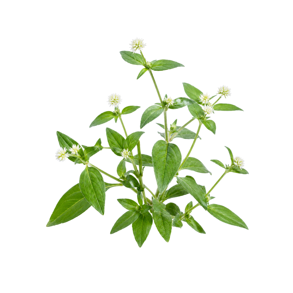 StrongStrongSumaPfaffia paniculataIts pfaffoside saponins arrest the cell cycle and trigger caspase-3 apoptosis, and powdered root has inhibited tumours and tumour blood-vessel growth in several mouse models.Mechanismcell-cycle arrest; caspase-3 apoptosis; anti-angiogenesisRead the evidenceShow lessMonograph →
The same São Paulo group has replicated tumour inhibition across several mouse models and cell lines. Powdered root cut ascitic tumour-cell counts in Ehrlich-tumour mice at 200 mg/kg 247Reference 247Effect of Pfaffia paniculata (Brazilian ginseng) on the Ehrlich tumor in its ascitic form — animal studyView study →, reduced preneoplastic liver lesions in a two-stage hepatocarcinogenesis model 248Reference 248AnimalInhibitory effects of Pfaffia paniculata on preneoplastic and neoplastic lesions in a mouse hepatocarcinogenesis model — animal studyView study →, and lowered corneal neovascularisation, a route to slowing tumour vascularisation, at 250–1000 mg/kg 249,
The active material tracks to the saponins. The butanolic residue extends survival in tumour-bearing mice 251Reference 251Antineoplastic effects of butanolic residue of Pfaffia paniculata — animal studyView study →, the purified pfaffosidic fraction arrests the cell cycle (cyclin E/CDK2 down, p27 up) and triggers caspase-3 apoptosis in HepG2 cells 252Reference 252In vitroPfaffosidic fraction from Hebanthe paniculata induces cell cycle arrest and caspase-3-induced apoptosis in HepG2 cells — in vitro studyView study →, and butanolic and methanolic extracts are cytotoxic to MCF-7, HCT116 and 4T1 lines 253,
Every result is a cell line or a mouse. There is no human oncology data, and the effective isolated-saponin doses correspond to impractically large amounts of raw root.
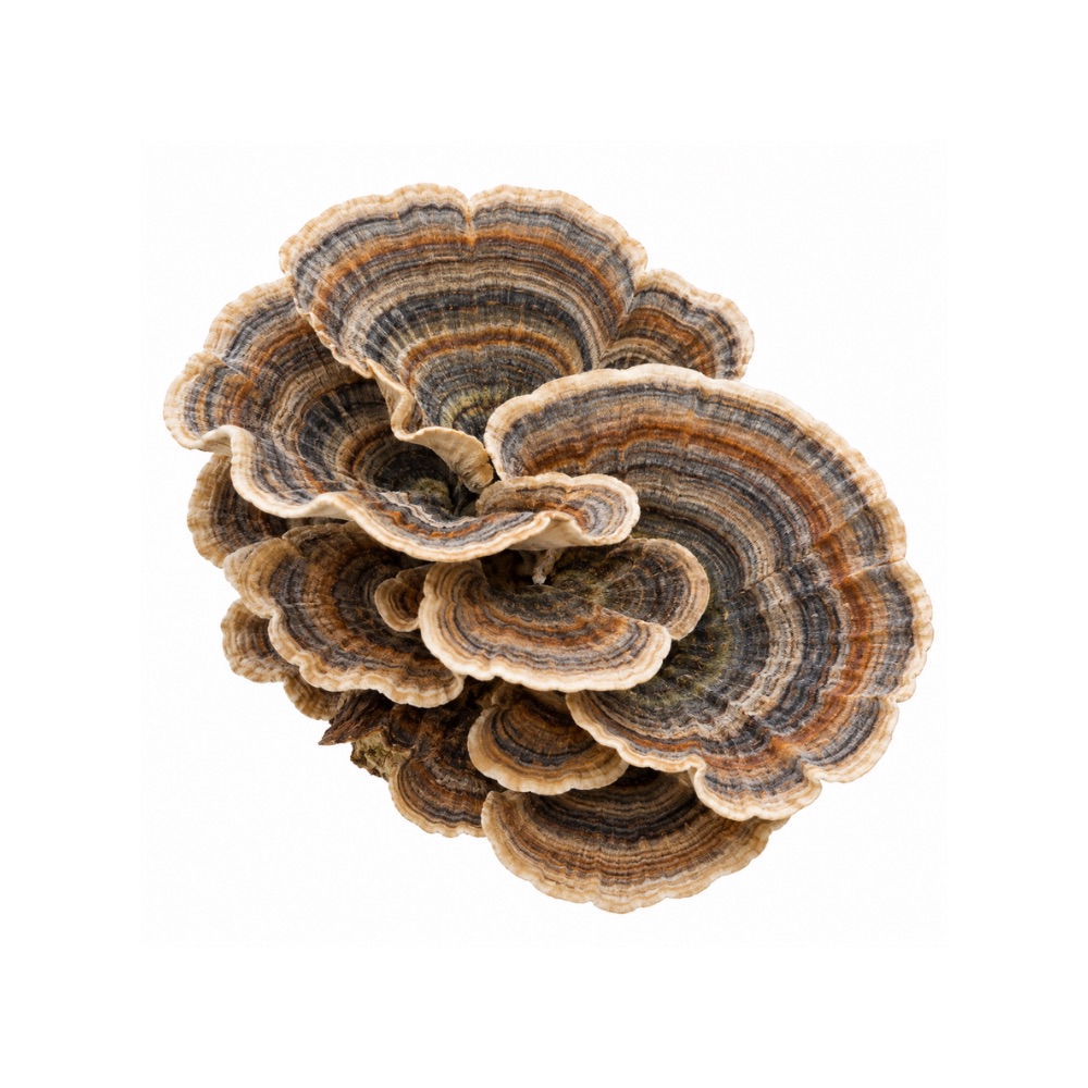 StrongStrongTurkey TailTrametes versicolorPSK, its purified protein-bound polysaccharide, added to chemotherapy improved survival in resected gastric and colorectal cancer across pooled randomised trials by restoring immune function after chemotherapy.Mechanismbiological response modifier; recovery from chemotherapy-induced immunosuppression; direct antimetastatic and pro-apoptotic actionRead the evidenceShow less
Given with chemotherapy after surgery, PSK improved overall survival in gastric cancer (HR 0.88, 8,009 patients) 258Reference 258Meta-analysisEfficacy of adjuvant immunochemotherapy with polysaccharide K for patients with curative resections of gastric cancer — meta-analysis of randomised controlled trialsView study → and colorectal cancer (overall survival RR 0.71, disease-free survival RR 0.72) 259,
PSK is a biological response modifier. It supports recovery from chemotherapy-induced immunosuppression, and it has direct antimetastatic and pro-apoptotic activity in laboratory models.
The effect is adjunctive to chemotherapy. A phase III trial could not verify non-inferiority of UFT/PSK to UFT/leucovorin 262Reference 262RCTPhase III trial comparing UFT + PSK to UFT + LV in stage IIB, III colorectal cancer (MCSGO-CCTG) — randomised trialView study →, the trials are decades old and concentrated in Japanese and Chinese gastrointestinal-cancer populations, and none of them used whole mushroom.
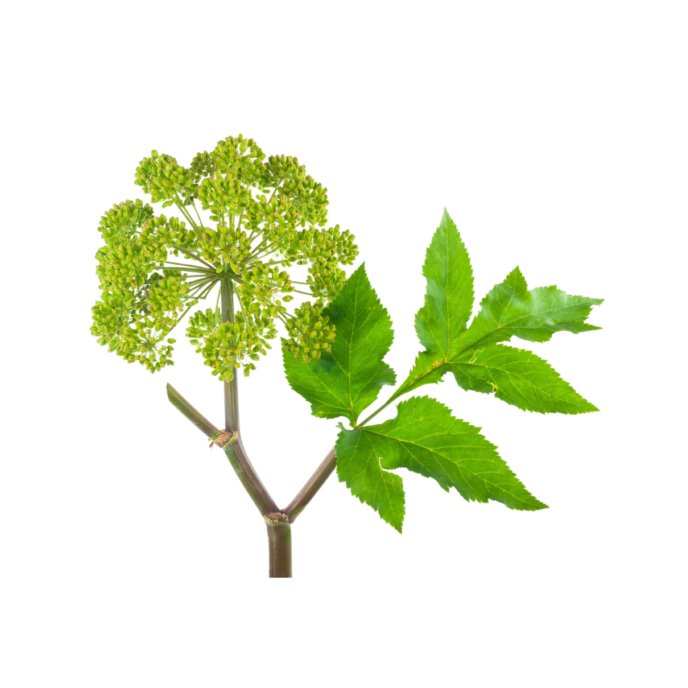 ModerateModerateAngelicaAngelica archangelicaAntiproliferative across every part of the plant: leaf, fruit, essential-oil and root extracts slow tumours in mice and kill cancer cells in culture, with imperatorin and angelicin the named constituents.MechanismG2/M cell-cycle arrest; MMP-2 ↓; direct tumor-cell cytotoxicity (high µM)Read the evidenceShow lessMonograph →
A leaf extract reduced tumour growth in a mouse sarcoma model 142Reference 142AnimalAntitumour activity of Angelica archangelica leaf extract — animal model (mouse)View study →, fruit extracts and two distinct essential-oil chemotypes were cytotoxic to cancer cell lines 143,
At constituent level the mechanisms are named. Imperatorin inhibited larynx cancer and rhabdomyosarcoma cells 146Reference 146In vitroImperatorin as a Promising Chemotherapeutic Agent Against Human Larynx Cancer and Rhabdomyosarcoma Cells — in vitroView study →, and angelicin induced G2/M arrest and suppressed migration in triple-negative breast cancer cells 147Reference 147In vitroAngelicin inhibits the growth and migration of triple-negative breast cancer cells — in vitroView study →.
Effects generally required high micromolar concentrations, no in-vivo dosing maps to achievable human exposure, and there is no clinical data. Provenance matters: the two fruit essential-oil chemotypes in 144Reference 144In vitroThe cytotoxic effect of two chemotypes of essential oils from the fruits of Angelica archangelica L — in vitroView study → were tested because coumarin and monoterpene ratios swing with origin.
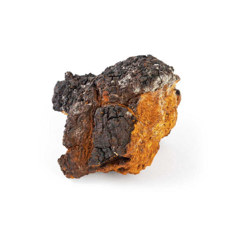 ModerateModerateChagaInonotus obliquusInotodiol, a lanostane triterpene of the fungus itself, is antiproliferative against lung adenocarcinoma cells; the betulin and betulinic acid it is often credited with come from the birch host.Mechanisminotodiol anti-proliferative in A549; betulin/betulinic-acid cytotoxicity; indirect immune-mediatedRead the evidenceShow lessMonograph →
The best fungal-origin signal is inotodiol, which is directly antiproliferative against A549 lung adenocarcinoma cells 175Reference 175In vitroEffects of inotodiol from Inonotus obliquus on the proliferation cycle and apoptotic genes of human lung adenocarcinoma A549 cells — in vitroView study → and is a chaga metabolite, at up to about 0.2% of the dried fungus 176Reference 176Chemical characterization and biological activity of Chaga (Inonotus obliquus), a medicinal “mushroom” — analytical study (inotodiol, betulin, betulinic acid, β-glucan content; host-tree variation)View study →.
Betulin and betulinic acid show inhibitory activity across cutaneous, ovarian, pulmonary, gastric and pancreatic lines 177Reference 177In vitroStudy of the betulin-enriched birch-bark extracts’ effects on human carcinoma cells and ear inflammation — in vitro / animal model178Reference 178ReviewBetulinic acid for cancer treatment and prevention — review179Reference 179In vitroComparison of the cytotoxic effects of birch-bark extract, betulin and betulinic acid towards human gastric and pancreatic carcinoma drug-sensitive and drug-resistant cell lines — in vitro, but they are host-derived, scavenged from the birch, present only at microgram-per-gram levels and varying more than 30-fold with the host tree 176Reference 176Chemical characterization and biological activity of Chaga (Inonotus obliquus), a medicinal “mushroom” — analytical study (inotodiol, betulin, betulinic acid, β-glucan content; host-tree variation)View study →. Two of the three studies are on birch bark extract rather than chaga, and the finding does not transfer to cultivated mycelium.
No clinical trials exist; everything is in vitro or animal. For a drinker of chaga tea the realistic mechanism is the indirect, immune-mediated one 180Reference 180In vitroImmuno-stimulating effect of the endo-polysaccharide from submerged culture of Inonotus obliquus — in vitroView study → rather than direct cytotoxicity.
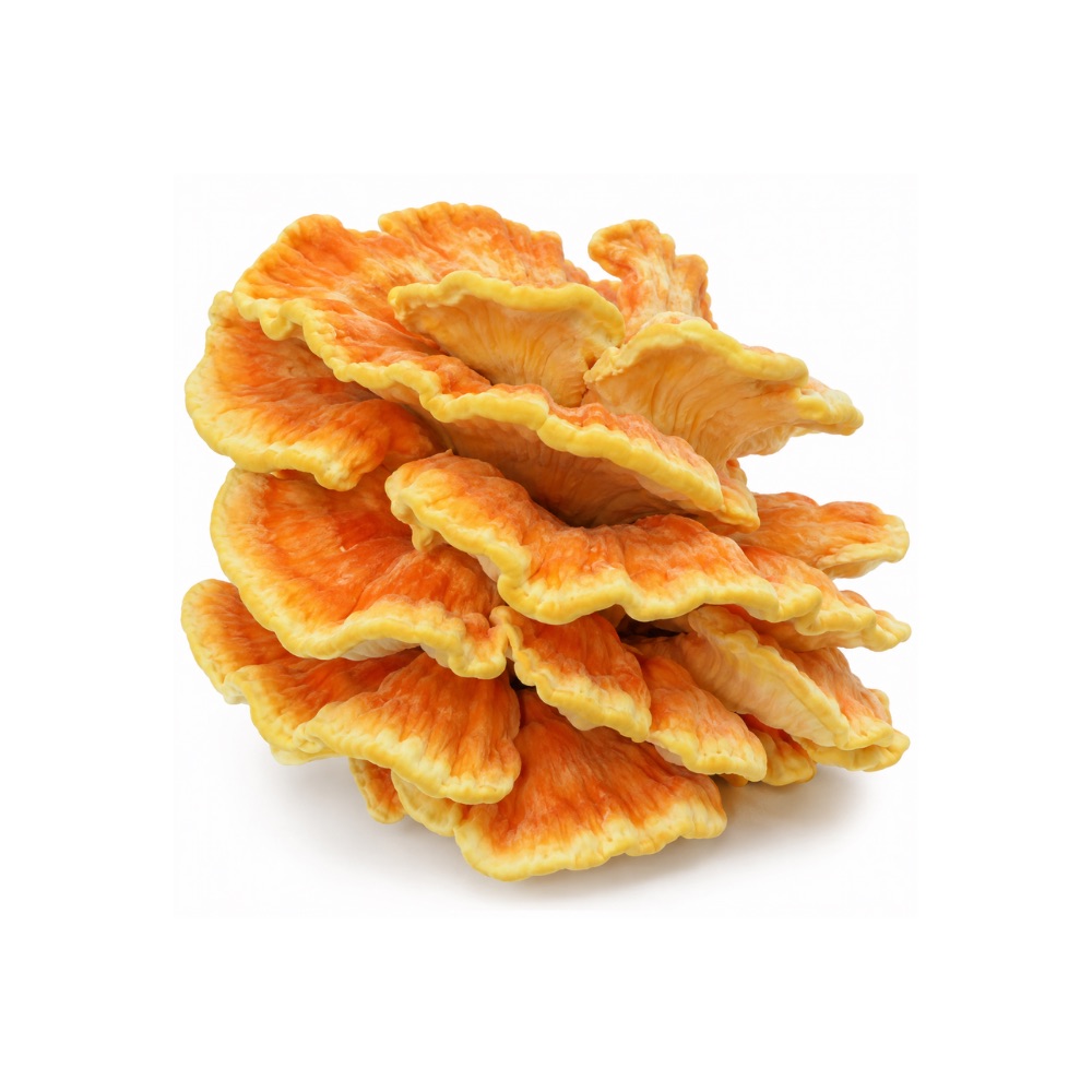 ModerateModerateChicken Of The WoodsLaetiporus sulphureusSulfated polysaccharides, a class rare in mushrooms, are antiproliferative against breast and lung cancer lines, and the LSL lectin is anti-angiogenic in zebrafish xenografts.Mechanismsulfated polysaccharides and the LSL lectin; anti-angiogenicRead the evidenceShow less
The F2 sulfated polysaccharide fraction suppressed triple-negative breast cancer proliferation and metastasis through EGFR signalling and arrested the cell cycle in breast cancer lines, and the LSL lectin inhibited angiogenesis and tumour development in zebrafish xenografts of colorectal carcinoma and melanoma 221,
There is no mammalian tumour model and no human data. LSL is a haemolytic pore-forming protein structurally related to bacterial toxins 224Reference 224In vitroMolecular cloning, expression and characterization of novel hemolytic lectins from Laetiporus sulphureus which show homology to bacterial toxins — in vitro studyView study →, and cytotoxicity from a haemolysin is not straightforwardly a therapeutic property.
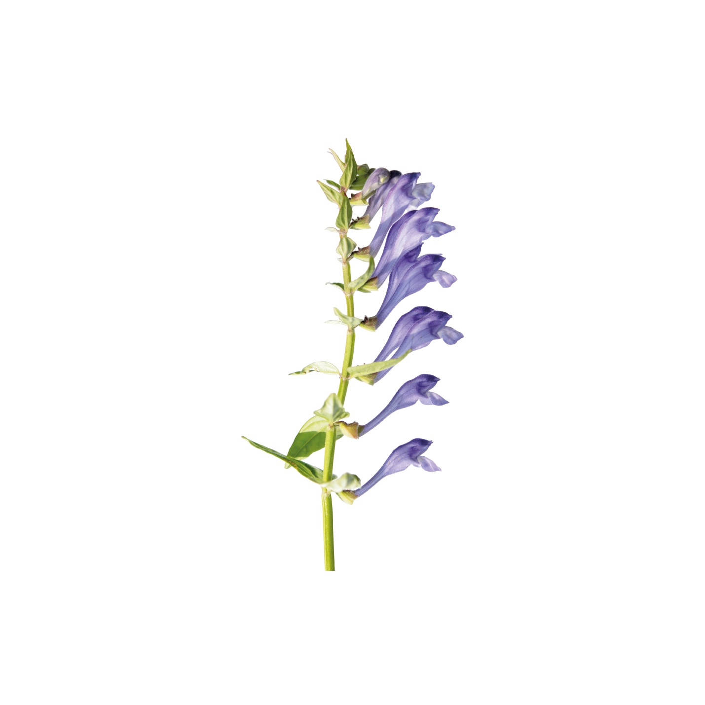 ModerateModerateChinese SkullcapScutellaria baicalensisBaicalein, wogonin and oroxylin A induce apoptosis and arrest the cell cycle across leukaemia, breast, colon, lung and cervical lines, largely by suppressing NF-κB and modulating p53.Mechanismpro-apoptotic; cell-cycle arrest; NF-κB ↓; p53 modulation; tumor-microenvironment modulationRead the evidenceShow lessMonograph →
Across leukaemia, breast, colon, lung and cervical lines the flavonoids, oroxylin A among them, suppress NF-κB and modulate p53 and the tumour microenvironment, inducing apoptosis, cell-cycle arrest and reduced proliferation 243,
Human exposure exists only through multi-herb formulas (PHY906 alongside chemotherapy) and ex-vivo work on patient leukocytes; skullcap has never been given to a person as an anticancer treatment. Oroxylin A has completed a phase I safety and pharmacokinetic study 245Reference 245RCTSafety and pharmacokinetics evaluation of oroxylin A in Chinese healthy volunteers — randomised, double-blind, placebo-controlled Phase I studyView study →, which establishes tolerability rather than efficacy.
The cell-line effect is real and reproducible, and nothing above rodent level yet supports it.
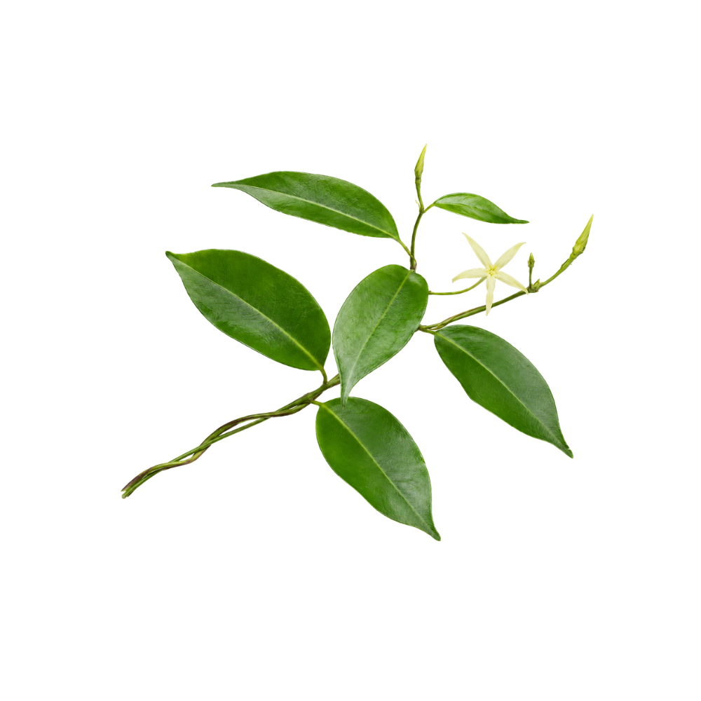 ModerateModerateCryptolepisCryptolepis sanguinolentaCryptolepine, its major alkaloid, intercalates DNA and poisons topoisomerase II; it suppresses colorectal cancer stemness through WNT/β-catenin and slowed breast and melanoma xenografts in mice, with genotoxicity as the same property seen from the other side.Mechanismtopoisomerase II poisoning / DNA intercalation; WNT/β-catenin ↓, IL-6/STAT3 ↓; HIF-1 glycolysis ↓, AMPKα-LKB1 ↑Read the evidenceShow lessMonograph →
As a DNA intercalator and topoisomerase II poison, cryptolepine suppresses colorectal cancer proliferation, stemness and metastatic behaviour through WNT/β-catenin inhibition, outperforming a reference WNT inhibitor 181Reference 181In vitroCryptolepine suppresses colorectal cancer cell proliferation, stemness, and metastatic processes by inhibiting WNT/β-catenin signaling — in vitroView study →; downregulates IL-6/STAT3 in hepatocellular carcinoma 182Reference 182In vitroCryptolepine inhibits hepatocellular carcinoma growth through inhibiting interleukin-6/STAT3 signalling — in vitroView study →; blocks HIF-1-mediated glycolysis and slowed a 4T1 breast xenograft 183Reference 183In vitroCryptolepine suppresses breast adenocarcinoma via inhibition of HIF-1 mediated glycolysis — in vitro and mouse xenograftView study →; and modulates mutant p53 to arrest and kill triple-negative breast cancer cells 184Reference 184In vitroMutant p53 modulation by cryptolepine through cell cycle arrest and apoptosis in triple negative breast cancer — in vitroView study →.
The most complete in-vivo work reduced A375 melanoma xenograft growth in nude mice through AMPKα-LKB1 activation and disrupted mitochondrial dynamics, sparing normal melanocytes in vitro 185Reference 185In vitroCryptolepine inhibits melanoma cell growth through coordinated changes in mitochondrial biogenesis, dynamics and the metabolic tumor suppressor AMPKα1/2-LKB1 — in vitro and mouse xenograftView study →. Reviews treat it as a lead compound 186Reference 186ReviewA review of the anticancer potential of the antimalarial herbal Cryptolepis sanguinolenta and its major alkaloid cryptolepine — reviewView study →.
All of it is preclinical and uses isolated cryptolepine rather than the root. The cytotoxicity is non-selective against mammalian cells 187Reference 187In vitroThe popular herbal antimalarial, extract of Cryptolepis sanguinolenta, is potently cytotoxic — in vitroView study →, and no therapeutic window in humans has been established.
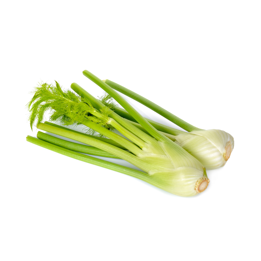 ModerateModerateFennelFoeniculum vulgareProtects liver and kidney tissue from chemical injury in animals through free-radical scavenging and antioxidant-enzyme induction; seed extract has also induced apoptosis in liver and lung cancer cells and slowed their xenografts in mice.Mechanismfree-radical scavenging; endogenous antioxidant-enzyme induction; COX/LOX inhibition; survivin ↓ / Bcl-2 ↓ apoptosis (seed extract)Read the evidenceShow lessMonograph →
Essential oil and phenolic extracts show consistent free-radical-scavenging and lipid-peroxidation-inhibiting activity in vitro, driven by phenolic acids and flavonoids rather than the volatile oil alone 196Reference 196In vitroFoeniculum vulgare essential oils: chemical composition, antioxidant and antimicrobial activities — in vitroView study →; in rats, seed and sprout extracts raised endogenous antioxidant enzymes and limited oxidative organ damage 197Reference 197AnimalNephroprotective effect of fennel seeds and sprouts on CCl₄-induced nephrotoxicity in rats — animal modelView study →.
The protection is organ-specific in animal models. Fennel essential oil protected rat liver against carbon-tetrachloride injury, with delta-limonene and beta-myrcene proposed as the active fraction 198,
A COX/LOX-mediated anti-inflammatory action runs underneath. Fruit extracts are anti-inflammatory and analgesic in animal models through both pathways 201Reference 201AnimalAntiinflammatory, analgesic and antioxidant activities of the fruit of Foeniculum vulgare — animal modelView study →, and isolated fennel coumarins suppressed pro-inflammatory mediators in LPS-stimulated macrophages and reduced mouse paw oedema 202Reference 202In vitroAnti-inflammatory and antioxidant effects of coumarins isolated from Foeniculum vulgare — in vitro / mouseView study →.
An ethanol seed extract reduced viability, induced apoptosis and inhibited migration in hepatocellular carcinoma cells by lowering survivin, without the same toxicity in normal liver cells, and inhibited xenograft growth in nude mice 270Reference 270In vitroFoeniculum vulgare seed extract exerts anti-cancer effects on hepatocellular carcinoma — in vitro and mouse xenograftView study →; the same extract induced Bcl-2-dependent apoptosis in lung cancer lines and slowed their xenografts 271Reference 271In vitroFoeniculum vulgare seed extract induces apoptosis in lung cancer cells partly through the down-regulation of Bcl-2 — in vitro and mouse xenograftView study →.
All of it is rodent and in-vitro work. There is no human antioxidant-biomarker trial, no evidence in human liver or kidney disease, and the effective animal doses used concentrated extracts well above culinary intake.
ModerateIpecacuanhaCarapichea ipecacuanhaEmetine, a protein-synthesis inhibitor, is potent against breast, lung, gastric and bladder cancer lines, and the same property makes it cardiotoxic.MechanismWnt/β-catenin and ERK suppression; protein-synthesis inhibitionRead the evidenceShow less
Emetine is potent in vitro across breast, lung, gastric and bladder lines 213,
Potency in cell culture is expected for a protein-synthesis inhibitor, because dividing cells are especially vulnerable, and the same property makes it toxic to heart muscle, a tissue that never stops turning protein over. The evidence is cell lines only.
 ModerateModerateIsatisIsatis indigoticaIndirubin, the active principle of the classical anti-leukaemia formula Danggui Longhui Wan, is a direct CDK2 inhibitor confirmed by co-crystal structure, and its semi-synthetic derivatives treat CML in China.MechanismCDK inhibition by indirubin (CDK2 co-crystal, G2/M arrest); GSK-3β/CDK5-p25 inhibition; c-Src/Abl and Stat5 blockade by 6-bromoindirubin derivativesRead the evidenceShow lessMonograph →
The classical anti-leukaemia formula Danggui Longhui Wan gave up indirubin as its active principle. It is a direct cyclin-dependent-kinase inhibitor confirmed by a CDK2 co-crystal structure, arresting cells at G2/M 217Reference 217In vitroIndirubin, the active constituent of a Chinese antileukaemia medicine, inhibits cyclin-dependent kinases — in vitroView study →, and it and its derivatives inhibit GSK-3β and CDK5/p25 at IC₅₀ 5–50 nM 218Reference 218In vitroIndirubins inhibit glycogen synthase kinase-3β and CDK5/p25 — in vitroView study →.
Natural 6-bromoindirubin analogues kill imatinib-resistant T315I-mutant CML cells at submicromolar potency through c-Src/Abl (BCR-ABL) inhibition 219Reference 219In vitroNatural-based indirubins display potent cytotoxicity toward wild-type and T315I-resistant leukemia cell lines — in vitroView study →, and the derivative E804 drives apoptosis in resistant CML lines and primary patient cells by blocking Stat5 220Reference 220In vitroIndirubin derivatives induce apoptosis of chronic myelogenous leukemia cells involving inhibition of Stat5 signaling — in vitroView study →. The semi-synthetic derivative meisoindigo has been used clinically for CML in China.
This is a drug-discovery lineage. Indirubin is present in the herb only in trace amounts, the active agents are semi-synthetic analogues at pharmaceutical doses, and the herb itself has never been trialled for cancer.
Show 22 more herbs with a mild anticancer action
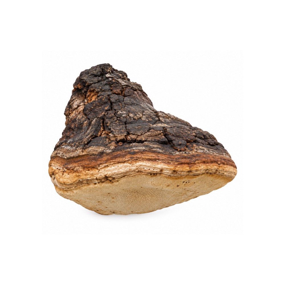 MildMildAgarikonFomitopsis officinalisPolysaccharides and triterpenoids are antiproliferative in cell lines and anti-angiogenic in zebrafish.Mechanismanti-proliferative and anti-angiogenic activity of polysaccharides and triterpenoidsRead the evidenceShow less
A fungal polysaccharide from Fomitopsis officinalis activated immunity and inhibited angiogenesis in zebrafish, and the triterpenoids are antiproliferative in cell lines 140,
There is no mammalian tumour model and no human data.
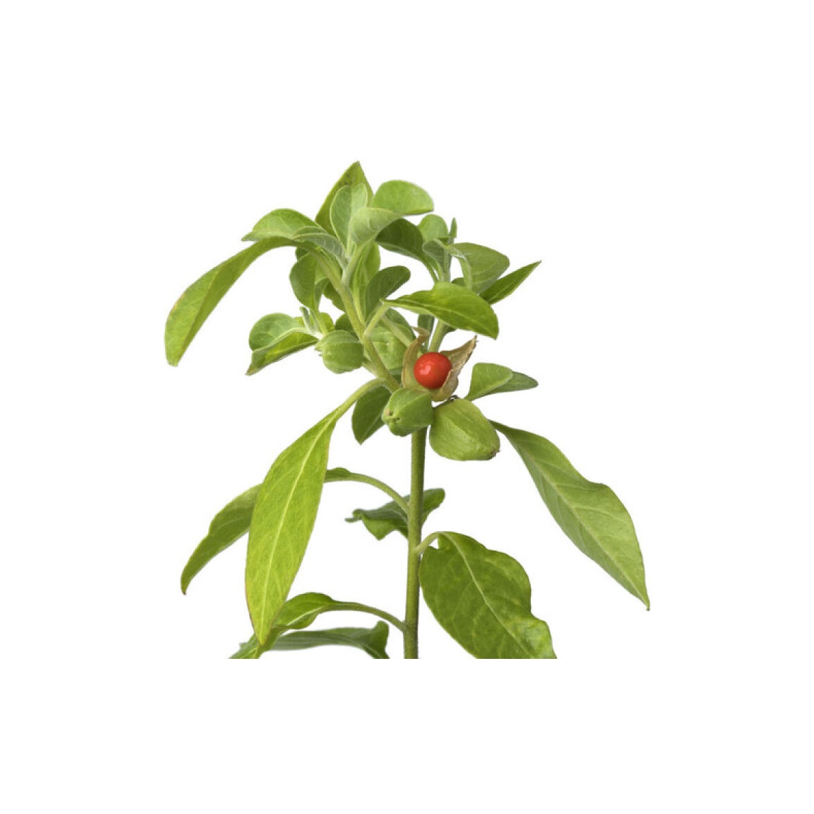 MildMildAshwagandhaWithania somniferaWithaferin A, its principal withanolide, suppresses NF-κB, targets HSP90 to degrade client oncoproteins and inhibits angiogenesis in preclinical models; oral root delivers little of it.MechanismNF-κB ↓; HSP90 inhibition; pro-apoptotic / anti-angiogenicRead the evidenceShow lessMonograph →
Withanolides potentiate apoptosis, inhibit invasion and abolish osteoclastogenesis by suppressing NF-κB 148Reference 148In vitroWithanolides potentiate apoptosis, inhibit invasion and abolish osteoclastogenesis through suppression of NF-κB — in vitroView study →. Withaferin A targets HSP90 to degrade client oncoproteins in pancreatic cancer cells 149Reference 149In vitroWithaferin A targets heat-shock protein 90 in pancreatic cancer cells — in vitroView study →, is a potent angiogenesis inhibitor 150Reference 150In vitroWithaferin A is a potent inhibitor of angiogenesis — in vitro/animal modelView study →, and produces cytotoxic and cytostatic effects on human T-leukaemia cells 151Reference 151In vitroWithania somnifera induces cytotoxic and cytostatic effects on human T-leukaemia cells — in vitroView study →, reviewed comprehensively at 152Reference 152ReviewMolecular targets and mechanisms of cancer prevention and treatment by withaferin A — reviewView study →.
There is no human efficacy data. Every result is purified withaferin A at concentrations unrelated to oral dosing, and oral root barely delivers the molecule.
The finding belongs to the compound rather than to the herb: withaferin A is a drug-discovery lead, and ashwagandha is not a cancer treatment.
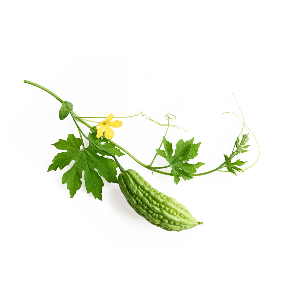 MildMildBitter MelonMomordica charantiaThe seed proteins MAP30 and BG-4, rather than the fruit, carry the activity: MAP30 induced apoptosis in liver cancer cells and acted synergistically with cisplatin in a mouse ovarian-cancer model.Mechanismribosome inactivation (rRNA N-glycosidase); Bax/Bcl-2 → caspase-3 apoptosis; p53/p21 G2–M arrestRead the evidenceShow lessMonograph →
Whole-fruit extract inhibited proliferation and induced apoptosis in MCF-7 and MDA-MB-231 breast cancer cells via p53/p21 and G2–M arrest 153Reference 153In vitroBitter melon (Momordica charantia) extract inhibits breast cancer cell proliferation by modulating cell-cycle regulatory genes and promotes apoptosis — in vitroView study →.
The translational interest sits on seed proteins. The peptide BG-4 triggered Bax/Bcl-2-mediated caspase-3 apoptosis in HCT-116 and HT-29 colon cells 154Reference 154In vitroBG-4, a novel anticancer peptide from bitter gourd (Momordica charantia), promotes apoptosis in human colon cancer cells — in vitroView study →, and the ribosome-inactivating protein MAP30 induced apoptosis in hepatocellular carcinoma cells 155Reference 155In vitroRibosome-inactivating protein MAP30 isolated from Momordica charantia L. induces apoptosis in hepatocellular carcinoma cells — in vitroView study → and, in a mouse ovarian-cancer model, acted synergistically with cisplatin through AMPK activation and ferroptosis without renal or hepatic toxicity in the treated animals 156Reference 156AnimalMAP30 protein from Momordica charantia is therapeutic and has synergic activity with cisplatin against ovarian cancer in vivo by altering metabolism and inducing ferroptosis — mouse modelView study →.
A widely cited 2016 paper on bitter-melon extract overcoming cisplatin resistance was retracted in 2025, and the active agents are purified proteins delivered parenterally in models rather than a food.
 MildMildCannabisCannabis sativaCBD and THC trigger cancer-cell death in cell and animal models through PPARγ and the cannabinoid receptors; the one human trial, THC infused into recurrent glioblastoma, showed no survival benefit.MechanismPPARγ activation; cannabinoid-receptor-mediated apoptosisRead the evidenceShow lessMonograph →
In cell and animal studies CBD and THC trigger cancer-cell death across several tumour types 157Reference 157ReviewMechanisms of Cell Death Induced by Cannabidiol Against Tumor Cells: A Review of Preclinical StudiesView study →, with PPARγ activation among the routes.
The one human test, a 2006 pilot infusing THC directly into recurrent glioblastoma in nine patients, established safety and showed no survival benefit 158Reference 158Clinical trialA pilot clinical study of delta-9-tetrahydrocannabinol in patients with recurrent glioblastoma multiforme — clinical trialView study →.
No controlled clinical data support cannabis as a cancer treatment. Its established oncology roles are supportive: nausea 159,
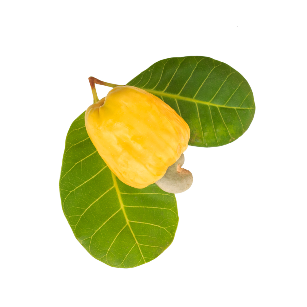 MildMildCashewAnacardium occidentaleAnacardic acid from the nut-shell liquid, not the leaf, inhibits histone acetyltransferase, modulates NF-κB and sensitises tumour cells to radiation, the basis of a traditional adjunctive use during radiotherapy.Mechanismhistone acetyltransferase (HAT) inhibition; NF-κB/IKKβ blockade; potentiation of TNF-induced apoptosisRead the evidenceShow lessMonograph →
The part is nut-shell liquid and isolated anacardic acid rather than the leaf. Anacardic acid is cytotoxic to lung, liver and gastric tumour cells through epigenetic HAT inhibition 164Reference 164In vitroCharacterization of cytotoxic activity of compounds derived from anacardic acid, cardanol and cardol in oral squamous cell carcinoma — in vitro, modulates NF-κB 165Reference 165In vitroAnacardic acid, an inhibitor of histone acetyltransferase, suppresses NF-κB–regulated gene products and potentiates apoptosis — in vitroView study →, potentiates TNF-induced apoptosis 165Reference 165In vitroAnacardic acid, an inhibitor of histone acetyltransferase, suppresses NF-κB–regulated gene products and potentiates apoptosis — in vitroView study →, and sensitises tumours to radiation 165Reference 165In vitroAnacardic acid, an inhibitor of histone acetyltransferase, suppresses NF-κB–regulated gene products and potentiates apoptosis — in vitroView study →, the last being the basis of the traditional adjunctive use during radiotherapy.
Natural (iCNSL, ≤45 °C) and technical (tCNSL, 195 °C) cashew nut-shell liquids were antimutagenic and reparative against micronuclei and chromosomal aberrations in Allium cepa and Artemia salina bioassays, with tCNSL non-toxic and non-mutagenic at every concentration tested 166Reference 166In vitroEvaluation of toxic, cytotoxic, mutagenic and antimutagenic activities of natural and technical cashew nut shell liquids — in vitro bioassaysView study →.
There is no animal tumour model and no patient data; the evidence is isolated-compound and brine-shrimp bioassay work.
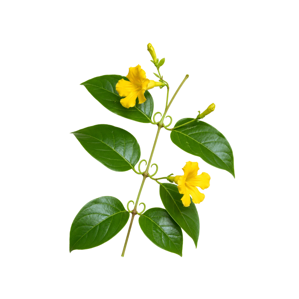 MildMildCat’s ClawUncaria tomentosaUsed against tumours in Amazonian tradition; its oxindole alkaloids induce apoptosis in leukaemia cells, mitraphylline is cytotoxic to sarcoma, breast, glioma and neuroblastoma lines, and the one human trial reduced chemotherapy toxicity.Mechanismapoptosis induction / G0–G1 arrest (mitraphylline, oxindole alkaloids); oxidative-stress modulationRead the evidenceShow lessMonograph →
Oxindole alkaloids from U. tomentosa induce apoptosis in acute lymphoblastic leukaemia cells, including bcl-2-expressing and G0/G1-arrested populations 167Reference 167In vitroOxindole alkaloids from Uncaria tomentosa induce apoptosis in proliferating, G0/G1-arrested and bcl-2-expressing acute lymphoblastic leukaemia cells — in vitroView study →; mitraphylline is cytotoxic to Ewing’s sarcoma, breast, glioma and neuroblastoma lines 168,
The only human trial is supportive: it reduced chemotherapy toxicity rather than tumour burden 172Reference 172Clinical trialUncaria tomentosa — adjuvant treatment for breast cancer — clinical trialView study →. Traditional Amazonian use against tumours is long documented 173Reference 173ReviewUncaria tomentosa — ethnomedicinal use and new pharmacological, toxicological and botanical results — reviewView study →.
Adjunct use during chemotherapy is the evidenced role, and it is a different thing from treating cancer.
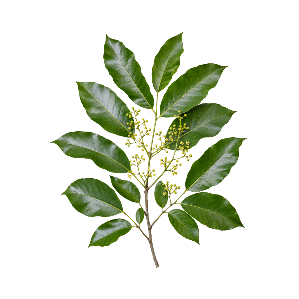 MildMildCatuabaTrichilia catiguaCrude extract and its ethyl-acetate fraction are cytotoxic to leukaemia, lymphoma and breast cancer lines, and isolated cinchonain Ib to K562 cells.Mechanismnot established — cell-line cytotoxicity (leukemia, lymphoma, breast)Read the evidenceShow lessMonograph →
Against HL-60 and TOLEDO lines the crude extract and the ethyl-acetate fraction were cytotoxic, the ethyl-acetate fraction was also active on MCF-7 breast cells, and isolated cinchonain Ib was cytotoxic to K562 cells 174Reference 174In vitroImmunomodulatory, antibacterial and antineoplastic potential of Trichilia catigua AView study →.
No apoptotic or signalling mechanism has been identified, no selectivity index against normal cells is reported, and there is no animal tumour model or clinical data. Cytotoxicity against a handful of cell lines is a screening signal.
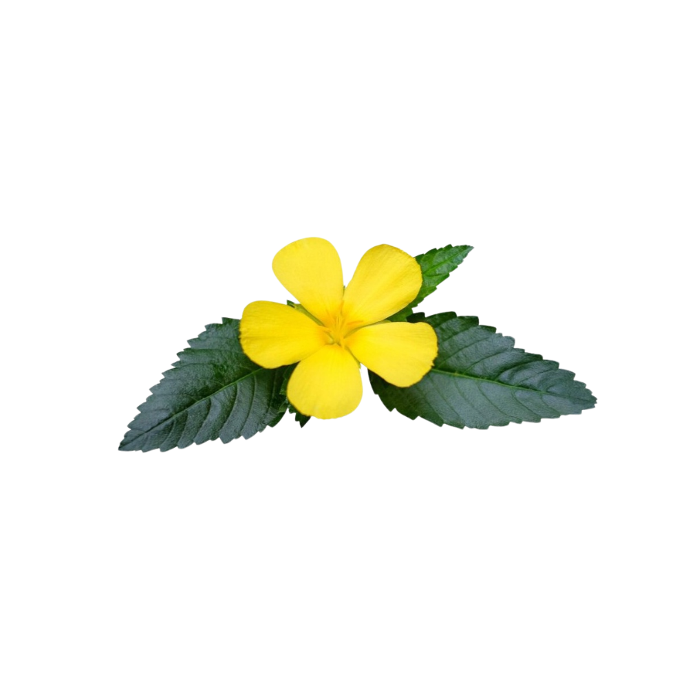 MildMildDamianaTurnera diffusaA methanolic extract is cytotoxic to several tumour lines, most strongly triple-negative breast cancer cells, with arbutin and apigenin isolated from the active fraction.Mechanismnot established (crude-extract cytotoxicity only)Read the evidenceShow lessMonograph →
The first report of cytotoxicity for the herb found a methanolic T. diffusa extract active against several tumour lines, most strongly MDA-MB-231 breast-cancer cells (IC₅₀ about 30.7 µg/mL), with arbutin and apigenin isolated from the most active fraction 188Reference 188In vitroCytotoxic activity of the methanolic extract of Turnera diffusa on breast cancer cells — in vitroView study →.
No mechanism was identified, there is no selectivity data against normal cells, and there is no animal tumour model or in-vivo confirmation.
The herb also has in-vitro estrogenic and aromatase-modulating activity 189,
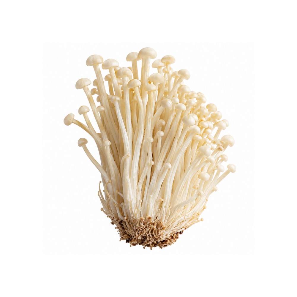 MildMildEnokiFlammulina velutipesErgosterol, the proteoglycan PGD1-1 and polysaccharides are each cytotoxic to cancer cell lines through mitochondrial apoptosis, with one in-vivo leukaemia model.Mechanismmitochondrial apoptosis — Bax/Bcl-2 ↑, caspase-3/-9 ↑Read the evidenceShow less
Three constituent types are cytotoxic in cell lines: ergosterol (HepG-2 IC₅₀ about 9.3 µg/mL, A549 about 20.4 µg/mL), the proteoglycan PGD1-1, and polysaccharides, with a single in-vivo P388 leukaemia model alongside.
Ergosterol has poor oral bioavailability, improved in the same paper only by liposomal encapsulation, so the cell-line potencies do not translate to the mushroom as eaten.
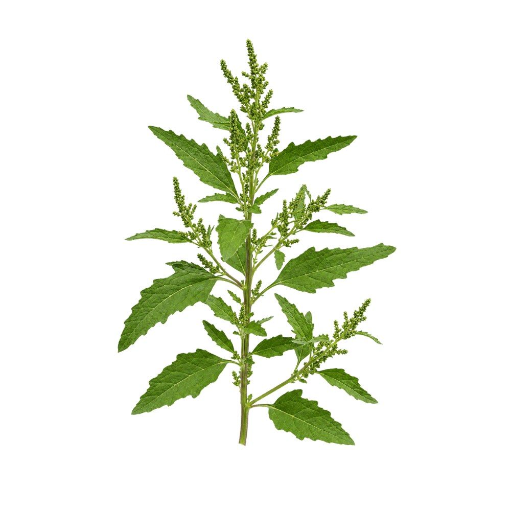 MildMildEpazoteDysphania ambrosioidesKaempferitrin, its major flavonol glycoside, shrank liver-cancer xenografts in mice through apoptosis and immune stimulation, and the essential oil killed the same cells while sparing normal hepatocytes.Mechanismcaspase-dependent apoptosis; G0/G1 arrest; immune stimulationRead the evidenceShow lessMonograph →
Kaempferitrin, the major flavonol glycoside of the ethanol extract, reduced tumour volume and mass in a human liver-cancer (SMMC-7721) mouse xenograft via apoptosis and immune stimulation 191Reference 191AnimalKaempferitrin, a major compound from ethanol extract of Chenopodium ambrosioides, exerts antitumour and hepatoprotective effects in the mice model of human liver cancer xenografts. — [in vivo mouse xenograft]View study →; the essential oil induced caspase-dependent apoptosis and G0/G1 arrest in the same line while sparing normal hepatocytes 192Reference 192In vitroEssential Oil of Chenopodium ambrosioides Induced Caspase-Dependent Apoptosis in SMMC-7721 Cells. — [in vitro]View study →; seed extract suppressed SMMC-7721 migration and invasion in vitro 193Reference 193In vitroPossible Mechanism of Dysphania ambrosioides (L.) Mosyakin & Clemants Seed Extract Suppresses the Migration and Invasion of Human Hepatocellular Carcinoma Cells SMMC-7721. — [in vitro]View study →; and whole-leaf extract inhibited Ehrlich ascitic and solid tumours in mice 194Reference 194AnimalAscitic and solid Ehrlich tumor inhibition by Chenopodium ambrosioides L. treatment. — [in vivo mouse model]View study →.
The only xenograft used an isolated compound rather than the herb, everything else is cell-line or non-clinical tumour work, and the essential oil’s own toxicity caps any therapeutic translation.
The herb’s ordinary infusion and decoction were genotoxic to human lymphocytes in vitro, with chromosomal aberrations and raised sister-chromatid exchange 195Reference 195In vitroIn vitro genotoxic evaluation of the medicinal plant Chenopodium ambrosioides L. — [in vitro genotoxicity]View study →.
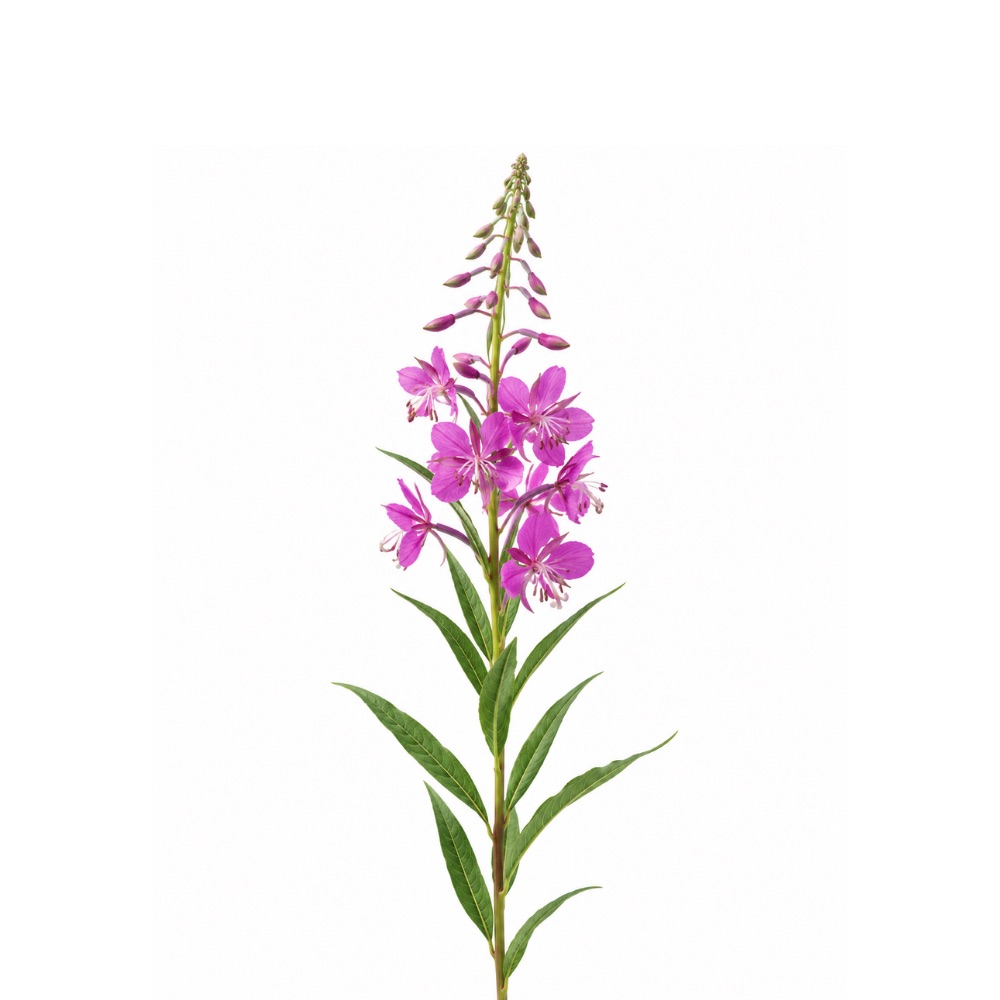 MildMildFireweedChamerion angustifoliumExtracts are antiproliferative across prostate, colon, breast, lung, liver and melanoma lines, and oral aqueous extract reduced prostatic adenoma in rats; the effect is not specific to cancer cells.Mechanismmitochondrial-pathway apoptosis (caspase-3 ↑, Δψm ↓)Read the evidenceShow less
Activity has been shown against LNCaP prostate, Caco-2 and HT-29 colon, breast, lung, liver and melanoma lines 203,
The antiproliferative effect is non-specific: extracts inhibited normal cells as strongly as cancer cells 204Reference 204In vitroCharacterization of the effect of Epilobium extracts on human cell proliferation — non-specific antiproliferation, oenothein B (in vitro)View study →. Oenothein B, the main ellagitannin, is poorly absorbed, which undercuts the in-vitro potencies 208Reference 208In vitroEvaluation of the effect of Epilobium angustifolium aqueous extract on LNCaP cell proliferation in in vitro and in vivo (rat) modelsView study →.
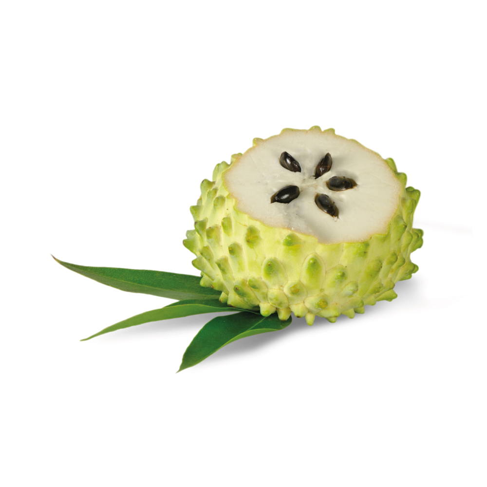 MildMildGraviolaAnnona muricataIts annonaceous acetogenins inhibit mitochondrial complex I and deplete ATP in high-demand cells; leaf extract slowed pancreatic tumours in mice, and no human tumour outcome has been measured.Mechanismannonaceous-acetogenin inhibition of mitochondrial complex I → ATP depletion in high-demand cells; mitochondrial-pathway apoptosis in tumor cell lines; altered cancer-cell metabolism (mouse pancreatic model)Read the evidenceShow lessMonograph →
The annonaceous acetogenins inhibit mitochondrial complex I, depleting ATP in cells with high energy demand 209Reference 209ReviewAnnona muricata (Annonaceae): a review of its traditional uses, isolated acetogenins and biological activities — reviewView study →. On that route a leaf extract induced mitochondrial-pathway apoptosis in A549 lung-cancer cells in vitro 210Reference 210In vitroAnnona muricata leaves induced apoptosis in A549 cells through the mitochondrial-mediated pathway — in vitroView study →, and in a pancreatic-cancer model graviola reduced tumorigenicity and metastasis in vitro and in mice by altering cancer-cell metabolism 211Reference 211In vitroGraviola: a natural-derived drug that inhibits tumorigenicity and metastasis of pancreatic cancer cells in vitro and in vivo — animal modelView study →; everything else is cell-line and rodent work 209Reference 209ReviewAnnona muricata (Annonaceae): a review of its traditional uses, isolated acetogenins and biological activities — reviewView study →.
The single controlled human study is a randomised, double-blind, placebo-controlled trial in 28 colorectal-cancer patients after tumour resection, which found that serum from the leaf-extract arm was more cytotoxic to colorectal cell lines than placebo serum 212Reference 212RCTThe effect of an Annona muricata leaf extract on nutritional status and cytotoxicity in colorectal cancer: a randomised controlled trialView study →. That is an ex-vivo laboratory surrogate rather than tumour shrinkage, survival or recurrence, and the trial itself called for clinically meaningful endpoints 212Reference 212RCTThe effect of an Annona muricata leaf extract on nutritional status and cytotoxicity in colorectal cancer: a randomised controlled trialView study →.
No tumour outcome has been measured in any human with any cancer, and the selectivity for tumour cells is a cell-culture observation rather than a demonstrated therapeutic window. Graviola is widely marketed for cancer, and the evidence does not support that use.
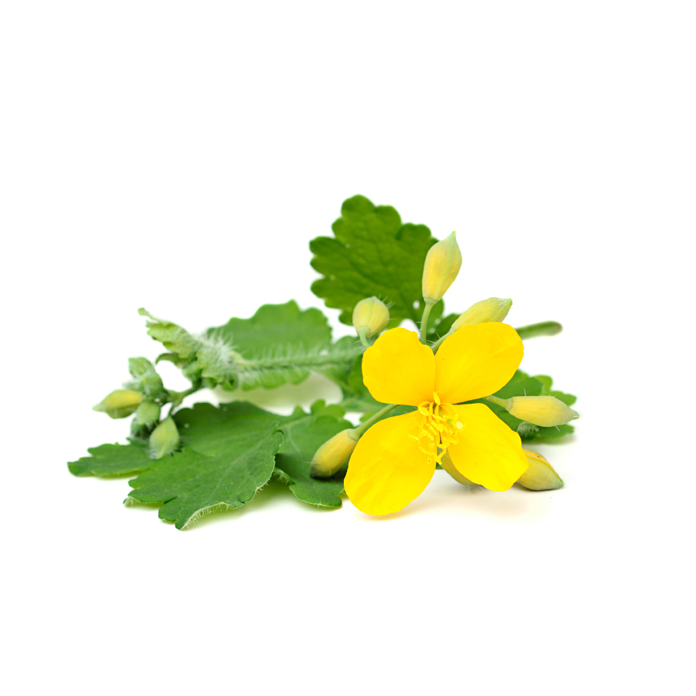 MildMildGreater CelandineChelidonium majusAlkaloid extracts induce apoptosis in carcinoma and leukaemia lines — but the impressive human data belong to a semisynthetic derivative, not the plant.Mechanismapoptosis via caspase activation and NF-\u03baB inhibition (A431 epidermoid carcinoma); benzophenanthridine alkaloids presumed driversRead the evidenceShow lessMonograph →
The cell-line data are broad and repeatable. Whole-herb and alkaloid extracts induce apoptosis in human epidermoid carcinoma through caspase activation and NF-κB inhibition, and in leukaemia lines, with the extract also scavenging DPPH radicals.
The individual benzophenanthridines are the presumed cytotoxic drivers.
The human trial evidence belongs to something else. Ukrain (NSC-631570) is a semisynthetic thiophosphoric derivative of celandine alkaloids, not the herb. A phase II pancreatic-cancer trial reported survival gains, but a systematic review found the supporting trials of poor methodological quality and urged independent replication.
Herb-level evidence is purely in vitro. Greater celandine is not a cancer treatment.
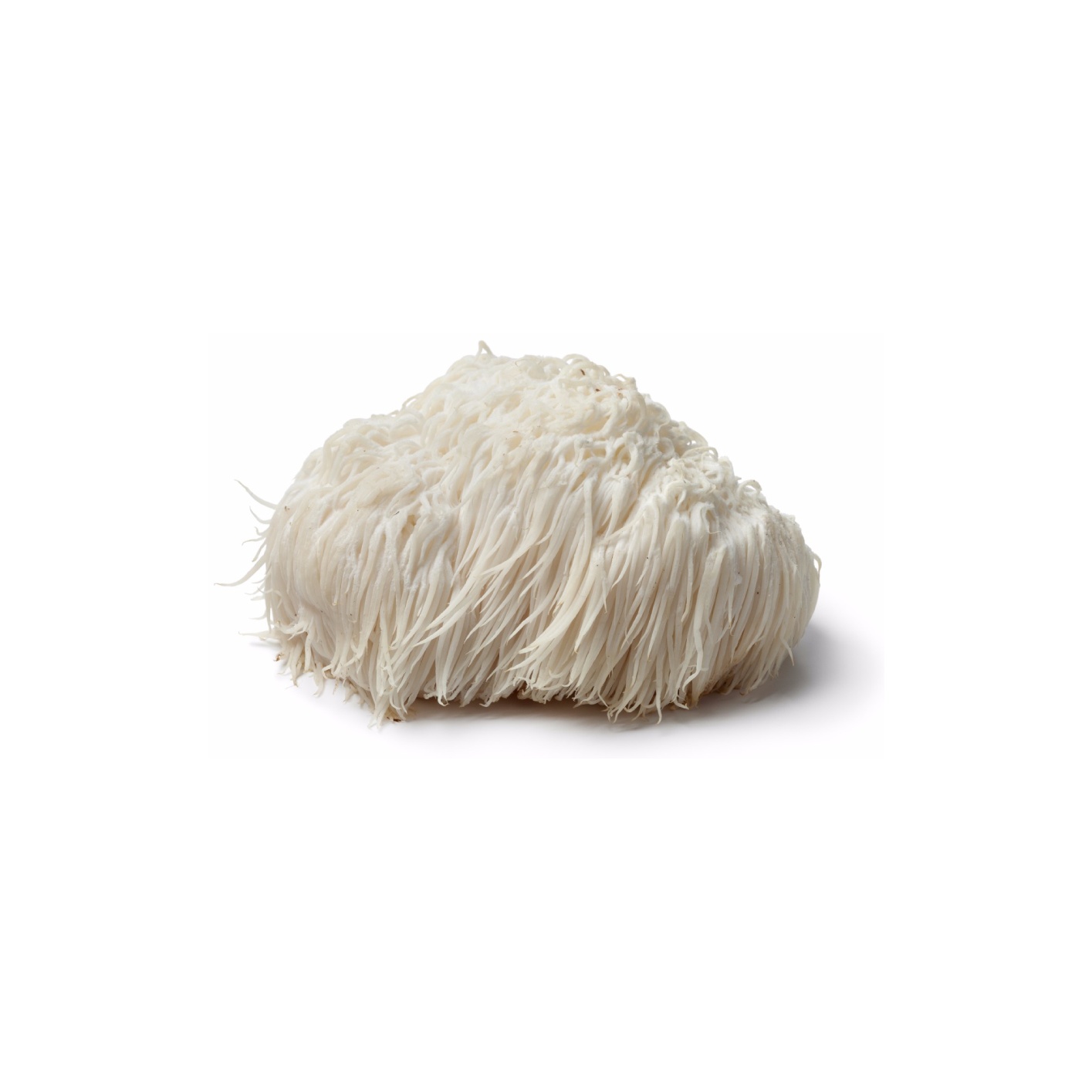 MildMildLion’s ManeHericium erinaceusAn ethanol fruiting-body extract induced apoptosis in prostate cancer cells through PI3K/AKT and RAS/MAPK, and the mycelial erinacines have laboratory antitumour activity.MechanismPI3K/AKT; RAS/MAPK; caspase activationRead the evidenceShow lessMonograph →
A 2024 in-vitro study found an ethanol fruiting-body extract induced apoptosis in prostate cancer cells via PI3K/AKT and RAS/MAPK signalling 225Reference 225In vitroHericium erinaceus extract induces apoptosis via PI3K/AKT and RAS/MAPK signaling in prostate cancer cells — in vitro studyView study →, in cell lines only, with no animal arm, no human data and no reported test of selectivity against non-malignant cells.
A 2020 review of erinacine A and related cyathane diterpenoids collects in-vitro and animal antitumour activity (reduced gastric-cancer-cell invasiveness, caspase activation) for isolated mycelial compounds 226Reference 226ReviewErinacine A and related cyathane diterpenoids: mechanisms underlying their neuroprotection and anticancer activities — reviewView study →, a different fraction from the ethanol extract and from the hot-water polysaccharide preparations that carry most of the mushroom’s other evidence.
The effective doses have no established relationship to oral supplementation, and the mushroom’s NGF and cognitive evidence does not transfer to this action.
 MildMildPau D’ArcoTabebuia impetiginosaBeta-lapachone is bioactivated by NQO1, an enzyme overexpressed in many solid tumours, into futile redox cycling that kills tumour cells selectively; the human trials used the isolated quinones, and both were abandoned.MechanismNQO1-mediated redox cycling (beta-lapachone); topoisomerase inhibition; IDO1 inhibitionRead the evidenceShow lessMonograph →
Beta-lapachone is bioactivated by the two-electron reductase NQO1, overexpressed in many solid tumours, producing futile redox cycling and selective tumour-cell death, and it also inhibits IDO1 and topoisomerase 227,
Translation has been tried twice. Lapachol was carried into NCI-sponsored human trials in the 1970s and abandoned, because the plasma levels needed for an antitumour effect produced unacceptable toxicity (nausea, vomiting, and anticoagulant effects with prolonged prothrombin time).
The modern oncology programme used the synthetic analogue ARQ 761 given intravenously, and the phase 1 in refractory solid tumours found only modest single-agent activity, best response stable disease, with dose-limiting anaemia and methaemoglobinaemia 231Reference 231Clinical trialPhase 1 study of ARQ 761, a β-lapachone analogue that promotes NQO1-mediated programmed cancer cell necrosis — clinical trialView study →.
The one whole-herb in-vivo cancer study was null (azoxymethane colon carcinogenesis in mice) 232Reference 232AnimalStudy of the antineoplastic action of Tabebuia avellanedae in azoxymethane-induced carcinogenesis in mice — animal model (null result)View study →, the in-vitro work that shows cytotoxicity also shows genotoxicity (DNA damage, micronuclei) 233Reference 233In vitroAnticancer potential and safety profile of β-lapachone in vitro — genotoxicity assessedView study →, and lapachol promoted metastasis at high oral doses in B16 melanoma through vitamin-K antagonism 234Reference 234AnimalPromotion or suppression of experimental metastasis of B16 melanoma cells after oral administration of lapachol — mouse model; vitamin K antagonismView study →. No bark tea or extract has been shown to deliver an anticancer dose safely.
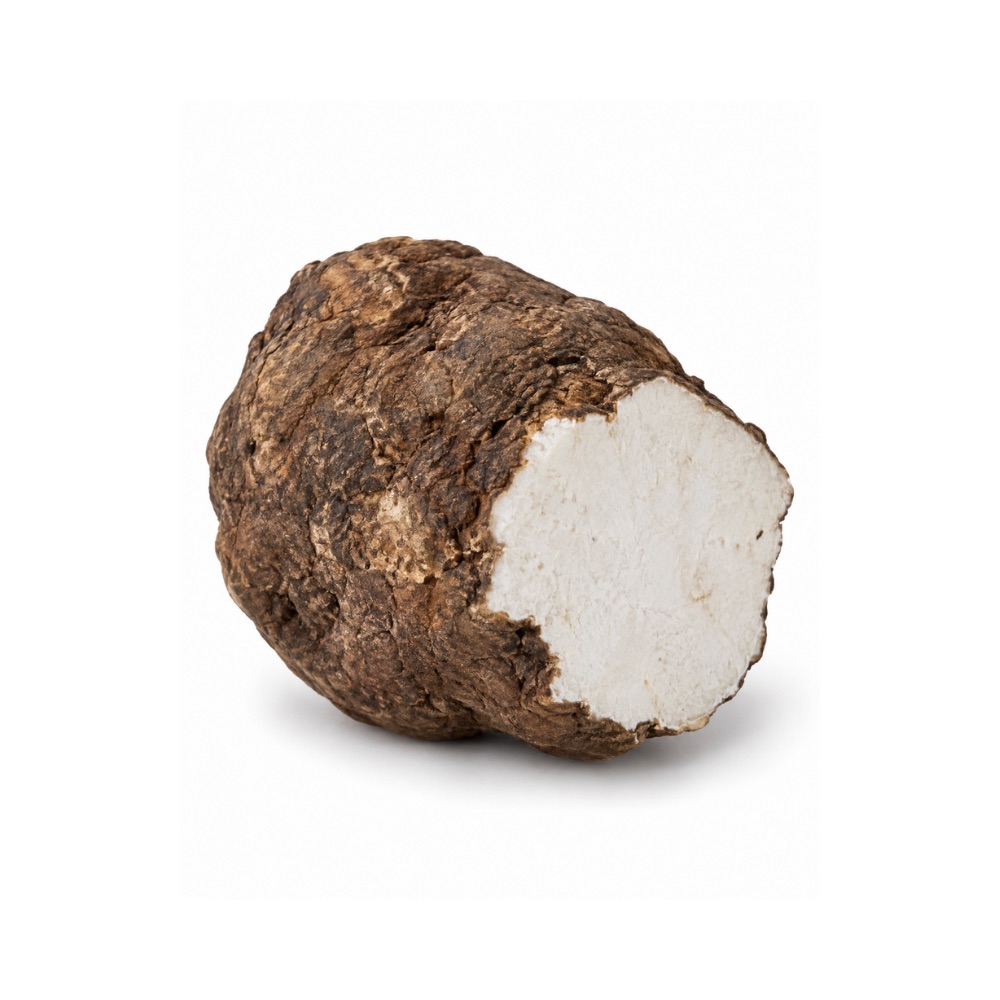 MildMildPoriaWolfiporia extensaPachymic acid drives caspase-9/-3 apoptosis and AKT downregulation in cell lines, and a poria polysaccharide is an approved adjuvant in China.Mechanismmitochondrial apoptosis (caspase-9/-3), AKT ↓Read the evidenceShow less
A large preclinical body sits alongside the Chinese-approved polysaccharide adjuvant 265,
Several pachymic-acid papers in this area have been retracted, and no Western randomised trial exists, so the citation base is less reliable than its volume suggests.
 MildMildSamambaiaPolypodium leucotomosCalagualine inhibits NF-κB, and oral extract blocked UVB-induced skin tumours in animals by reducing COX-2 and activating p53; the evidence is skin chemoprevention.MechanismNF-κB inhibition; p53 activation; anti-photocarcinogenesisRead the evidenceShow lessMonograph →
Calagualine inhibits NF-κB across multiple cell lines in vitro 235Reference 235In vitroCalagualine inhibits nuclear transcription factor-κB activated by various inflammatory and tumour-promoting agents — in vitroView study →, and in animal models oral extract blocked UVB-induced skin-tumour formation, reduced COX-2 and activated p53 236Reference 236AnimalPolypodium leucotomos extract decreases UV-induced Cox-2 expression and inflammation, enhances DNA repair in Xpc+/− mice — animal modelView study →. These are the same pathways behind its anti-inflammatory and photoprotective actions.
There are no human oncology trials and no clinical outcome data. The evidence is cell-line and rodent skin only, and it describes prevention of photocarcinogenesis rather than treatment of established disease.
 MildMildSarsaparillaSmilax spp.Steroidal saponins from commercial sarsaparilla are antiproliferative across six tumour lines, and Smilax china polyphenols and extracts inhibit NF-κB and trigger death-receptor apoptosis in ovarian and oral cancer cells.Mechanismcaspase/DR5 apoptosis induction; ERK inhibition; NF-κB ↓; antiestrogenic receptor bindingRead the evidenceShow lessMonograph →
A new furostanol saponin and other steroidal saponins from commercial S. ornata sarsaparilla were antiproliferative across six tumour lines and most active against HT29 colon cancer 237Reference 237In vitroSteroidal saponins from the roots of Smilax sp.: structure and bioactivity — in vitroView study →; polyphenols from S. china were cytotoxic to breast tumour cells partly as antiestrogenic phytoestrogens 238Reference 238In vitroCytotoxic polyphenols against breast tumor cell in Smilax china L. — in vitroView study →; S. china rhizome extract inhibited NF-κB and induced apoptosis in ovarian cancer cells 239Reference 239In vitroSmilax china L. rhizome extract inhibits nuclear factor-κB and induces apoptosis in ovarian cancer cells — in vitroView study →; and a methanol extract triggered ERK-dependent, death-receptor-5-mediated apoptosis in oral carcinoma cells 240Reference 240In vitroExtracellular signal-regulated kinase inhibition is required for methanol extract of Smilax china L. induced apoptosis through death receptor 5 in human oral mucoepidermoid carcinoma cells — in vitroView study →.
All of it is in-vitro cytotoxicity. There is no animal tumour model, no pharmacokinetics showing these concentrations are reachable in tissue, and no human data.
The traditional use for cancer has not been tested.
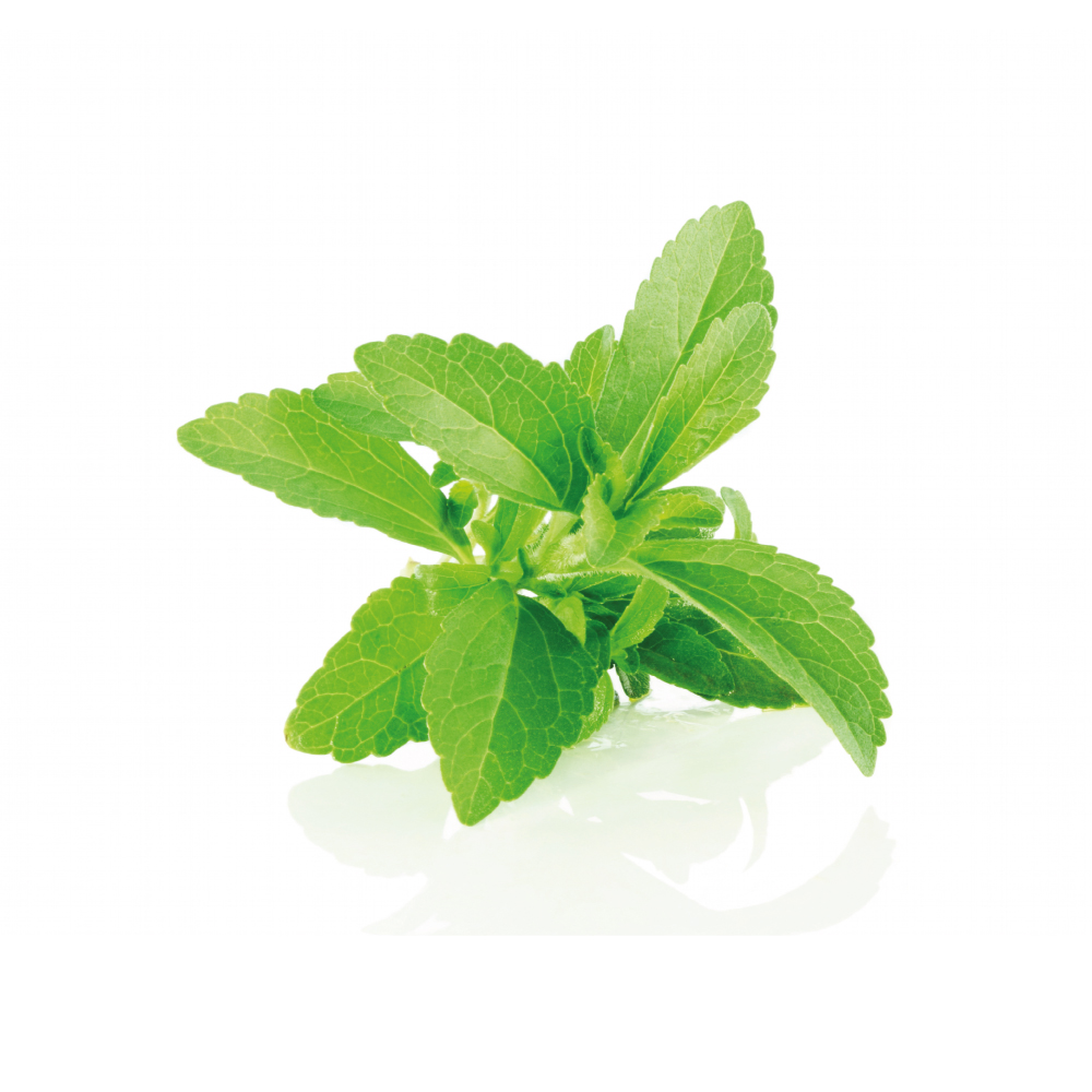 MildMildSteviaStevia rebaudianaSteviol and isosteviol inhibit proliferation and induce apoptosis across several tumour cell lines, with scattered rodent data and no established pathway.Mechanismantiproliferative / apoptosis induction in tumor cell lines (pathway not established)Read the evidenceShow lessMonograph →
Reviews collate cell-line evidence that stevia derivatives, steviol and isosteviol most often, inhibit proliferation and induce apoptosis across several tumour types, with scattered supporting rodent data 246Reference 246In vitroAnti-cancer properties of Stevia rebaudiana; more than a sweetener — review (in vitro and in vivo)View study →.
No target pathway is established and there are no human studies of any kind. The reviewers’ own conclusion is that extrapolating cell-line cytotoxicity to clinical use “is not currently justified”.
 MildMildTeaCamellia sinensisIts catechins inhibit cancer-cell proliferation in the laboratory, and high green-tea intake is associated with lower liver-cancer risk in Asian cohorts; the human evidence is suggestive and inconsistent.Mechanismcatechin antiproliferative; antioxidantRead the evidenceShow lessMonograph →
An umbrella meta-analysis of 113 studies across 26 cancer sites found site-varying, mostly low-certainty associations 255Reference 255Meta-analysisTea drinking and risk of cancer incidence: a meta-analysis of prospective cohort studies and evidence evaluationView study →, and Asian cohorts linked the highest green-tea intake to lower liver-cancer risk (RR about 0.88, not seen at one cup a day) 256Reference 256Meta-analysisGreen tea and liver cancer risk: a meta-analysis of prospective cohort studies in Asian populationsView study →.
Cochrane concluded that the epidemiological evidence is insufficient and inconsistent and that trial evidence does not establish prevention 257Reference 257Systematic reviewGreen tea (Camellia sinensis) for the prevention of cancer — systematic review (Cochrane)View study →. Cohort findings are confounded by smoking, diet and hot-beverage temperature.
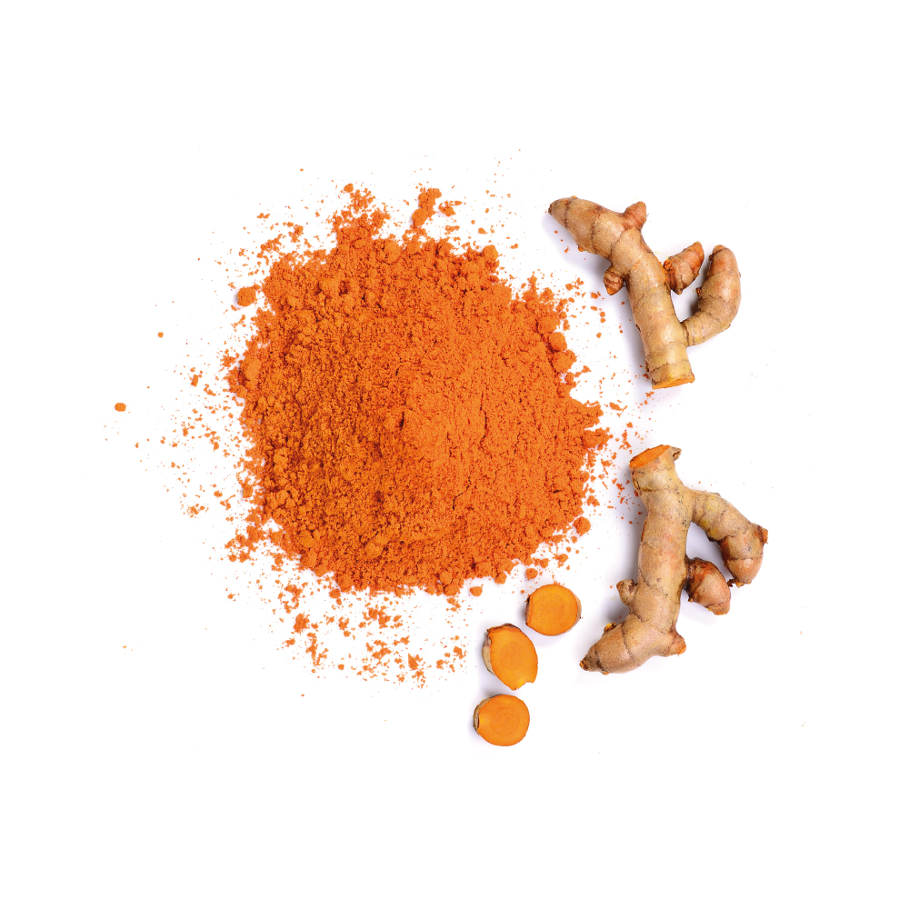 MildMildTurmericCurcuma longaCurcumin is antiproliferative and pro-apoptotic against leukaemia and prostate-cancer cells and modulates many carcinogenic pathways; the only human data are early-phase tolerability.Mechanismpro-apoptotic; antiproliferative; NF-κB modulationRead the evidenceShow lessMonograph →
Preclinical work is extensive. In vitro, curcumin is antiproliferative and pro-apoptotic against leukaemia and prostate-cancer cell lines and modulates numerous carcinogenic pathways 263Reference 263In vitroColorectal cancer: chemopreventive role of curcumin and resveratrol — review of mechanisms (in vitro and animal model evidence)View study →.
The only human data are early-phase: a Theracurmin plus gemcitabine dose-escalation study established tolerability and measurable plasma curcumin, not efficacy 264Reference 264A phase I study investigating the safety and pharmacokinetics of highly bioavailable curcumin (Theracurmin) in cancer patients on gemcitabine-based chemotherapyView study →.
There is no phase III or robust phase II efficacy trial, and curcumin’s low systemic bioavailability is a fundamental obstacle.
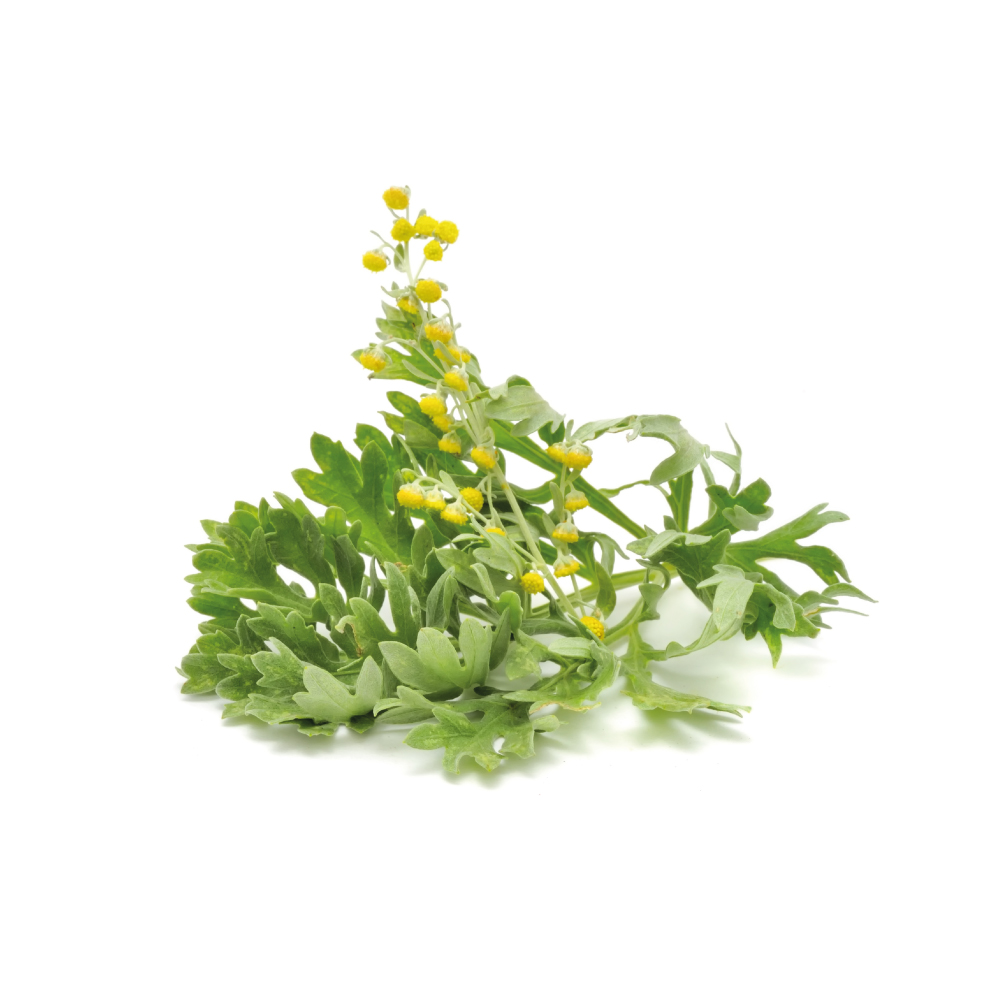 MildMildWormwoodArtemisia absinthiumExtract triggers apoptosis in breast cancer lines and thujone disrupts choriocarcinoma cell metabolism; the artemisinin literature belongs to sweet wormwood, a different species.Mechanismapoptosis induction (pathway not specified); metabolic disruption (thujone); multidrug-resistance transporter modulationRead the evidenceShow lessMonograph →
In human breast cancer lines the extract triggered apoptosis through multiple pathways 267Reference 267In vitroArtemisia absinthium (AA): a novel potential complementary and alternative medicine for breast cancer — in vitroView study →, isolated α,β-thujone suppressed placental choriocarcinoma cells through metabolic disruption 268Reference 268In vitroLee, J. Y., et al. (2020). α,β-Thujone suppresses human placental choriocarcinoma cells via metabolic disruption — in vitro. Reproduction. https://pubmed.ncbi.nlm.nih.gov/32240978/View study →, and a 2025 characterisation reported antiproliferative activity against ovarian tumour lines linked to apoptosis and multidrug-resistance modulation 269Reference 269In vitroFlowers and leaves of Artemisia absinthium and Artemisia annua: phytochemical characterization, anti-inflammatory, antioxidant and anti-proliferative activities — in vitro and rat modelView study →.
There is no named molecular target, no animal tumour model and no human data.
Two points of attribution. The 2025 paper is a comparative study of Artemisia absinthium and Artemisia annua together, and it does not state which plant produced the ovarian result; A. annua is the artemisinin plant, whose endoperoxide, iron-dependent anticancer literature belongs to sweet wormwood, and wormwood makes no artemisinin.
The thujone result comes from an isolated GABA-A-antagonist convulsant, so its cytotoxicity is the same general toxicity that caps the herb’s dose, read out in a dish.
No herbs match these filters.
Safety
- No anticancer herb replaces oncology treatment, and delaying conventional care in favour of a botanical worsens survival.
- Many chemotherapy and targeted drugs are CYP3A4 substrates with narrow therapeutic windows, and CYP-active herbs can raise or lower their levels.
- Whether high-dose antioxidants blunt radiotherapy and oxidative chemotherapy is unresolved, which is a reason for caution during those regimens.
- Immune-stimulating polysaccharides are inappropriate alongside immunosuppressive protocols and after transplant, and unstudied with checkpoint immunotherapy.
- A herb that damages DNA in tumour cells damages DNA generally, so the genotoxic herbs are not for long-term use.
- Anticoagulant and antiplatelet herbs matter more against chemotherapy-induced thrombocytopenia.
- Herbs with estrogenic or antiestrogenic activity are unstudied alongside hormonal therapy for hormone-sensitive cancers.
- Chemoprevention in healthy tissue and treatment of established disease are different claims with different evidence.
References
- Zhang X, Chen J, Xi B, Liu Y, Wang S, Gu L, Zhao H, Tao L, Hua Y, Wang Y, Chen M (2023). Agrimoniin is a dual inhibitor of AKT and ERK pathways that inhibit pancreatic cancer cell proliferation — in vitro and in vivo. Phytotherapy Research. https://pubmed.ncbi.nlm.nih.gov/37156642/
- Zhu X, Zhang Y, Wang Y, Zhang H, Wang X, Tang H, Huang H, Zhou Z, Chen B, Sun L (2022). Agrimoniin sensitizes pancreatic cancer to apoptosis through ROS-mediated energy metabolism dysfunction — in vitro and nude mouse study. Phytomedicine. https://pubmed.ncbi.nlm.nih.gov/34785107/
- Fedotcheva TA, Sheichenko OP, Fedotcheva NI (2021). New properties and mitochondrial targets of polyphenol agrimoniin as a natural anticancer and preventive agent — in vitro. Pharmaceutics. https://pubmed.ncbi.nlm.nih.gov/34959369/
- Wang L, Chen Q, Song H, Xing W, Shi J, Li Y, Lv Y, Wang Z, Chen J, Zhao W (2024). The anti-colorectal cancer effect and metabolites of Agrimonia pilosa Ledeb. — AOM/DSS mouse model. Journal of Ethnopharmacology. https://pubmed.ncbi.nlm.nih.gov/38604512/
- Miyamoto K, Kishi N, Koshiura R (1987). Antitumor effect of agrimoniin, a tannin of Agrimonia pilosa Ledeb., on transplantable rodent tumors — mouse study. Japanese Journal of Pharmacology. https://pubmed.ncbi.nlm.nih.gov/3573425/
- Molnár J, Szebeni GJ, Csupor-Löffler B, Hajdú Z, Szekeres T, Saiko P, Ocsovszki I, Puskás LG, Hohmann J, Zupkó I (2016). Investigation of the antiproliferative properties of natural sesquiterpenes from Artemisia asiatica and Onopordum acanthium on HL-60 cells — in vitro. International Journal of Molecular Sciences 17(2):83. https://pubmed.ncbi.nlm.nih.gov/26901188/
- Hajdú Z, Hohmann J, Forgo P, Máthé I, Molnár J, Zupkó I (2014). Antiproliferative activity of Artemisia asiatica extract and its constituents on human tumor cell lines — in vitro. Planta Medica 80(18):1692–1697. https://pubmed.ncbi.nlm.nih.gov/25295671/
- Liu Q, Beyraghdar Kashkooli A, Manzano D, et al. (2018). Kauniolide synthase is a P450 with unusual hydroxylation and cyclization-elimination activity — in vitro enzyme characterisation. Nature Communications 9:4657. https://pubmed.ncbi.nlm.nih.gov/30405138/
- Kwok BH, Koh B, Ndubuisi MI, Elofsson M, Crews CM (2001). The anti-inflammatory natural product parthenolide from the medicinal herb feverfew directly binds to and inhibits IκB kinase — in vitro. Chemistry & Biology 8(8):759–766. https://pubmed.ncbi.nlm.nih.gov/11514225/
- Johnson JJ, Syed DN, Suh Y, et al. (2010). Disruption of androgen and estrogen receptor activity in prostate cancer by a novel dietary diterpene carnosol — implications for chemoprevention. Cancer Prevention Research, 3(9), 1112–1123. https://pubmed.ncbi.nlm.nih.gov/20736335/
- Johnson JJ, Syed DN, Heren CR, Suh Y, Adhami VM, Mukhtar H. (2008). Carnosol, a dietary diterpene, displays growth inhibitory effects in human prostate cancer PC3 cells leading to G2-phase cell cycle arrest and targets the 5’-AMP-activated protein kinase (AMPK) pathway. Pharmaceutical Research, 25(9), 2125–2134. https://pubmed.ncbi.nlm.nih.gov/18286356/
- Al Dhaheri Y, Attoub S, Ramadan G, et al. (2014). Carnosol induces ROS-mediated beclin1-independent autophagy and apoptosis in triple negative breast cancer. PLoS One, 9(10), e109630. https://pubmed.ncbi.nlm.nih.gov/25299698/
- Alsamri H, Alneyadi A, Muhammad K, Ayoub MA, Eid A, Iratni R. (2022). Carnosol induces p38-mediated ER stress response and autophagy in human breast cancer cells. Frontiers in Oncology, 12, 911615. https://pubmed.ncbi.nlm.nih.gov/35712465/
- Park KW, Kundu J, Chae IG, et al. (2014). Carnosol induces apoptosis through generation of ROS and inactivation of STAT3 signaling in human colon cancer HCT116 cells. International Journal of Oncology, 44(4), 1309–1315. https://pubmed.ncbi.nlm.nih.gov/24481553/
- Dörrie J, Sapala K, Zunino SJ. (2001). Carnosol-induced apoptosis and downregulation of Bcl-2 in B-lineage leukemia cells. Cancer Letters, 170(1), 33–39. https://pubmed.ncbi.nlm.nih.gov/11448532/
- Gassib N, Issa H, Loubaki L, et al. (2024). Cellular mechanisms mediating the anti-cancer effects of carnosol on gingiva carcinoma. Scientific Reports, 14(1), 12266. https://pubmed.ncbi.nlm.nih.gov/38806527/
- López-Jiménez A, García-Caballero M, Medina MÁ, Quesada AR. (2013). Anti-angiogenic properties of carnosol and carnosic acid, two major dietary compounds from rosemary. European Journal of Nutrition, 52(1), 85–95. https://pubmed.ncbi.nlm.nih.gov/22173778/
- Vemu B, Tocmo R, Nauman MC, Flowers SA, Veenstra JP, Johnson JJ. (2021). Pharmacokinetic characterization of carnosol from rosemary (Salvia rosmarinus) in male C57BL/6 mice and inhibition profile in human cytochrome P450 enzymes. Toxicology and Applied Pharmacology, 431, 115729. https://pubmed.ncbi.nlm.nih.gov/34592323/
- Zunino SJ, Storms DH. (2009). Carnosol delays chemotherapy-induced DNA fragmentation and morphological changes associated with apoptosis in leukemic cells. Nutrition and Cancer, 61(1), 94–102. https://pubmed.ncbi.nlm.nih.gov/19116879/
- Kamada Y, et al. (2018). Castalagin and vescalagin purified from leaves of Syzygium samarangense (Blume) Merrill & L.M. Perry: dual inhibitory activity against PARP1 and DNA topoisomerase II. Fitoterapia. https://pubmed.ncbi.nlm.nih.gov/29928967/
- Kashiwada Y, Nonaka G, Nishioka I, Chang JJ, Lee KH (1992). Antitumor agents, 129. Tannins and related compounds as selective cytotoxic agents. Journal of Natural Products. https://pubmed.ncbi.nlm.nih.gov/1431932/
- Yang LL, et al. (2000). Induction of apoptosis by hydrolyzable tannins from Eugenia jambos L. on human leukemia cells. Cancer Letters. https://pubmed.ncbi.nlm.nih.gov/10893444/
- Messaoudene M, Pidgeon R, Richard C, et al. (2022). A natural polyphenol exerts antitumor activity and circumvents anti-PD-1 resistance through effects on the gut microbiota. Cancer Discovery. https://pubmed.ncbi.nlm.nih.gov/35031549/
- Alghamdi HA, Alghamdi SS, Al-Zahrani MH, et al. (2025). Reticuline and coclaurine exhibit vitamin D receptor-dependent anticancer and pro-apoptotic activities in the colorectal cancer cell line HCT116 — in vitro. Current Issues in Molecular Biology, 47(10), 810. https://pubmed.ncbi.nlm.nih.gov/41150758/
- Hu SY, Lin TH, Chen CY, et al. (2024). Stephania tetrandra and its active compound coclaurine sensitize NSCLC cells to cisplatin through EFHD2 inhibition — in vitro. Pharmaceuticals, 17(10), 1356. https://pubmed.ncbi.nlm.nih.gov/39458997/
- Lee MG, Lee KT, Chi SG, Park JH. (2001). Costunolide induces apoptosis by ROS-mediated mitochondrial permeability transition and cytochrome C release — in vitro. Biological & Pharmaceutical Bulletin. https://pubmed.ncbi.nlm.nih.gov/11256490/
- Choi JH, Lee KT. (2009). Costunolide-induced apoptosis in human leukemia cells — involvement of c-jun N-terminal kinase activation — in vitro. Biological & Pharmaceutical Bulletin. https://pubmed.ncbi.nlm.nih.gov/19801848/
- Kanno S, Kitajima Y, Kakuta M, Osanai Y, Kurauchi K, Ujibe M, Ishikawa M. (2008). Costunolide-induced apoptosis is caused by receptor-mediated pathway and inhibition of telomerase activity in NALM-6 cells — in vitro. Biological & Pharmaceutical Bulletin. https://pubmed.ncbi.nlm.nih.gov/18451540/
- Choi SH, Im E, Kang HK, Lee JH, Kwak HS, Bae YT, Park HJ, Kim ND. (2005). Inhibitory effects of costunolide on the telomerase activity in human breast carcinoma cells — in vitro. Cancer Letters. https://pubmed.ncbi.nlm.nih.gov/16112418/
- Ahmad F, Dixit D, Sharma V, Kumar A, Joshi SD, Sarkar C, Sen E. (2016). Nrf2-driven TERT regulates pentose phosphate pathway in glioblastoma — in vitro and mouse xenograft. Cell Death & Disease. https://pubmed.ncbi.nlm.nih.gov/27148686/
- Jeong SJ, Itokawa T, Shibuya M, Kuwano M, Ono M, Higuchi R, Miyamoto T. (2002). Costunolide, a sesquiterpene lactone from Saussurea lappa, inhibits the VEGFR KDR/Flk-1 signaling pathway — in vitro and mouse corneal micropocket assay. Cancer Letters. https://pubmed.ncbi.nlm.nih.gov/12359360/
- Whipple RA, Vitolo MI, Boggs AE, Charpentier MS, Thompson K, Martin SS. (2013). Parthenolide and costunolide reduce microtentacles and tumor cell attachment by selectively targeting detyrosinated tubulin independent from NF-κB inhibition — in vitro. Breast Cancer Research. https://pubmed.ncbi.nlm.nih.gov/24028602/
- Choi YK, Cho SG, Woo SM, Yun YJ, Jo J, Kim W, Shin YC, Ko SG. (2013). Saussurea lappa Clarke-derived costunolide prevents TNFα-induced breast cancer cell migration and invasion by inhibiting NF-κB activity — in vitro and orthotopic mouse model. Evidence-Based Complementary and Alternative Medicine. https://pubmed.ncbi.nlm.nih.gov/23997800/
- Ohnishi M, Yoshimi N, Kawamori T, Ino N, Hirose Y, Tanaka T, Yamahara J, Miyata H, Mori H. (1997). Inhibitory effects of dietary protocatechuic acid and costunolide on 7,12-dimethylbenz[a]anthracene-induced hamster cheek pouch carcinogenesis — animal model. Japanese Journal of Cancer Research. https://pubmed.ncbi.nlm.nih.gov/9119738/
- Merfort I. (2011). Perspectives on sesquiterpene lactones in inflammation and cancer — review. Current Drug Targets. https://pubmed.ncbi.nlm.nih.gov/21561425/
- Zhang J, Hu X, Gao W, Qu Z, Guo H, Liu Z, Liu C. (2014). Pharmacokinetic study on costunolide and dehydrocostuslactone after oral administration of traditional medicine Aucklandia lappa Decne. by LC/MS/MS — animal model in rats. Journal of Ethnopharmacology. https://pubmed.ncbi.nlm.nih.gov/24216164/
- Shin DS, Kim HN, Shin KD, et al. (2009). Cryptotanshinone inhibits constitutive signal transducer and activator of transcription 3 function through blocking the dimerization in DU145 prostate cancer cells — in vitro study. Cancer Research. https://pubmed.ncbi.nlm.nih.gov/19118003/
- Li H, Gao C, Liu C, et al. (2021). A review of the biological activity and pharmacology of cryptotanshinone, an important active constituent in Danshen — review. Biomedicine & Pharmacotherapy. https://pubmed.ncbi.nlm.nih.gov/33548911/
- Wu YH, Wu YR, Li B, Yan ZY (2020). Cryptotanshinone: a review of its pharmacology activities and molecular mechanisms — review. Fitoterapia. https://pubmed.ncbi.nlm.nih.gov/32445662/
- Lu L, Zhang S, Li C, et al. (2017). Cryptotanshinone inhibits human glioma cell proliferation in vitro and in vivo through SHP-2-dependent inhibition of STAT3 activation — in vitro and mouse xenograft study. Cell Death & Disease. https://pubmed.ncbi.nlm.nih.gov/28492557/
- Wang H, Liu Z, Guan L, et al. (2020). LYW-6, a novel cryptotanshinone derived STAT3 targeting inhibitor, suppresses colorectal cancer growth and metastasis — medicinal chemistry and in vivo study. Pharmacological Research. https://pubmed.ncbi.nlm.nih.gov/31982491/
- Chen W, Luo Y, Liu L, et al. (2010). Cryptotanshinone inhibits cancer cell proliferation by suppressing mammalian target of rapamycin-mediated cyclin D1 expression and Rb phosphorylation — in vitro study. Cancer Prevention Research. https://pubmed.ncbi.nlm.nih.gov/20628002/
- Jeoung NH, Jeong JY, Kang BS (2021). Cryptotanshinone prevents the binding of S6K1 to mTOR/Raptor leading to the suppression of mTORC1-S6K1 signaling activity and neoplastic cell transformation — in vitro study. Journal of Cancer Prevention. https://pubmed.ncbi.nlm.nih.gov/34258253/
- Chen W, Pan Y, et al. (2017). Cryptotanshinone activates AMPK-TSC2 axis leading to inhibition of mTORC1 signaling in cancer cells — in vitro study. BMC Cancer. https://pubmed.ncbi.nlm.nih.gov/28061838/
- Pan Y, Shi J, Ni W, et al. (2017). Cryptotanshinone inhibition of mammalian target of rapamycin pathway is dependent on oestrogen receptor alpha in breast cancer — in vitro study. Journal of Cellular and Molecular Medicine. https://pubmed.ncbi.nlm.nih.gov/28272775/
- Hou Y, Zhong B, et al. (2025). A small molecule cryptotanshinone induces non-enzymatic NQO1-dependent necrosis in cancer cells through the JNK1/2/iron/PARP/calcium pathway — in vitro and in vivo study. Acta Pharmaceutica Sinica B. https://pubmed.ncbi.nlm.nih.gov/40177544/
- Lee DS, Lee SH, Noh JG, Hong SD (1999). Antibacterial activities of cryptotanshinone and dihydrotanshinone I from a medicinal herb, Salvia miltiorrhiza Bunge — in vitro study. Bioscience, Biotechnology, and Biochemistry. https://pubmed.ncbi.nlm.nih.gov/10664860/
- Bissinger R, Lupescu A, Zelenak C, Jilani K, Lang F (2014). Stimulation of eryptosis by cryptotanshinone — human erythrocyte in vitro study. Cellular Physiology and Biochemistry. https://pubmed.ncbi.nlm.nih.gov/25095724/
- Uche FI, Drijfhout FP, McCullagh J, Richardson A, Li WW (2016). Cytotoxicity effects and apoptosis induction by bisbenzylisoquinoline alkaloids from Triclisia subcordata. Phytotherapy Research. https://pubmed.ncbi.nlm.nih.gov/27270992/
- Uche FI, McCullagh J, Claridge TWD, Richardson A, Li WW (2018). Synthesis of (aminoalkyl)cycleanine analogues: cytotoxicity, cellular uptake, and apoptosis induction in ovarian cancer cells. Bioorganic & Medicinal Chemistry Letters. https://pubmed.ncbi.nlm.nih.gov/29588214/
- Nwaefulu ON, Al-Shar’i NA, Owolabi JO, Sagineedu SR, Woei LC, Wai LK, Islam MK, Jayanthi S, Stanslas J (2022). The impact of cycleanine in cancer research: a computational study. Journal of Molecular Modeling. https://pubmed.ncbi.nlm.nih.gov/36194315/
- Tian H, Pan OC (1997). Modulation of multidrug resistance by three bisbenzyl-isoquinolines in comparison with verapamil. Zhongguo Yao Li Xue Bao. https://pubmed.ncbi.nlm.nih.gov/10322941/
- D’Agostino VG, Lal P, Mantelli B, et al. (2015). Dihydrotanshinone-I interferes with the RNA-binding activity of HuR affecting its post-transcriptional function — in vitro and cell study. Scientific Reports. https://pubmed.ncbi.nlm.nih.gov/26553968/
- Lal P, Cerofolini L, D’Agostino VG, et al. (2017). Regulation of HuR structure and function by dihydrotanshinone-I — NMR, molecular dynamics, RIP-chip and xenograft study. Nucleic Acids Research. https://pubmed.ncbi.nlm.nih.gov/28934484/
- Manzoni L, Zucal C, Di Maio D, et al. (2018). Interfering with HuR-RNA interaction: design, synthesis and biological characterization of tanshinone mimics as novel, effective HuR inhibitors — medicinal chemistry study. Journal of Medicinal Chemistry. https://pubmed.ncbi.nlm.nih.gov/29313684/
- Dev AJR, Malhotra L, Kashyap A, et al. (2025). Biophysical and molecular characterization of HuR inhibitors, CMLD-2 and dihydrotanshinone I (DHTS), in triple negative breast cancer — in vitro study. International Journal of Biological Macromolecules. https://pubmed.ncbi.nlm.nih.gov/40639505/
- Duan P, Huang Y, Chen K, Cheng C, Wu Z, Wu Y (2021). 15,16-dihydrotanshinone I inhibits EOMA cells proliferation by interfering in posttranscriptional processing of hypoxia-inducible factor 1 — in vitro study. International Journal of Medical Sciences. https://pubmed.ncbi.nlm.nih.gov/34400891/
- Nie Y, Yan J, Huang X, Jiang T, Zhang S, Zhang G (2024). Dihydrotanshinone I targets ESR1 to induce DNA double-strand breaks and proliferation inhibition in hepatocellular carcinoma — in vitro and animal study. Phytomedicine. https://pubmed.ncbi.nlm.nih.gov/38833789/
- Zhao H, Liang Y, Sun C, et al. (2022). Dihydrotanshinone I inhibits the lung metastasis of breast cancer by suppressing neutrophil extracellular traps formation — mouse study. International Journal of Molecular Sciences. https://pubmed.ncbi.nlm.nih.gov/36499502/
- Wang L, Hu T, Shen J, et al. (2015). Dihydrotanshinone I induced apoptosis and autophagy through caspase dependent pathway in colon cancer — in vitro study. Phytomedicine. https://pubmed.ncbi.nlm.nih.gov/26547530/
- Cao Y, Huang B, Gao C (2017). Salvia miltiorrhiza extract dihydrotanshinone induces apoptosis and inhibits proliferation of glioma cells — in vitro study. Bosnian Journal of Basic Medical Sciences. https://pubmed.ncbi.nlm.nih.gov/28485251/
- Cheng R, Chen J, Wang Y, Ge Y, Huang Z, Zhang G (2016). Dihydrotanshinone induces apoptosis of SGC7901 and MGC803 cells via activation of JNK and p38 signalling pathways — in vitro study. Pharmaceutical Biology. https://pubmed.ncbi.nlm.nih.gov/27431278/
- Allegri L, Domenis R, Navarra M, et al. (2021). Dihydrotanshinone exerts antitumor effects and improves the effects of cisplatin in anaplastic thyroid cancer cells — in vitro study. Oncology Reports. https://pubmed.ncbi.nlm.nih.gov/34318905/
- Kumar V, Radin D, Leonardi D (2019). Studies examining the synergy between dihydrotanshinone and temozolomide against MGMT+ glioblastoma cells in vitro — in vitro study. Biomedicine & Pharmacotherapy. https://pubmed.ncbi.nlm.nih.gov/30399573/
- Irtegun Kandemir S, Fidan HS, Yener I, Mete N, Ertas A, Topcu G, Kolak U (2022). Investigation of cytotoxic and apoptotic effects of 63 compounds obtained from Salvia species — in vitro screen including a healthy cell line. Journal of Food Biochemistry. https://pubmed.ncbi.nlm.nih.gov/35608363/
- Aumsuwan P, Khan SI, Khan IA, Ali Z, Avula B, Walker LA, Shariat-Madar Z, Helferich WG, Katzenellenbogen BS, Dasmahapatra AK (2016). The anticancer potential of steroidal saponin, dioscin, isolated from wild yam (Dioscorea villosa) root extract in invasive human breast cancer cell line MDA-MB-231 in vitro. Archives of Biochemistry and Biophysics. https://pubmed.ncbi.nlm.nih.gov/26682631/
- Zhang YS, Ma YL, Thakur K, Hussain SS, Wang J, Zhang Q, Zhang JG, Wei ZJ (2018). Molecular mechanism and inhibitory targets of dioscin in HepG2 cells. Food and Chemical Toxicology. https://pubmed.ncbi.nlm.nih.gov/29990575/
- Li W, Qiu Z, Wang Y, Zhang Y, Li M, Yu J, Zhang L, Zhu Z, Yu B (2007). Synthesis, cytotoxicity, and hemolytic activity of 6′-O-substituted dioscin derivatives. Carbohydrate Research. https://pubmed.ncbi.nlm.nih.gov/17945208/
- Mazzio E, Almalki A, Darling-Reed SF, Soliman KFA (2021). Effects of wild yam root (Dioscorea villosa) extract on the gene expression profile of triple-negative breast cancer cells. Cancer Genomics & Proteomics. https://pubmed.ncbi.nlm.nih.gov/34697066/
- Tao X, Xu L, Yin L, Han X, Qi Y, Xu Y, Song S, Zhao Y, Peng J (2017). Dioscin induces prostate cancer cell apoptosis through activation of estrogen receptor-β. Cell Death & Disease. RETRACTED — retraction notice Cell Death & Disease 2021;12:800, https://doi.org/10.1038/s41419-021-04083-y. https://pubmed.ncbi.nlm.nih.gov/28796245/
- Darias V, Bravo L, Rabanal R, Sánchez-Mateo CC, Martín-Herrera DA. (1990). Cytostatic and antibacterial activity of some compounds isolated from several Lamiaceae species from the Canary Islands — in vitro. Planta Medica, 56(1), 70–72. https://pubmed.ncbi.nlm.nih.gov/17221371/
- Min HY, Jang HJ, Park KH, Hyun SY, Park SJ, Kim JH, Son J, Kang SS, Lee HY (2019). The natural compound gracillin exerts potent antitumor activity by targeting mitochondrial complex II. Cell Death & Disease. https://pubmed.ncbi.nlm.nih.gov/31649278/
- Min HY, Pei H, Hyun SY, Boo HJ, Jang HJ, Cho J, Kim JH, Son J, Lee HY (2020). Potent anticancer effect of the natural steroidal saponin gracillin is produced by inhibiting glycolysis and oxidative phosphorylation-mediated bioenergetics. Cancers (Basel). https://pubmed.ncbi.nlm.nih.gov/32276500/
- Su JX, Zhou HX, Zhang ZJ, et al. (2025). Gracillin suppresses cancer progression through inducing Merlin/LATS protein–protein interaction and activating Hippo signaling pathway. Acta Pharmacologica Sinica. https://pubmed.ncbi.nlm.nih.gov/40055528/
- Liao J, Liang Q, Wu X, Li D, Liu Y, Han S, Wang R, Guo P, Yi M, Li W (2026). Gracillin overcomes osimertinib resistance in NSCLC via dual inhibition of EGFR and Mcl-1. Cancer Gene Therapy. https://pubmed.ncbi.nlm.nih.gov/41857145/
- Lee W, Song G, Bae H (2025). Gracillin induces mitochondria-mediated apoptosis on pancreatic ductal adenocarcinoma through disruption of redox homeostasis via inhibiting NRF2/HO-1 antioxidant axis. Bioorganic Chemistry. https://pubmed.ncbi.nlm.nih.gov/40505322/
- Li JK, Zhu PL, Wang Y, Jiang XL, Zhang Z, Zhang Z, Yung KK (2023). Gracillin exerts anti-melanoma effects in vitro and in vivo: role of DNA damage, apoptosis and autophagy. Phytomedicine. https://pubmed.ncbi.nlm.nih.gov/36334389/
- Yang L, Zhu T, Ye H, et al. (2021). Gracillin shows potent efficacy against colorectal cancer through inhibiting the STAT3 pathway. Journal of Cellular and Molecular Medicine. https://pubmed.ncbi.nlm.nih.gov/33259114/
- Li Y, Liu H, Liu X, Xiao B, Zhang M, Luo Y, Li M, Yang J (2022). Gracillin shows potential efficacy against non-small cell lung cancer through inhibiting the mTOR pathway. Frontiers in Oncology. https://pubmed.ncbi.nlm.nih.gov/35392243/
- Huang S, Lin W, Ding X, Shi J, Liu F, Wang G, Gao C, Su X (2026). CCL24 recruits CCR3+ TAMs to promote immunosuppression via YAP1 activation and serves as a therapeutic target for gracillin in colorectal cancer. International Journal of Biological Sciences. https://pubmed.ncbi.nlm.nih.gov/41694595/
- Cui A, Tian Y, Chen H, et al. (2026). Study on the anti-lung cancer effects of gracillin from Reineckia carnea by targeting STC1. BMC Cancer. https://pubmed.ncbi.nlm.nih.gov/42135683/
- Liu W, Wang Y, Chen J, Lin Z, Lin M, Lin X, Fan Y (2021). Beneficial effects of gracillin from Rhizoma Paridis against gastric carcinoma via the potential TIPE2-mediated induction of endogenous apoptosis and inhibition of migration in BGC823 cells. Frontiers in Pharmacology. https://pubmed.ncbi.nlm.nih.gov/34630074/
- Hu K, Yao X (2003). The cytotoxicity of methyl protoneogracillin (NSC-698793) and gracillin (NSC-698787), two steroidal saponins from the rhizomes of Dioscorea collettii var. hypoglauca, against human cancer cells in vitro. Phytotherapy Research. https://pubmed.ncbi.nlm.nih.gov/12820229/
- Hoang LTA, Pham VC, Ngo VD, et al. (2025). Diosgenin glycosides from Paris vietnamensis (Takht) H.Li with cytotoxic and inhibitory NO production activities. Natural Product Research. https://pubmed.ncbi.nlm.nih.gov/41021975/
- Lim CB, Fu PY, Ky N, et al. (2012). NF-κB p65 repression by the sesquiterpene lactone, helenalin, contributes to the induction of autophagy cell death — in vitro. BMC Complementary and Alternative Medicine. https://pubmed.ncbi.nlm.nih.gov/22784363/
- Dirsch VM, Stuppner H, Vollmar AM. (2001). Helenalin triggers a CD95 death receptor-independent apoptosis that is not affected by overexpression of Bcl-x(L) or Bcl-2 — in vitro. Cancer Research. https://pubmed.ncbi.nlm.nih.gov/11479221/
- Woerdenbag HJ, Merfort I, Passreiter CM, et al. (1994). Cytotoxicity of flavonoids and sesquiterpene lactones from Arnica species against the GLC4 and the COLO 320 cell lines — in vitro. Planta Medica. https://pubmed.ncbi.nlm.nih.gov/7997472/
- Grippo AA, Hall IH, Kiyokawa H, Muraoka O, Shen YC, Lee KH. (1992). The cytotoxicity of helenalin, its mono and difunctional esters, and related sesquiterpene lactones in murine and human tumor cells — in vitro. Drug Design and Discovery. https://pubmed.ncbi.nlm.nih.gov/1525302/
- Dirsch VM, Stuppner H, Vollmar AM. (2001). Cytotoxic sesquiterpene lactones mediate their death-inducing effect in leukemia T cells by triggering apoptosis — in vitro. Planta Medica. https://pubmed.ncbi.nlm.nih.gov/11509981/
- Witzel DA, Ivie GW, Dollahite JW. (1976). Mammalian toxicity of helenalin, the toxic principle of Helenium microcephalum DC (smallhead sneezeweed) — animal model, five species. American Journal of Veterinary Research. https://pubmed.ncbi.nlm.nih.gov/937811/
- Guzman ML, Rossi RM, Karnischky L, et al. (2005). The sesquiterpene lactone parthenolide induces apoptosis of human acute myelogenous leukemia stem and progenitor cells — in vitro / animal model. Blood. https://pubmed.ncbi.nlm.nih.gov/15687234/
- Ghantous A, Sinjab A, Herceg Z, Darwiche N. (2013). Parthenolide: from plant shoots to cancer roots — review. Drug Discovery Today. https://pubmed.ncbi.nlm.nih.gov/23688583/
- Guzman ML, Rossi RM, Neelakantan S, et al. (2007). An orally bioavailable parthenolide analog selectively eradicates acute myelogenous leukemia stem and progenitor cells — in vitro / animal model. Blood. https://pubmed.ncbi.nlm.nih.gov/17804695/
- Neelakantan S, Nasim S, Guzman ML, Jordan CT, Crooks PA. (2009). Aminoparthenolides as novel anti-leukemic agents: discovery of the NF-κB inhibitor, DMAPT (LC-1) — in vitro. Bioorganic & Medicinal Chemistry Letters. https://pubmed.ncbi.nlm.nih.gov/19505822/
- Hewamana S, Lin TT, Jenkins C, et al. (2008). The novel nuclear factor-κB inhibitor LC-1 is equipotent in poor prognostic subsets of chronic lymphocytic leukemia and shows strong synergy with fludarabine — in vitro. Clinical Cancer Research. https://pubmed.ncbi.nlm.nih.gov/19088025/
- Tai CT, Hsieh YH, Lee HL, et al. (2025). Targeting LC3B/MCL-1 expression by protodioscin induces autophagy and apoptosis in human glioblastoma cells in vitro and in vivo — [animal model]. International Journal of Medical Sciences. https://pubmed.ncbi.nlm.nih.gov/41049425/
- Bouchmaa N, Ben Mrid R, Bouargalne Y, Ajouaoi S, Cacciola F, El Fatimy R, Nhiri M, Zyad A (2023). In vitro evaluation of dioscin and protodioscin against ER-positive and triple-negative breast cancer — [in vitro]. PLoS One. https://pubmed.ncbi.nlm.nih.gov/36757991/
- Hibasami H, Moteki H, Ishikawa K, et al. (2003). Protodioscin isolated from fenugreek (Trigonella foenumgraecum L.) induces cell death and morphological change indicative of apoptosis in leukemic cell line H-60, but not in gastric cancer cell line KATO III — [in vitro]. International Journal of Molecular Medicine. https://pubmed.ncbi.nlm.nih.gov/12469212/
- Hwang JT, Park KS, Ryuk JA, Kim HJ, Ko BS (2019). Development of an oriental medicine discrimination method through analysis of steroidal saponins in Dioscorea nipponica Makino and their anti-osteosarcoma effects. Molecules. https://pubmed.ncbi.nlm.nih.gov/31698850/
- Hu K, Dong A, Yao X, Kobayashi H, Iwasaki S (1997). Antineoplastic agents. II. Four furostanol glycosides from rhizomes of Dioscorea collettii var. hypoglauca. Planta Medica. https://pubmed.ncbi.nlm.nih.gov/17252340/
- Podolak I, Galanty A, Sobolewska D (2010). Saponins as cytotoxic agents: a review. Phytochemistry Reviews. https://pubmed.ncbi.nlm.nih.gov/20835386/
- Liao M, Dai C, Liu M, Chen J, Chen Z, Xie Z, Yao M (2016). Simultaneous determination of four furostanol glycosides in rat plasma by UPLC-MS/MS and its application to PK study after oral administration of Dioscorea nipponica extracts. Journal of Pharmaceutical and Biomedical Analysis. https://pubmed.ncbi.nlm.nih.gov/26433169/
- Tang YN, Pang YX, He XC, Zhang YZ, Zhang JY, Zhao ZZ, Yi T, Chen HB (2015). UPLC-QTOF-MS identification of metabolites in rat biosamples after oral administration of Dioscorea saponins: a comparative study. Journal of Ethnopharmacology. https://pubmed.ncbi.nlm.nih.gov/25698242/
- Sturgeon CM, Craig K, Brown C, Rundle NT, Andersen RJ, Roberge M (2005). Modulation of the G2 cell cycle checkpoint by sesquiterpene lactones psilostachyins A and C isolated from the common ragweed Ambrosia artemisiifolia — in vitro. Planta Medica. https://pubmed.ncbi.nlm.nih.gov/16254826/
- Kovács B, Szemerédi N, Kúsz N, Kiss T, Csupor-Löffler B, Tsai YC, Rácz B, Spengler G, Csupor D (2022). Antiproliferative and cytotoxic effects of sesquiterpene lactones isolated from Ambrosia artemisiifolia on human adenocarcinoma and normal cell lines — in vitro. Pharmaceutical Biology. https://pubmed.ncbi.nlm.nih.gov/35952383/
- Martino R, Beer MF, Elso O, Donadel O, Sülsen V, Anesini C (2015). Sesquiterpene lactones from Ambrosia spp. are active against a murine lymphoma cell line by inducing apoptosis and cell cycle arrest — in vitro. Toxicology in Vitro. https://pubmed.ncbi.nlm.nih.gov/26086122/
- Adaramoye O, Erguen B, Nitzsche B, Höpfner M, Jung K, Rabien A (2017). Punicalagin, a polyphenol from pomegranate fruit, induces growth inhibition and apoptosis in human PC-3 and LNCaP cells. Chemico-Biological Interactions, 274, 100–106. https://pubmed.ncbi.nlm.nih.gov/28709945/
- Chen LG, Huang WT, Lee LT, Wang CC (2009). Ellagitannins from Terminalia calamansanai induced apoptosis in HL-60 cells. Toxicology in Vitro, 23(4), 603–609. https://pubmed.ncbi.nlm.nih.gov/19490839/
- Kasimsetty SG, Bialonska D, Reddy MK, Thornton C, Willett KL, Ferreira D (2009). Effects of pomegranate chemical constituents/intestinal microbial metabolites on CYP1B1 in 22Rv1 prostate cancer cells. Journal of Agricultural and Food Chemistry, 57(22), 10636–10644. https://pubmed.ncbi.nlm.nih.gov/19919114/
- Cheng AC, Lee MF, Tsai ML, Lai CS, Lee JH, Ho CT, Pan MH. (2011). Rosmanol potently induces apoptosis through both the mitochondrial apoptotic pathway and death receptor pathway in human colon adenocarcinoma COLO 205 cells — in vitro. Food and Chemical Toxicology. https://pubmed.ncbi.nlm.nih.gov/21112365/
- Jiang D, Xu J, Liu S, Nasser MI, Wei W, Mao T, et al. (2021). Rosmanol induces breast cancer cells apoptosis by regulating PI3K/AKT and STAT3/JAK2 signaling pathways — in vitro. Oncology Letters. https://pubmed.ncbi.nlm.nih.gov/34267823/
- Acunha T, García-Cañas V, Valdés A, Cifuentes A, Simó C. (2018). Metabolomics study of early metabolic changes in hepatic HepaRG cells in response to rosemary diterpenes exposure — in vitro. Analytica Chimica Acta. https://pubmed.ncbi.nlm.nih.gov/30292288/
- Wang L, Gan C, Wang Z, Liu L, Gao M, Li Q, Yang C. (2017). Determination and pharmacokinetic study of three diterpenes in rat plasma by UHPLC-ESI-MS/MS after oral administration of Rosmarinus officinalis L. extract — rats. Molecules. https://pubmed.ncbi.nlm.nih.gov/28587218/
- Chang J, Xiaoran W, Lisha Z, Shiran Z, Hui Z. (2026). Protective effects of rosmanol on DEN-induced lung cancer via targeting Nrf2/NF-κB/PI3K/Akt and COX-2/CYP2E1/VEGF pathways — rats. Journal of Molecular Histology. https://pubmed.ncbi.nlm.nih.gov/41729379/
- Tao W, Jiang C, Velu P, Lv C, Niu Y. (2025). Rosmanol suppresses nasopharyngeal carcinoma cell proliferation and enhances apoptosis, the regulation of MAPK/NF-κB signaling pathway — in vitro. Biotechnology and Applied Biochemistry. https://pubmed.ncbi.nlm.nih.gov/40170441/
- Niu YB, Velu P, Khalid A, Zyoud SH, Kazi M, Li Y. (2025). Rosmanol triggers apoptosis against MNNG-induced gastric carcinogenesis in rats through attenuation of the PI3K/AKT/HMGB1 signaling pathway. Combinatorial Chemistry & High Throughput Screening. https://pubmed.ncbi.nlm.nih.gov/38910269/
- Bastow KF, Bori ID, Fukushima Y, Kashiwada Y, Tanaka T, Nonaka G, Nishioka I, Lee KH (1993). Inhibition of DNA topoisomerases by sanguiin H-6, a cytotoxic dimeric ellagitannin from Sanguisorba officinalis — in vitro. Planta Medica. https://pubmed.ncbi.nlm.nih.gov/8391144/
- Lee SJ, Lee HK (2005). Sanguiin H-6 blocks endothelial cell growth through inhibition of VEGF binding to VEGF receptor — in vitro. Archives of Pharmacal Research. https://pubmed.ncbi.nlm.nih.gov/16350854/
- Park EH, Park JY, Yoo HS, Yoo JE, Lee HL (2016). Assessment of the anti-metastatic properties of sanguiin H-6 in HUVECs and MDA-MB-231 human breast cancer cells — in vitro. Bioorganic & Medicinal Chemistry Letters. https://pubmed.ncbi.nlm.nih.gov/27237777/
- Park EJ, et al. (2017). Cytotoxic effect of sanguiin H-6 on MCF-7 and MDA-MB-231 human breast carcinoma cells — in vitro. Bioorganic & Medicinal Chemistry Letters. https://pubmed.ncbi.nlm.nih.gov/28835347/
- Lee D, et al. (2016). Inhibition of A2780 human ovarian carcinoma cell proliferation by a Rubus component, sanguiin H-6 — in vitro. Journal of Agricultural and Food Chemistry. https://pubmed.ncbi.nlm.nih.gov/26725849/
- Ko H, Jeon H, Lee D, Choi HK, Kang KS, Choi KC (2015). Sanguiin H6 suppresses TGF-β induction of the epithelial-mesenchymal transition and inhibits migration and invasion in A549 lung cancer — in vitro. Bioorganic & Medicinal Chemistry Letters. https://pubmed.ncbi.nlm.nih.gov/26508552/
- González-Barrio R, Borges G, Mullen W, Crozier A (2010). Bioavailability of anthocyanins and ellagitannins following consumption of raspberries by healthy human volunteers and subjects with an ileostomy. Journal of Agricultural and Food Chemistry. https://pubmed.ncbi.nlm.nih.gov/20218618/
- Chatterjee A, Roy T, Kumar Mishra V, Swarnakar S (2024). Shatavarin-IV, a steroidal saponin from Asparagus racemosus, inhibits cell cycle progression and epithelial-to-mesenchymal transition in AGS cells under hyperglycemic conditions. Steroids (in vitro). https://pubmed.ncbi.nlm.nih.gov/39106908/
- Mitra SK, Prakash NS, Sundaram R (2012). Shatavarins (containing Shatavarin IV) with anticancer activity from the roots of Asparagus racemosus. Indian Journal of Pharmacology (in vitro and animal model). https://pubmed.ncbi.nlm.nih.gov/23248403/
- Gong Y, Li Y, Abdolmaleky HM, Li L, Zhou JR (2012). Tanshinones inhibit the growth of breast cancer cells through epigenetic modification of Aurora A expression and function. PLoS One. https://pubmed.ncbi.nlm.nih.gov/22485147/
- Li Y, Gong Y, Li L, Abdolmaleky HM, Zhou JR (2013). Bioactive tanshinone I inhibits the growth of lung cancer in part via downregulation of Aurora A function. Molecular Carcinogenesis. https://pubmed.ncbi.nlm.nih.gov/22389266/
- Lu M, Wang C, Wang J (2016). Tanshinone I induces human colorectal cancer cell apoptosis: the potential roles of Aurora A-p53 and survivin-mediated signaling pathways. International Journal of Oncology. https://pubmed.ncbi.nlm.nih.gov/27279458/
- Lee CY, Sher HF, Chen HW, Liu CC, Chen CH, Lin CS, Yang PC, Tsay HS, Chen JJ (2008). Anticancer effects of tanshinone I in human non-small cell lung cancer. Molecular Cancer Therapeutics. https://pubmed.ncbi.nlm.nih.gov/19001436/
- Nizamutdinova IT, Lee GW, Lee JS, Cho MK, Son KH, Jeon SJ, et al. (2008). Tanshinone I suppresses growth and invasion of human breast cancer cells, MDA-MB-231, through regulation of adhesion molecules. Carcinogenesis. https://pubmed.ncbi.nlm.nih.gov/18586687/
- Wang W, Li J, Ding Z, Li Y, Wang J, Chen S, Miao J (2019). Tanshinone I inhibits the growth and metastasis of osteosarcoma via suppressing JAK/STAT3 signalling pathway. Journal of Cellular and Molecular Medicine. https://pubmed.ncbi.nlm.nih.gov/31293090/
- Cui S, Chen T, Wang M, Chen Y, Zheng Q, Feng X, Li S, Wang J (2022). Tanshinone I inhibits metastasis of cervical cancer cells by inducing BNIP3/NIX-mediated mitophagy and reprogramming mitochondrial metabolism. Phytomedicine. https://pubmed.ncbi.nlm.nih.gov/35124382/
- Miyamoto K, Nomura M, Murayama T, Furukawa T, Hatano T, Yoshida T, Koshiura R, Okuda T (1993). Antitumor activities of ellagitannins against sarcoma-180 in mice. Biological & Pharmaceutical Bulletin. https://pubmed.ncbi.nlm.nih.gov/8358389/
- Fan W, Zhang Y, Zhao R (2025). Tellimagrandin II stimulates inflammasomes by causing an accumulation of 3-aminopropanal, which promotes apoptosis of endometriotic cells while inhibiting invasion. Journal of Inflammation Research. https://pubmed.ncbi.nlm.nih.gov/41487967/
- Wei S, Fukuhara H, Chen G, Kawada C, Kurabayashi A, Furihata M, Inoue K, Shuin T (2014). Terrestrosin D, a steroidal saponin from Tribulus terrestris L., inhibits growth and angiogenesis of human prostate cancer in vitro and in vivo — [animal model]. Pathobiology. https://pubmed.ncbi.nlm.nih.gov/24642631/
- Jia LL, Wu CJ, Ye PW, Zhang Q, Liu H, Li TP, Hu XL (2025). Terrestrosin D promotes autophagy and apoptosis of breast cancer cells through PSMD1-dependent activation of the P53 pathway — [animal model]. Phytomedicine. https://pubmed.ncbi.nlm.nih.gov/40412055/
- Auzanneau C, Montaudon D, Jacquet R, Puyo S, Pouységu L, Deffieux D, Elkaoukabi-Chaibi A, De Giorgi F, Ichas F, Quideau S, Pourquier P (2012). The polyphenolic ellagitannin vescalagin acts as a preferential catalytic inhibitor of the α isoform of human DNA topoisomerase II — in vitro. Molecular Pharmacology. https://pubmed.ncbi.nlm.nih.gov/22528119/
- Orabi MAA, Sakagami H, Umemura N, Alyami HS, Hatano T (2021). Two new C-glycosidic ellagitannins and accompanying tannins from Lawsonia inermis leaves and their cytotoxic effects — in vitro. Fitoterapia. https://pubmed.ncbi.nlm.nih.gov/33984438/
- Piwowarski JP, Granica S, Zwierzyńska M, Stefańska J, Schopohl P, Melzig MF, Kiss AK (2014). Role of human gut microbiota metabolism in the anti-inflammatory effect of traditionally used ellagitannin-rich plant materials — ex vivo human faecal incubation and in vitro macrophage assay. Journal of Ethnopharmacology. https://pubmed.ncbi.nlm.nih.gov/24969824/
- Shen, Y., Hou, J., Liu, W., et al. (2024). An antitumor fungal polysaccharide from Fomitopsis officinalis by activating immunity and inhibiting angiogenesis — [in vivo, zebrafish]. International Journal of Biological Macromolecules. https://pubmed.ncbi.nlm.nih.gov/38569989/
- Liu, W., Shen, Y., Hou, J., et al. (2024). A fungal polysaccharide from Fomitopsis officinalis as a multi-target molecule to combat cancer — [in vivo, zebrafish]. International Journal of Biological Macromolecules. https://pubmed.ncbi.nlm.nih.gov/38788870/
- Sigurdsson S, Ogmundsdottir HM, Hallgrimsson J, Gudbjarnason S (2005). Antitumour activity of Angelica archangelica leaf extract — animal model (mouse). In Vivo. https://pubmed.ncbi.nlm.nih.gov/15796173/
- Sigurdsson S, Ogmundsdottir HM, Gudbjarnason S (2004). Antiproliferative effect of Angelica archangelica fruits — in vitro. Z Naturforsch C J Biosci. https://pubmed.ncbi.nlm.nih.gov/15813373/
- Sigurdsson S, Ogmundsdottir HM, Gudbjarnason S (2005). The cytotoxic effect of two chemotypes of essential oils from the fruits of Angelica archangelica L — in vitro. Anticancer Res. https://pubmed.ncbi.nlm.nih.gov/16158920/
- Oliveira CR, Spindola DG, Garcia DM, Erustes A, et al. (2019). Medicinal properties of Angelica archangelica root extract: Cytotoxicity in breast cancer cells and its protective effects against in vivo tumor development — in vitro and animal model (mouse). J Integr Med. https://pubmed.ncbi.nlm.nih.gov/30799248/
- Grabarska A, Skalicka-Woźniak K, Kiełbus M, Dmoszyńska-Graniczka M, et al. (2020). Imperatorin as a Promising Chemotherapeutic Agent Against Human Larynx Cancer and Rhabdomyosarcoma Cells — in vitro. Molecules. https://pubmed.ncbi.nlm.nih.gov/32353989/
- I-Hsiao C, Chien-Yu C, Yu-Hsuan L, I-Fen C (2023). Angelicin inhibits the growth and migration of triple-negative breast cancer cells — in vitro. Pak J Pharm Sci. https://pubmed.ncbi.nlm.nih.gov/36967496/
- Ichikawa H, et al. (2006). Withanolides potentiate apoptosis, inhibit invasion and abolish osteoclastogenesis through suppression of NF-κB — in vitro. Molecular Cancer Therapeutics. https://pubmed.ncbi.nlm.nih.gov/16818501/
- Yu Y, et al. (2010). Withaferin A targets heat-shock protein 90 in pancreatic cancer cells — in vitro. Biochemical Pharmacology. https://pubmed.ncbi.nlm.nih.gov/19769945/
- Mohan R, et al. (2004). Withaferin A is a potent inhibitor of angiogenesis — in vitro/animal model. Angiogenesis. https://pubmed.ncbi.nlm.nih.gov/15516832/
- Turrini E, et al. (2016). Withania somnifera induces cytotoxic and cytostatic effects on human T-leukaemia cells — in vitro. Toxins (Basel). https://pubmed.ncbi.nlm.nih.gov/27187469/
- Vyas AR, Singh SV. (2014). Molecular targets and mechanisms of cancer prevention and treatment by withaferin A — review. AAPS Journal. https://pubmed.ncbi.nlm.nih.gov/24046237/
- Ray, R. B., Raychoudhuri, A., Steele, R., & Nerurkar, P. (2010). Bitter melon (Momordica charantia) extract inhibits breast cancer cell proliferation by modulating cell-cycle regulatory genes and promotes apoptosis — in vitro. Cancer Research. https://pubmed.ncbi.nlm.nih.gov/20179194/
- Dia, V. P., & Krishnan, H. B. (2016). BG-4, a novel anticancer peptide from bitter gourd (Momordica charantia), promotes apoptosis in human colon cancer cells — in vitro. Scientific Reports. https://pubmed.ncbi.nlm.nih.gov/27628414/
- Zhou, Y., Yang, D., Qiang, Z., et al. (2024). Ribosome-inactivating protein MAP30 isolated from Momordica charantia L. induces apoptosis in hepatocellular carcinoma cells — in vitro. Recent Patents on Anti-Cancer Drug Discovery. https://pubmed.ncbi.nlm.nih.gov/36330636/
- Chan, D. W., Yung, M. M., Chan, Y. S., et al. (2020). MAP30 protein from Momordica charantia is therapeutic and has synergic activity with cisplatin against ovarian cancer in vivo by altering metabolism and inducing ferroptosis — mouse model. Pharmacological Research. https://pubmed.ncbi.nlm.nih.gov/32814169/
- Melo ESA, Asevedo EA, Duarte-Almeida JM, et al. (2025). Mechanisms of Cell Death Induced by Cannabidiol Against Tumor Cells: A Review of Preclinical Studies. Plants. https://pubmed.ncbi.nlm.nih.gov/40006844/
- Guzmán, M., Duarte, M. J., Blázquez, C., et al. (2006). A pilot clinical study of delta-9-tetrahydrocannabinol in patients with recurrent glioblastoma multiforme — clinical trial. British Journal of Cancer. https://pubmed.ncbi.nlm.nih.gov/16804518/
- Smith, L. A., Azariah, F., Lavender, V. T., et al. (2015). Cannabinoids for nausea and vomiting in adults with cancer receiving chemotherapy — Cochrane systematic review. Cochrane Database of Systematic Reviews. https://pubmed.ncbi.nlm.nih.gov/26561338/
- Braun, I. M., Bohlke, K., Abrams, D. I., et al. (2024). Cannabis and Cannabinoids in Adults With Cancer: ASCO Guideline — practice guideline. Journal of Clinical Oncology. https://pubmed.ncbi.nlm.nih.gov/38478773/
- Häuser W, Welsch P, Radbruch L, et al. (2023). Cannabis-based medicines and medical cannabis for adults with cancer pain — Cochrane systematic review. Cochrane Database of Systematic Reviews. https://pubmed.ncbi.nlm.nih.gov/37283486/
- Mücke, M., Weier, M., Carter, C., et al. (2018). Systematic review and meta-analysis of cannabinoids in palliative medicine. Journal of Cachexia, Sarcopenia and Muscle. https://pubmed.ncbi.nlm.nih.gov/29400010/
- Razmovski-Naumovski V, Luckett T, Amgarth-Duff I, et al. (2022). Efficacy of medicinal cannabis for appetite-related symptoms in people with cancer — systematic review and meta-analysis. Palliative Medicine. https://pubmed.ncbi.nlm.nih.gov/35360989/
- Lucio Neto, M. P., Matos, N., Gonzaga, W., et al. (2014). Characterization of cytotoxic activity of compounds derived from anacardic acid, cardanol and cardol in oral squamous cell carcinoma — in vitro. BMC Proceedings, 8(Suppl 4), P30.
- Sung, B., et al. (2008). Anacardic acid, an inhibitor of histone acetyltransferase, suppresses NF-κB–regulated gene products and potentiates apoptosis — in vitro. Blood. https://pubmed.ncbi.nlm.nih.gov/18349320/
- Leite, A. de S., et al. (2015). Evaluation of toxic, cytotoxic, mutagenic and antimutagenic activities of natural and technical cashew nut shell liquids — in vitro bioassays. BioMed Research International. https://pubmed.ncbi.nlm.nih.gov/25861638/
- Bacher, N., Tiefenthaler, M., Sturm, S., et al. (2006). Oxindole alkaloids from Uncaria tomentosa induce apoptosis in proliferating, G0/G1-arrested and bcl-2-expressing acute lymphoblastic leukaemia cells — in vitro. British Journal of Haematology. https://pubmed.ncbi.nlm.nih.gov/16445836/
- García Giménez, D., García Prado, E., Sáenz Rodríguez, T., et al. (2010). Cytotoxic effect of the pentacyclic oxindole alkaloid mitraphylline on human Ewing’s sarcoma and breast cancer cell lines — in vitro. Planta Medica. https://pubmed.ncbi.nlm.nih.gov/19724995/
- García Prado, E., García Giménez, M. D., De la Puerta Vázquez, R., et al. (2007). Antiproliferative effects of mitraphylline on human glioma and neuroblastoma cell lines — in vitro. Phytomedicine. https://pubmed.ncbi.nlm.nih.gov/17296291/
- Dreifuss, A. A., Bastos-Pereira, A. L., Fabossi, I. A., et al. (2013). Uncaria tomentosa exerts extensive anti-neoplastic effects against the Walker-256 tumour by modulating oxidative stress and not by alkaloid activity — animal model. PLoS ONE. https://pubmed.ncbi.nlm.nih.gov/23408945/
- Dreifuss, A. A., Bastos-Pereira, A. L., Ávila, T. V., et al. (2010). Antitumoral and antioxidant effects of a hydroalcoholic extract of cat’s claw — animal model. Journal of Ethnopharmacology. https://pubmed.ncbi.nlm.nih.gov/20435132/
- Santos Araújo, M. C., Farias, I. L., Gutierres, J., et al. (2012). Uncaria tomentosa — adjuvant treatment for breast cancer — clinical trial. Evidence-Based Complementary and Alternative Medicine. https://pubmed.ncbi.nlm.nih.gov/22811748/
- Keplinger, K., Laus, G., Wurm, M., et al. (1999). Uncaria tomentosa — ethnomedicinal use and new pharmacological, toxicological and botanical results — review. Journal of Ethnopharmacology. https://pubmed.ncbi.nlm.nih.gov/10075119/
- Cordeiro, M. F., et al. (2026). Immunomodulatory, antibacterial and antineoplastic potential of Trichilia catigua A. Juss. (Meliaceae) bark extracts and isolated compound cinchonain Ib — in vitro. Brazilian Journal of Biology. https://pubmed.ncbi.nlm.nih.gov/41538575/
- Zhong, X. H., Wang, L. B., & Sun, D. Z. (2011). Effects of inotodiol from Inonotus obliquus on the proliferation cycle and apoptotic genes of human lung adenocarcinoma A549 cells — in vitro. Chinese Journal of Integrative Medicine. https://pubmed.ncbi.nlm.nih.gov/21359924/
- Glamočlija, J., Ćirić, A., Nikolić, M., et al. (2015). Chemical characterization and biological activity of Chaga (Inonotus obliquus), a medicinal “mushroom” — analytical study (inotodiol, betulin, betulinic acid, β-glucan content; host-tree variation). Journal of Ethnopharmacology. https://pubmed.ncbi.nlm.nih.gov/25576897/
- Dehelean, C. A., Şoica, C., Ledeţi, I., et al. (2012). Study of the betulin-enriched birch-bark extracts’ effects on human carcinoma cells and ear inflammation — in vitro / animal model. Chemistry Central Journal, 6(1), 137.
- Fulda, S. (2008). Betulinic acid for cancer treatment and prevention — review. International Journal of Molecular Sciences, 9(6), 1096–1107.
- Drag, M., Surowiak, P., Drag-Zalesinska, M., et al. (2009). Comparison of the cytotoxic effects of birch-bark extract, betulin and betulinic acid towards human gastric and pancreatic carcinoma drug-sensitive and drug-resistant cell lines — in vitro. Molecules, 14(4), 1639–1651.
- Kim, Y. O., Han, S. B., Lee, H. W., et al. (2005). Immuno-stimulating effect of the endo-polysaccharide from submerged culture of Inonotus obliquus — in vitro. Life Sciences. https://pubmed.ncbi.nlm.nih.gov/15970296/
- Quarshie, J. T., et al. (2023). Cryptolepine suppresses colorectal cancer cell proliferation, stemness, and metastatic processes by inhibiting WNT/β-catenin signaling — in vitro. Pharmaceuticals. https://pubmed.ncbi.nlm.nih.gov/37513937/
- Domfeh, S. A., et al. (2021). Cryptolepine inhibits hepatocellular carcinoma growth through inhibiting interleukin-6/STAT3 signalling — in vitro. BMC Complementary Medicine and Therapies. https://pubmed.ncbi.nlm.nih.gov/34078370/
- Zheng, Z., et al. (2022). Cryptolepine suppresses breast adenocarcinoma via inhibition of HIF-1 mediated glycolysis — in vitro and mouse xenograft. Biomedicine & Pharmacotherapy. https://pubmed.ncbi.nlm.nih.gov/35753261/
- Qayoom, H., & Mir, M. A. (2024). Mutant p53 modulation by cryptolepine through cell cycle arrest and apoptosis in triple negative breast cancer — in vitro. Biomedicine & Pharmacotherapy. https://pubmed.ncbi.nlm.nih.gov/39216450/
- Pal, H. C., et al. (2017). Cryptolepine inhibits melanoma cell growth through coordinated changes in mitochondrial biogenesis, dynamics and the metabolic tumor suppressor AMPKα1/2-LKB1 — in vitro and mouse xenograft. Scientific Reports. https://pubmed.ncbi.nlm.nih.gov/28473727/
- Ansah, C., & Mensah, K. B. (2013). A review of the anticancer potential of the antimalarial herbal Cryptolepis sanguinolenta and its major alkaloid cryptolepine — review. Ghana Medical Journal. https://pubmed.ncbi.nlm.nih.gov/24391229/
- Ansah, C., & Gooderham, N. J. (2002). The popular herbal antimalarial, extract of Cryptolepis sanguinolenta, is potently cytotoxic — in vitro. Toxicological Sciences. https://pubmed.ncbi.nlm.nih.gov/12441369/
- Avelino-Flores, M. C., et al. (2015). Cytotoxic activity of the methanolic extract of Turnera diffusa on breast cancer cells — in vitro. J Med Food. https://pubmed.ncbi.nlm.nih.gov/25299247/
- Zhao, J., et al. (2008). Anti-aromatase activity of the constituents from damiana (Turnera diffusa) — in vitro. J Ethnopharmacol. https://pubmed.ncbi.nlm.nih.gov/18948180/
- Powers, C. N., & Setzer, W. N. (2015). A molecular docking study of phytochemical estrogen mimics from dietary herbal supplements — in silico. In Silico Pharmacol. https://pubmed.ncbi.nlm.nih.gov/25878948/
- Su M, et al. (2023). Kaempferitrin, a major compound from ethanol extract of Chenopodium ambrosioides, exerts antitumour and hepatoprotective effects in the mice model of human liver cancer xenografts. — [in vivo mouse xenograft]. J Pharm Pharmacol. https://pubmed.ncbi.nlm.nih.gov/37203217/
- Wang YN, et al. (2016). Essential Oil of Chenopodium ambrosioides Induced Caspase-Dependent Apoptosis in SMMC-7721 Cells. — [in vitro]. Zhong Yao Cai. https://pubmed.ncbi.nlm.nih.gov/30133197/
- Huang J, et al. (2023). Possible Mechanism of Dysphania ambrosioides (L.) Mosyakin & Clemants Seed Extract Suppresses the Migration and Invasion of Human Hepatocellular Carcinoma Cells SMMC-7721. — [in vitro]. Chem Biodivers. https://pubmed.ncbi.nlm.nih.gov/36694378/
- Nascimento FR, et al. (2006). Ascitic and solid Ehrlich tumor inhibition by Chenopodium ambrosioides L. treatment. — [in vivo mouse model]. Life Sci. https://pubmed.ncbi.nlm.nih.gov/16307762/
- Gadano A, et al. (2002). In vitro genotoxic evaluation of the medicinal plant Chenopodium ambrosioides L. — [in vitro genotoxicity]. J Ethnopharmacol. https://pubmed.ncbi.nlm.nih.gov/12020922/
- Miguel, M. G., et al. (2010). Foeniculum vulgare essential oils: chemical composition, antioxidant and antimicrobial activities — in vitro. Natural Product Communications. https://pubmed.ncbi.nlm.nih.gov/20334152/
- Barakat, H., et al. (2023). Nephroprotective effect of fennel seeds and sprouts on CCl₄-induced nephrotoxicity in rats — animal model. Antioxidants. https://pubmed.ncbi.nlm.nih.gov/36829884/
- Badgujar, S. B., et al. (2014). Foeniculum vulgare Mill: a review of its botany, phytochemistry, pharmacology and toxicology — review. BioMed Research International. https://pubmed.ncbi.nlm.nih.gov/25162032/
- Ozbek, H., et al. (2003). Hepatoprotective effect of Foeniculum vulgare essential oil — rat in vivo. Fitoterapia. https://pubmed.ncbi.nlm.nih.gov/12727504/
- Zhang, C., et al. (2017). Protective effects of fennel root bark extract against CCl₄-induced hepatic fibrosis in mice — animal model. World Journal of Gastroenterology. https://pubmed.ncbi.nlm.nih.gov/28883697/
- Choi, E. M., & Hwang, J. K. (2004). Antiinflammatory, analgesic and antioxidant activities of the fruit of Foeniculum vulgare — animal model. Fitoterapia. https://pubmed.ncbi.nlm.nih.gov/15351109/
- Yang, I. J., et al. (2015). Anti-inflammatory and antioxidant effects of coumarins isolated from Foeniculum vulgare — in vitro / mouse. Immunopharmacology and Immunotoxicology. https://pubmed.ncbi.nlm.nih.gov/25990850/
- Stolarczyk, M., Naruszewicz, M., & Kiss, A. K. (2013). Extracts from Epilobium sp. herbs induce apoptosis in human hormone-dependent prostate cancer (LNCaP) cells by activating the mitochondrial pathway (in vitro). Journal of Pharmacy and Pharmacology. https://pubmed.ncbi.nlm.nih.gov/23738732/
- Vitalone, A., McColl, J., Thome, D., et al. (2003). Characterization of the effect of Epilobium extracts on human cell proliferation — non-specific antiproliferation, oenothein B (in vitro). Pharmacology. https://pubmed.ncbi.nlm.nih.gov/12928581/
- Vitalone, A., Bordi, F., Baldazzi, C., et al. (2001). Anti-proliferative effect on a prostatic epithelial cell line (PZ-HPV-7) by Epilobium angustifolium L. (in vitro). Il Farmaco. https://pubmed.ncbi.nlm.nih.gov/11482783/
- Uogintaite, D., Baniene, R., Jasukaitiene, A., et al. (2025). Effect of fireweed (Chamerion angustifolium L.) extracts and oenothein B on colon cancer cells: impact of leaf fermentation on viability and mitochondrial function — Caco-2 (in vitro). Medicina. https://pubmed.ncbi.nlm.nih.gov/41303794/
- Perużyńska, M., Nowak, A., Birger, R., et al. (2023). Anticancer properties of bacterial cellulose membrane containing ethanolic extract of Epilobium angustifolium L. — HT-29 and other lines (in vitro). Frontiers in Bioengineering and Biotechnology. https://pubmed.ncbi.nlm.nih.gov/36890919/
- Piwowarski, J. P., Bobrowska-Korczak, B., Stanisławska, I., et al. (2017). Evaluation of the effect of Epilobium angustifolium aqueous extract on LNCaP cell proliferation in in vitro and in vivo (rat) models. Planta Medica. https://pubmed.ncbi.nlm.nih.gov/28454190/
- Moghadamtousi, S. Z., Fadaeinasab, M., Nikzad, S., et al. (2015). Annona muricata (Annonaceae): a review of its traditional uses, isolated acetogenins and biological activities — review. International Journal of Molecular Sciences, 16(7), 15625–15658. https://pubmed.ncbi.nlm.nih.gov/26184167/
- Moghadamtousi, S. Z., Kadir, H. A., Paydar, M., et al. (2014). Annona muricata leaves induced apoptosis in A549 cells through the mitochondrial-mediated pathway — in vitro. BMC Complementary and Alternative Medicine, 14, 299. https://pubmed.ncbi.nlm.nih.gov/25127718/
- Torres, M. P., Rachagani, S., Purohit, V., et al. (2012). Graviola: a natural-derived drug that inhibits tumorigenicity and metastasis of pancreatic cancer cells in vitro and in vivo — animal model. Cancer Letters, 323(1), 29–40. https://pubmed.ncbi.nlm.nih.gov/22475682/
- Indrawati, L., Ascobat, P., Bela, B., Abdullah, M., & Surono, I. S. (2017). The effect of an Annona muricata leaf extract on nutritional status and cytotoxicity in colorectal cancer: a randomised controlled trial. Asia Pacific Journal of Clinical Nutrition, 26(4), 606–612. https://pubmed.ncbi.nlm.nih.gov/28582808/
- Sun Q., et al. (2019). Emetine exhibits anticancer activity in breast cancer cells as an antagonist of Wnt/β-catenin signaling — in vitro. Oncol Rep. https://pubmed.ncbi.nlm.nih.gov/31436297/
- Emetine inhibits migration and invasion of human non-small-cell lung cancer cells via regulation of ERK (2015) — in vitro. Chem Biol Interact. https://pubmed.ncbi.nlm.nih.gov/26332055/
- Peng X., et al. (2023). Emetine, a small molecule natural product, displays potent anti-gastric cancer activity via regulation of multiple signaling pathways — in vitro. Cancer Chemother Pharmacol. https://pubmed.ncbi.nlm.nih.gov/36941385/
- Emetine dihydrochloride: a novel therapy for bladder cancer (2014) — in vitro/in vivo. Urol Oncol. https://pubmed.ncbi.nlm.nih.gov/24045224/
- Hoessel, R., et al. (1999). Indirubin, the active constituent of a Chinese antileukaemia medicine, inhibits cyclin-dependent kinases — in vitro. Nature Cell Biology. https://pubmed.ncbi.nlm.nih.gov/10559866/
- Leclerc, S., et al. (2001). Indirubins inhibit glycogen synthase kinase-3β and CDK5/p25 — in vitro. Journal of Biological Chemistry. https://pubmed.ncbi.nlm.nih.gov/11013232/
- Gaboriaud-Kolar, N., et al. (2016). Natural-based indirubins display potent cytotoxicity toward wild-type and T315I-resistant leukemia cell lines — in vitro. Journal of Natural Products. https://pubmed.ncbi.nlm.nih.gov/27726390/
- Nam, S., et al. (2012). Indirubin derivatives induce apoptosis of chronic myelogenous leukemia cells involving inhibition of Stat5 signaling — in vitro. Molecular Oncology. https://pubmed.ncbi.nlm.nih.gov/22387217/
- Jen CI et al. (2025). F2-sulfated polysaccharides of Laetiporus sulphureus suppress triple-negative breast cancer proliferation and metastasis via EGFR signalling — in vitro study. Int J Biol Macromol. https://pubmed.ncbi.nlm.nih.gov/39993674/
- Jen CI et al. (2024). Sulfated polysaccharides of Laetiporus sulphureus fruiting bodies exhibit anti-breast cancer activity through cell-cycle arrest, apoptosis and inhibiting cell migration — in vitro study. J Ethnopharmacol. https://pubmed.ncbi.nlm.nih.gov/38061441/
- Petrović J et al. (2020). Lectin from Laetiporus sulphureus effectively inhibits angiogenesis and tumor development in zebrafish xenograft models of colorectal carcinoma and melanoma — in vivo (zebrafish) study. Int J Biol Macromol. https://pubmed.ncbi.nlm.nih.gov/31935408/
- Tateno H et al. (2003). Molecular cloning, expression and characterization of novel hemolytic lectins from Laetiporus sulphureus which show homology to bacterial toxins — in vitro study. J Biol Chem. https://pubmed.ncbi.nlm.nih.gov/12900403/
- Atmaca, H., et al. (2024). Hericium erinaceus extract induces apoptosis via PI3K/AKT and RAS/MAPK signaling in prostate cancer cells — in vitro study. Chem Biodivers. https://pubmed.ncbi.nlm.nih.gov/39183463/
- Bailly, C. (2020). Erinacine A and related cyathane diterpenoids: mechanisms underlying their neuroprotection and anticancer activities — review. Pharmacol Res. https://pubmed.ncbi.nlm.nih.gov/32485283/
- Mosquera-Morales, L., Orozco, L. M., Veloza, L. A., & Sepúlveda-Arias, J. C. (2025). In vitro antiproliferative activity in plants of the genus Tabebuia: A systematic review. Molecules, 30(11), 2315. https://pubmed.ncbi.nlm.nih.gov/40509203/
- Flick, H. E., LaLonde, J. M., Malachowski, W. P., & Muller, A. J. (2013). The tumor-selective cytotoxic agent β-lapachone is a potent inhibitor of IDO1 — in vitro. International Journal of Tryptophan Research, 6, 35-45. https://pubmed.ncbi.nlm.nih.gov/24023520/
- Kee, J. Y., Han, Y. H., Park, J., et al. (2017). β-Lapachone inhibits lung metastasis of colorectal cancer by inducing apoptosis of CT26 cells — in vitro and mouse model. Integrative Cancer Therapies, 16(4), 585-596. https://pubmed.ncbi.nlm.nih.gov/27923905/
- Telang, N., Nair, H. B., & Wong, G. Y. C. (2019). Growth inhibitory efficacy and anti-aromatase activity of Tabebuia avellanedae in a post-menopausal Luminal A breast cancer model — in vitro. Biomedical Reports, 11(5), 222-229. https://pubmed.ncbi.nlm.nih.gov/31632670/
- Gerber, D. E., Beg, M. S., Fattah, F., et al. (2018). Phase 1 study of ARQ 761, a β-lapachone analogue that promotes NQO1-mediated programmed cancer cell necrosis — clinical trial. British Journal of Cancer, 119(8), 928-936. https://pubmed.ncbi.nlm.nih.gov/30318513/
- Higa, R. A., Aydos, R. D., Silva, I. S., Ramalho, R. T., & Souza, A. S. (2011). Study of the antineoplastic action of Tabebuia avellanedae in azoxymethane-induced carcinogenesis in mice — animal model (null result). Acta Cirúrgica Brasileira, 26(2), 125-128. https://pubmed.ncbi.nlm.nih.gov/21445475/
- Lima, K. M. M., Calandrini de Azevedo, L. F., Rissino, J. D., et al. (2024). Anticancer potential and safety profile of β-lapachone in vitro — genotoxicity assessed. Molecules, 29(6), 1395. https://pubmed.ncbi.nlm.nih.gov/38543031/
- Maeda, M., Murakami, M., Takegami, T., & Ota, T. (2008). Promotion or suppression of experimental metastasis of B16 melanoma cells after oral administration of lapachol — mouse model; vitamin K antagonism. Toxicology and Applied Pharmacology, 229(2), 232-238. https://pubmed.ncbi.nlm.nih.gov/18294668/
- Manna, S. K., et al. (2003). Calagualine inhibits nuclear transcription factor-κB activated by various inflammatory and tumour-promoting agents — in vitro. Cancer Lett. https://pubmed.ncbi.nlm.nih.gov/12565172/
- Zattra, E., et al. (2009). Polypodium leucotomos extract decreases UV-induced Cox-2 expression and inflammation, enhances DNA repair in Xpc+/− mice — animal model. Am J Pathol. https://doi.org/10.2353/ajpath.2009.090351
- Challinor VL, et al. (2012). Steroidal saponins from the roots of Smilax sp.: structure and bioactivity — in vitro. Steroids. https://pubmed.ncbi.nlm.nih.gov/22285850/
- Wu LS, et al. (2010). Cytotoxic polyphenols against breast tumor cell in Smilax china L. — in vitro. J Ethnopharmacol. https://pubmed.ncbi.nlm.nih.gov/20669365/
- Hu LL, et al. (2015). Smilax china L. rhizome extract inhibits nuclear factor-κB and induces apoptosis in ovarian cancer cells — in vitro. Chin J Integr Med. https://pubmed.ncbi.nlm.nih.gov/25511428/
- Yu HJ, et al. (2014). Extracellular signal-regulated kinase inhibition is required for methanol extract of Smilax china L. induced apoptosis through death receptor 5 in human oral mucoepidermoid carcinoma cells — in vitro. Mol Med Rep. https://pubmed.ncbi.nlm.nih.gov/24270523/
- Oba K, Kobayashi M, Matsui T, et al. (2009). Individual patient based meta-analysis of lentinan for unresectable/recurrent gastric cancer — meta-analysis. Anticancer Res. https://pubmed.ncbi.nlm.nih.gov/19596954/
- Ng ML, Yap AT (2002). Inhibition of human colon carcinoma development by lentinan from shiitake mushrooms (Lentinus edodes) — animal model. J Altern Complement Med. https://pubmed.ncbi.nlm.nih.gov/12470439/
- Banik, K., et al. (2022). Wogonin and its analogs for the prevention and treatment of cancer — systematic review. Phytotherapy Research. https://pubmed.ncbi.nlm.nih.gov/35102626/
- Zieniuk, B., et al. (2025). The therapeutic potential of baicalin and baicalein in breast cancer — systematic review. Current Issues in Molecular Biology. https://pubmed.ncbi.nlm.nih.gov/40136435/
- Yang, F., et al. (2026). Safety and pharmacokinetics evaluation of oroxylin A in Chinese healthy volunteers — randomised, double-blind, placebo-controlled Phase I study. Clinical and Translational Oncology. https://pubmed.ncbi.nlm.nih.gov/40903694/
- Iatridis, N. et al. (2022). Anti-cancer properties of Stevia rebaudiana; more than a sweetener — review (in vitro and in vivo). Molecules. https://pubmed.ncbi.nlm.nih.gov/35209150/
- Matsuzaki, P., et al. (2003). Effect of Pfaffia paniculata (Brazilian ginseng) on the Ehrlich tumor in its ascitic form — animal study. Life Sciences. https://pubmed.ncbi.nlm.nih.gov/14623028/
- da Silva, T. C., et al. (2005). Inhibitory effects of Pfaffia paniculata on preneoplastic and neoplastic lesions in a mouse hepatocarcinogenesis model — animal study. Cancer Letters. https://pubmed.ncbi.nlm.nih.gov/16039950/
- Carneiro, C. S., et al. (2007). Pfaffia paniculata methanolic extract reduces angiogenesis in mice — animal study. Experimental and Toxicologic Pathology. https://pubmed.ncbi.nlm.nih.gov/17481871/
- da Silva, T. C., et al. (2010). Pfaffia paniculata roots decrease proliferation and increase apoptosis but do not affect cell communication in murine hepatocarcinogenesis — animal study. Experimental and Toxicologic Pathology. https://pubmed.ncbi.nlm.nih.gov/19427770/
- Matsuzaki, P., et al. (2006). Antineoplastic effects of butanolic residue of Pfaffia paniculata — animal study. Cancer Letters. https://pubmed.ncbi.nlm.nih.gov/16051424/
- da Silva, T. C., et al. (2015). Pfaffosidic fraction from Hebanthe paniculata induces cell cycle arrest and caspase-3-induced apoptosis in HepG2 cells — in vitro study. Evidence-Based Complementary and Alternative Medicine. https://pubmed.ncbi.nlm.nih.gov/26075002/
- Nagamine, M. K., et al. (2009). Cytotoxic effects of butanolic extract from Pfaffia paniculata on the human breast cancer cell line MCF-7 — in vitro study. Experimental and Toxicologic Pathology. https://pubmed.ncbi.nlm.nih.gov/18485683/
- Rahamouz-Haghighi, S., & Sharafi, A. (2024). Antiproliferative assay of suma (Hebanthe eriantha) methanolic extract on HCT116 and 4T1 cancer cell lines, in vitro toxicity, and antibacterial activity — in vitro study. Natural Product Research. https://pubmed.ncbi.nlm.nih.gov/37337697/
- Zhao, L. G., Li, Z. Y., Feng, G. S., et al. (2021). Tea drinking and risk of cancer incidence: a meta-analysis of prospective cohort studies and evidence evaluation. Adv Nutr, 12(4), 1200-1210. https://pubmed.ncbi.nlm.nih.gov/33002099/
- Huang, Y. Q., Lu, X., Min, H., et al. (2016). Green tea and liver cancer risk: a meta-analysis of prospective cohort studies in Asian populations. Nutrition, 32(1), 3-8. https://pubmed.ncbi.nlm.nih.gov/26412579/
- Filippini, T., Malavolti, M., Borrelli, F., et al. (2020). Green tea (Camellia sinensis) for the prevention of cancer — systematic review (Cochrane). Cochrane Database Syst Rev, 3(3), CD005004. https://pubmed.ncbi.nlm.nih.gov/32118296/
- Oba K, Teramukai S, Kobayashi M, et al. (2007). Efficacy of adjuvant immunochemotherapy with polysaccharide K for patients with curative resections of gastric cancer — meta-analysis of randomised controlled trials. Cancer Immunology, Immunotherapy. https://pubmed.ncbi.nlm.nih.gov/17106715/
- Sakamoto J, Morita S, Oba K, et al. (2006). Efficacy of adjuvant immunochemotherapy with polysaccharide K for patients with curatively resected colorectal cancer — meta-analysis of centrally randomised controlled clinical trials. Cancer Immunology, Immunotherapy. https://pubmed.ncbi.nlm.nih.gov/16133112/
- Ohwada S, Ikeya T, Yokomori T, et al. (2004). Adjuvant immunochemotherapy with oral tegafur/uracil plus PSK in patients with stage II or III colorectal cancer — a randomised controlled study. British Journal of Cancer. https://pubmed.ncbi.nlm.nih.gov/14997197/
- Ma Y, Wu X, Yu J, et al. (2017). Can polysaccharide K improve therapeutic efficacy and safety in gastrointestinal cancer? A systematic review and network meta-analysis. Oncotarget. https://pubmed.ncbi.nlm.nih.gov/29179503/
- Miyake Y, Nishimura J, Kato T, et al. (2018). Phase III trial comparing UFT + PSK to UFT + LV in stage IIB, III colorectal cancer (MCSGO-CCTG) — randomised trial. Surgery Today. https://pubmed.ncbi.nlm.nih.gov/28634730/
- Patel VB, Misra S, Patel BB, Majumdar APN. (2010). Colorectal cancer: chemopreventive role of curcumin and resveratrol — review of mechanisms (in vitro and animal model evidence). Nutr Cancer. https://pubmed.ncbi.nlm.nih.gov/20924971/
- Kanai M, Otsuka Y, Otsuka K, et al. (2013). A phase I study investigating the safety and pharmacokinetics of highly bioavailable curcumin (Theracurmin) in cancer patients on gemcitabine-based chemotherapy. Cancer Chemother Pharmacol. https://pubmed.ncbi.nlm.nih.gov/23543271/
- Tian, H., et al. (2019). Immunomodulatory effects exerted by Poria cocos polysaccharides via TLR4/TRAF6/NF-κB signaling in vitro and in vivo — animal model. Biomedicine & Pharmacotherapy. https://pubmed.ncbi.nlm.nih.gov/30970514/
- Li, X., et al. (2019). Molecular basis for Poria cocos mushroom polysaccharide used as an antitumor drug in China — review. Progress in Molecular Biology and Translational Science. https://pubmed.ncbi.nlm.nih.gov/31030751/
- Shafi, G., et al. (2012). Artemisia absinthium (AA): a novel potential complementary and alternative medicine for breast cancer — in vitro. Molecular Biology Reports. https://pubmed.ncbi.nlm.nih.gov/22311047/
- Lee, J. Y., et al. (2020). α,β-Thujone suppresses human placental choriocarcinoma cells via metabolic disruption — in vitro. Reproduction. https://pubmed.ncbi.nlm.nih.gov/32240978/
- Țicolea, M., et al. (2025). Flowers and leaves of Artemisia absinthium and Artemisia annua: phytochemical characterization, anti-inflammatory, antioxidant and anti-proliferative activities — in vitro and rat model. Plants. https://pubmed.ncbi.nlm.nih.gov/40219097/
- Ke, W., Wang, H., Zhao, X., & Lu, Z. (2021). Foeniculum vulgare seed extract exerts anti-cancer effects on hepatocellular carcinoma — in vitro and mouse xenograft. Food & Function, 12(4), 1482–1497. https://pubmed.ncbi.nlm.nih.gov/33502415/
- Ke, W., Zhao, X., & Lu, Z. (2021). Foeniculum vulgare seed extract induces apoptosis in lung cancer cells partly through the down-regulation of Bcl-2 — in vitro and mouse xenograft. Biomedicine & Pharmacotherapy, 135, 111213. https://pubmed.ncbi.nlm.nih.gov/33395604/
Keep Exploring